Download to read offline

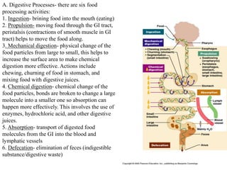
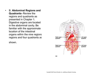
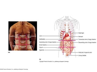
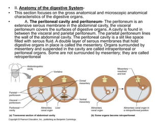
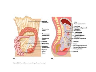
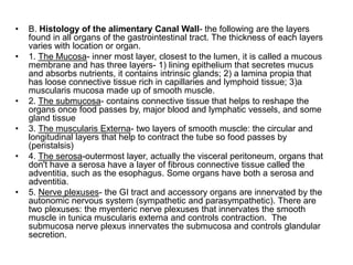
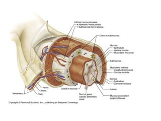
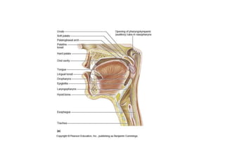
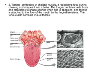
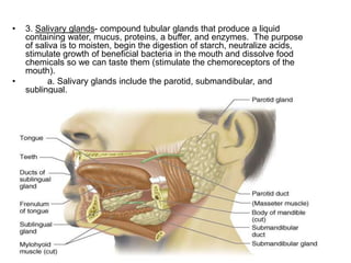
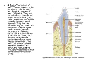
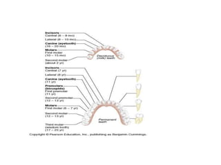
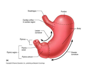
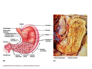
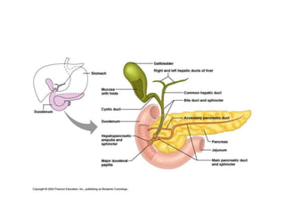
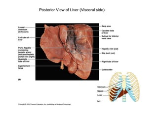
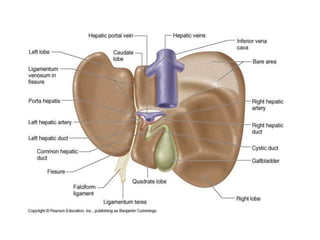
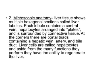
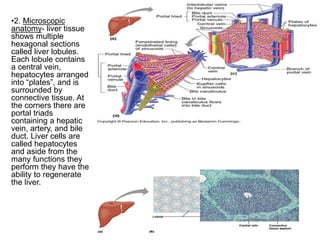
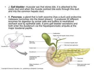
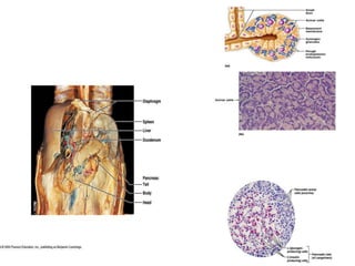
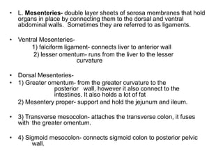
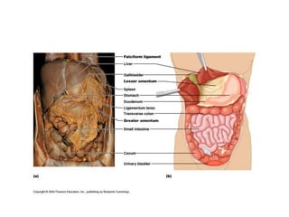
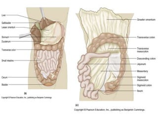
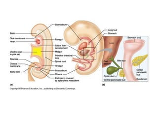

Chapter 22 of human anatomy discusses the digestive system, detailing its organs which are categorized as part of the gastrointestinal tract or accessory organs, and outlines the six digestive processes: ingestion, propulsion, mechanical digestion, chemical digestion, absorption, and defecation. It covers the anatomical and histological structures of the digestive organs, including the mouth, esophagus, stomach, small intestine, large intestine, liver, gallbladder, pancreas, and associated mesenteries. The chapter emphasizes the relationships among different components of the digestive system and their roles in digestion and absorption.


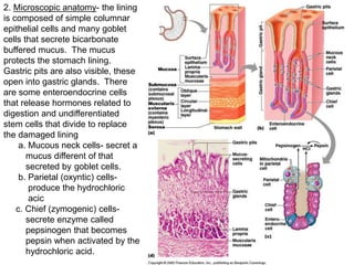
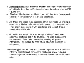
![CTEV [ clubfoot] DR ARUN LAL ,DR MOHAMED ASHRAF travancore medical college k...](https://cdn.slidesharecdn.com/ss_thumbnails/ctevclubfootdrarunlaldrmohamedashraftravancoremedicalcollegekollamkeralaindia-260208063247-18fc466c-thumbnail.jpg?width=640&height=640&fit=bounds)


![ONFH[AVN HIP] -TRIPLE REGIME -A NOVAL SURGICAL CONCEPT .pptx](https://cdn.slidesharecdn.com/ss_thumbnails/onfhavnhip2026koaconcalicutdrgokuldevdrmashraf-260210064517-213ec005-thumbnail.jpg?width=640&height=640&fit=bounds)


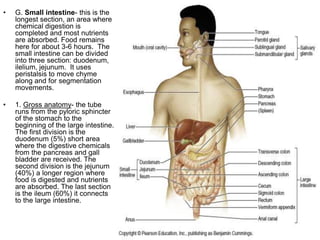
![PERI-PROSTHETIC FRACTURE NAIL-PLATE CONSTRUCT [NPC].pptx](https://cdn.slidesharecdn.com/ss_thumbnails/drarunkumardrmohamedashrafperiprostheticfrasturenail-plateconstructnpc-260209164459-7e9d15a1-thumbnail.jpg?width=640&height=640&fit=bounds)

