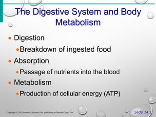
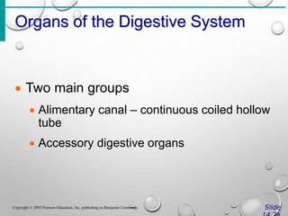
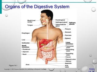
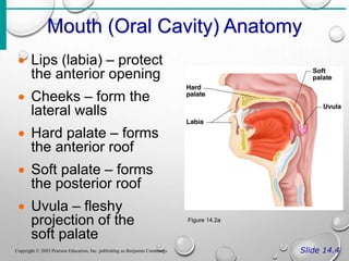
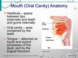
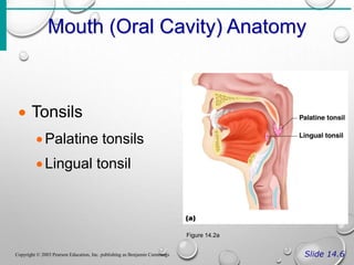
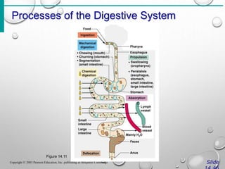
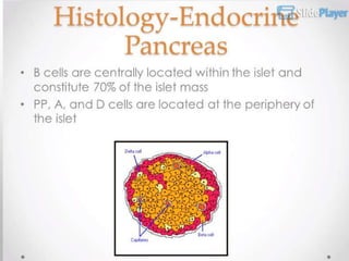
DIGESTIVE SYSTEM
AP 1.2 K. LAGAT
2022
The Digestive System and Body
Metabolism
Slide 14.1
Copyright © 2003 Pearson Education, Inc. publishing as Beamin Cings
 Digestion
Breakdown of ingested food
 Absorption
Passage of nutrients into the blood
 Metabolism
Production of cellular energy (ATP)
Organs of the Digestive System
Slide
Copyright © 2003 Pearson Education, Inc. publishing as Benjamin Cummings
 Two main groups
 Alimentary canal – continuous coiled hollow
tube
 Accessory digestive organs
Organs of the Digestive System
Slide
Copyright © 2003 Pearson Education, Inc. publishing as Benjamin Cummings
Figure 14.1
Organs of the Alimentary Canal
Slide 14.3
Copyright © 2003 Pearson Education, Inc. publishing as Benjamin Cummings
 Mouth
 Pharynx
 Esophagus
 Stomach
 Small intestine
 Large intestine
 Anus
Mouth (Oral Cavity) Anatomy
Slide 14.4
Copyright © 2003 Pearson Education, Inc. publishing as Benjamin Cummings
 Lips (labia) – protect
the anterior opening
 Cheeks – form the
lateral walls
 Hard palate – forms
the anterior roof
 Soft palate – forms
the posterior roof
 Uvula – fleshy
projection of the
soft palate
Figure 14.2a
Mouth (Oral Cavity) Anatomy
Slide 14.5
Copyright © 2003 Pearson Education, Inc. publishing as Benjamin Cummings
 Vestibule – space
between lips
externally and teeth
and gums internally
 Oral cavity – area
contained by the
teeth
 Tongue – attached at
hyoid and styloid
processes of the
skull, and by the
lingual frenulum Figure 14.2a
Mouth (Oral Cavity) Anatomy
Slide 14.6
Copyright © 2003 Pearson Education, Inc. publishing as Benjamin Cummings
 Tonsils
Palatine tonsils
Lingual tonsil
Figure 14.2a
Processes of the Mouth
Slide 14.7
Copyright © 2003 Pearson Education, Inc. publishing as Benjamin Cummings
 Mastication (chewing) of food
 Mixing masticated food with saliva
 Initiation of swallowing by the tongue
 Allowing for the sense of taste
Pharynx Anatomy
Slide 14.8
Copyright © 2003 Pearson Education, Inc. publishing as Benjamin Cummings
 Nasopharynx –
not part of the
digestive system
 Oropharynx –
posterior to oral
cavity
 Laryngopharynx –
below the oropharynx
and connected to
the esophagus Figure 14.2a
Pharynx Function
Slide 14.9
Copyright © 2003 Pearson Education, Inc. publishing as Benjamin Cummings
 Serves as a passageway for air and
food
 Food is propelled to the esophagus by
two muscle layers
circular inner layer
Lonitudinal outer layer
 Food movement is by alternating
contractions of the muscle layers
(peristalsis)
Esophagus
Slide
Copyright © 2003 Pearson Education, Inc. publishing as Benjamin Cummings
 Runs from pharynx to stomach through
the diaphragm
 Conducts food by peristalsis
(slow rhythmic squeezing)
 Passageway for food only (respiratory
system branches off after the pharynx)
Layers of Alimentary Canal Organs
Slide
Copyright © 2003 Pearson Education, Inc. publishing as Benjamin Cummings
 Mucosa
Innermost layer
Moist membrane
Surface epithelium
Small amount of connective tissue
(lamina propria)
Small smooth muscle layer
Layers of Alimentary Canal Organs
Slide
Copyright © 2003 Pearson Education, Inc. publishing as Benjamin Cummings
 Submucosa
Just beneath the mucosa
Soft connective tissue with blood vessels,
nerve endings, and lymphatics
Layers of Alimentary Canal Organs
Slide
Copyright © 2003 Pearson Education, Inc. publishing as Benjamin Cummings
 Muscularis externa – smooth muscle
Inner circular layer
Outer longitudinal layer
 Serosa
Outermost layer – visceral peritoneum
Layer of serous fluid-producing cells
Layers of Alimentary Canal Organs
Slide
Copyright © 2003 Pearson Education, Inc. publishing as Benjamin Cummings
Figure 14.3
PERITONEUM
• the peritoneum is the largest serous membrane of the body
• it is a single membrane, forming a closed sac and containing a
small amount of serous fluid. within the abdominal cavity. it is
richly supplied with blood and lymph vessels, and contains many
lymph nodes.
• it provides a physical barrier to local spread of infection and can
isolate an infective focus such as appendicitis, preventing
involvement of other abdominal structures. it has two layers:
1. the parietal peritoneum, which lines the abdominal wall
2. the visceral peritoneum, which covers the organs
• (viscera) within the abdominal and pelvic cavities.
.
• the parietal peritoneum lines the anterior
abdominal wall. the two layers of
peritoneum are in close contact, and
friction between them is prevented by the
presence of peritoneal fluid, a serous
fluid secreted by the peritoneal cells; the
peritoneal cavity is only a potential cavity.
the abdominal organs are covered to
varying degrees by the loops and folds of
the visceral peritoneum, which attaches
them firmly to the abdominal wall.
if the organ is almost completely covered by
the visceral peritoneum, it is said to be
intraperitoneal. if the organ is covered on
its anterior side only, it is said to be
retroperitoneal (meaning behind the
peritoneum). This means that:
• pelvic organs are covered only on their
superior surface.
• the stomach and intestines are almost
completely
surrounded by peritoneum and have a
double fold (the
mesentery) that attaches them to the
posterior abdominal.
.
Stomach Anatomy
Slide
Copyright © 2003 Pearson Education, Inc. publishing as Benjamin Cummings
 Located on the left side of the
abdominal cavity
 Food enters at the cardioesophageal
sphincter
Stomach Anatomy
Slide
Copyright © 2003 Pearson Education, Inc. publishing as Benjamin Cummings
 Regions of the stomach
Cardiac region – near the heart
Fundus
Body
Phylorus – funnel-shaped terminal end
 Food empties into the small intestine at
the pyloric sphincter
Stomach Anatomy
Slide
Copyright © 2003 Pearson Education, Inc. publishing as Benjamin Cummings
 Rugae – internal folds of the mucosa
 External regions
Lesser curvature
Greater curvature
Stomach Anatomy
Slide
Copyright © 2003 Pearson Education, Inc. publishing as Benjamin Cummings
 Layers of peritoneum attached to the
stomach
Lesser omentum – attaches the liver to the
lesser curvature
Greater omentum – attaches the greater
curvature to the posterior body wall
Contains fat to insulate, cushion, and
protect abdominal organs
Stomach Anatomy
Slide
Copyright © 2003 Pearson Education, Inc. publishing as Benjamin Cummings
Figure 14.4a
Stomach Functions
Slide
Copyright © 2003 Pearson Education, Inc. publishing as Benjamin Cummings
 Acts as a storage tank for food
 Site of food breakdown
 Chemical breakdown of protein begins
 Delivers chyme (processed food) to the
small intestine
Specialized Mucosa of the
Stomach
Slide
Copyright © 2003 Pearson Education, Inc. publishing as Benjamin Cummings
 Simple columnar epithelium
Mucous neck cells – produce a sticky
alkaline mucus
Gastric glands – secrete gastric juice
Chief cells – produce protein-digesting
enzymes (pepsinogens)
Parietal cells – produce hydrochloric acid
Endocrine cells – produce gastrin
Structure of the Stomach Mucosa
Slide
Copyright © 2003 Pearson Education, Inc. publishing as Benjamin Cummings
 Gastric pits formed by folded mucosa
 Glands and specialized cells are in the
gastric gland region
Structure of the Stomach Mucosa
Slide
Copyright © 2003 Pearson Education, Inc. publishing as Benjamin Cummings
Figure 14.4b, c
.
• .
.
.
• .
.
.
.
Small Intestine
Slide
Copyright © 2003 Pearson Education, Inc. publishing as Benjamin Cummings
 The body’s major digestive organ
 Site of nutrient absorption into the blood
 Muscular tube extending form the
pyloric sphincter to the ileocecal valve
 Suspended from the posterior
abdominal wall by the mesentery
Subdivisions of the Small Intestine
Slide
Copyright © 2003 Pearson Education, Inc. publishing as Benjamin Cummings
 Duodenum
Attached to the stomach
Curves around the head of the pancreas
 Jejunum
Attaches anteriorly to the duodenum
 Ileum
Extends from jejunum to large intestine
Chemical Digestion in the Small
Intestine
Slide
Copyright © 2003 Pearson Education, Inc. publishing as Benjamin Cummings
 Source of enzymes that are mixed with
chyme
Intestinal cells
Pancreas
 Bile enters from the gall bladder
Villi of the Small Intestine
Slide
Copyright © 2003 Pearson Education, Inc. publishing as Benjamin Cummings
 Fingerlike
structures formed
by the mucosa
 Give the small
intestine more
surface area
Figure 14.7a
Microvilli of the Small Intestine
Slide
Copyright © 2003 Pearson Education, Inc. publishing as Benjamin Cummings
 Small projections of the
plasma membrane
 Found on absorptive cells
Figure 14.7c
Structures Involved in Absorption of
Nutrients
Slide
Copyright © 2003 Pearson Education, Inc. publishing as Benjamin Cummings
 Absorptive cells
 Blood capillaries
 Lacteals (specialized
lymphatic capillaries)
Figure 14.7b
Folds of the Small Intestine
Slide
Copyright © 2003 Pearson Education, Inc. publishing as Benjamin Cummings
 Called circular folds or plicae circulares
 Deep folds of the mucosa and
submucosa
 Do not disappear when filled with food
 The submucosa has Peyer’s patches
(collections of lymphatic tissue)
Digestion in the Small Intestine
Slide
Copyright © 2003 Pearson Education, Inc. publishing as Benjamin Cummings
 Enzymes from the brush border
Break double sugars into simple sugars
Complete some protein digestion
 Pancreatic enzymes play the major
digestive function
Help complete digestion of starch
(pancreatic amylase)
Carry out about half of all protein digestion
(trypsin, etc.)
Digestion in the Small Intestine
Slide
Copyright © 2003 Pearson Education, Inc. publishing as Benjamin Cummings
 Pancreatic enzymes play the major
digestive function (continued)
Responsible for fat digestion (lipase)
Digest nucleic acids (nucleases)
Alkaline content neutralizes acidic chyme
Absorption in the Small Intestine
Slide
Copyright © 2003 Pearson Education, Inc. publishing as Benjamin Cummings
 Water is absorbed along the length of
the small intestine
 End products of digestion
Most substances are absorbed by active
transport through cell membranes
Lipids are absorbed by diffusion
 Substances are transported to the liver
by the hepatic portal vein or lymph
Propulsion in the Small Intestine
Slide
Copyright © 2003 Pearson Education, Inc. publishing as Benjamin Cummings
 Peristalsis is the major means of
moving food
 Segmental movements
Mix chyme with digestive juices
Aid in propelling food
Large Intestine
Slide
Copyright © 2003 Pearson Education, Inc. publishing as Benjamin Cummings
 Larger in diameter, but shorter than the
small intestine
 Frames the internal abdomen
Large Intestine
Slide
Copyright © 2003 Pearson Education, Inc. publishing as Benjamin Cummings
Figure 14.8
Functions of the Large Intestine
Slide
Copyright © 2003 Pearson Education, Inc. publishing as Benjamin Cummings
 Absorption of water
 Eliminates indigestible food from the
body as feces
 Does not participate in digestion of food
 Goblet cells produce mucus to act as a
lubricant
Structures of the Large Intestine
Slide
Copyright © 2003 Pearson Education, Inc. publishing as Benjamin Cummings
 Cecum – saclike first part of the large
intestine
 Appendix
Accumulation of lymphatic tissue that
sometimes becomes inflamed
(appendicitis)
Hangs from the cecum
Structures of the Large Intestine
Slide
Copyright © 2003 Pearson Education, Inc. publishing as Benjamin Cummings
 Colon
Ascending
Transverse
Descending
S-shaped sigmoidal
 Rectum
 Anus – external body opening
Structures of the Large Intestine
Slide
Copyright © 2003 Pearson Education, Inc. publishing as Benjamin Cummings
 Colon
Ascending
Transverse
Descending
S-shaped sigmoidal
 Rectum
 Anus – external body opening
Food Breakdown and Absorption in
the Large Intestine
Slide
Copyright © 2003 Pearson Education, Inc. publishing as Benjamin Cummings
 No digestive enzymes are produced
 Resident bacteria digest remaining
nutrients
Produce some vitamin K and B
Release gases
 Water and vitamins K and B are absorbed
 Remaining materials are eliminated via
feces
Propulsion in the Large Intestine
Slide
Copyright © 2003 Pearson Education, Inc. publishing as Benjamin Cummings
 Sluggish peristalsis
 Mass movements
Slow, powerful movements
Occur three to four times per day
 Presence of feces in the rectum causes
a defecation reflex
Internal anal sphincter is relaxed
Defecation occurs with relaxation of the
voluntary (external) anal sphincter
Accessory Digestive Organs
Slide
Copyright © 2003 Pearson Education, Inc. publishing as Benjamin Cummings
 Salivary glands
 Teeth
 Pancreas
 Liver
 Gall bladder
Salivary Glands
Slide
Copyright © 2003 Pearson Education, Inc. publishing as Benjamin Cummings
 Saliva-producing glands
Parotid glands – located anterior to ears
Submandibular glands
Sublingual glands
.
Saliva
Slide
Copyright © 2003 Pearson Education, Inc. publishing as Benjamin Cummings
 Mixture of mucus and serous fluids
 Helps to form a food bolus
 Contains salivary amylase to begin
starch digestion
 Dissolves chemicals so they can be
tasted
Teeth
Slide
Copyright © 2003 Pearson Education, Inc. publishing as Benjamin Cummings
 The role is to masticate (chew) food
 Humans have two sets of teeth
Deciduous (baby or milk) teeth
20 teeth are fully formed by age two
Teeth
Slide
Copyright © 2003 Pearson Education, Inc. publishing as Benjamin Cummings
 Permanent teeth
Replace deciduous teeth beginning
between the ages of 6 to 12
A full set is 32 teeth, but some people do
not have wisdom teeth
Regions of a Tooth
Slide
Copyright © 2003 Pearson Education, Inc. publishing as Benjamin Cummings
 Crown – exposed
part
 Outer enamel
 Dentin
 Pulp cavity
 Neck
 Region in contact
with the gum
 Connects crown to
root
Figure 14.10
Regions of a Tooth
Slide
Copyright © 2003 Pearson Education, Inc. publishing as Benjamin Cummings
 Root
Periodontal
membrane
attached to the
bone
Root canal carrying
blood vessels and
nerves
Figure 14.10
Liver
Slide
Copyright © 2003 Pearson Education, Inc. publishing as Benjamin Cummings
 Largest gland in the body about 1.5kg
 Located on the right side of the body
under the diaphragm
 Consists of four lobes suspended from
the diaphragm and abdominal wall by
the falciform ligament
 Connected to the gall bladder via the
common hepatic duct
LIVER
Role of the Liver in Metabolism
Slide
Copyright © 2003 Pearson Education, Inc. publishing as Benjamin Cummings
 Several roles in digestion
 Detoxifies drugs and alcohol
 Degrades hormones
 Produce cholesterol, blood proteins
(albumin and clotting proteins)
 Plays a central role in metabolism
Bile
Slide
Copyright © 2003 Pearson Education, Inc. publishing as Benjamin Cummings
 Produced by cells in the liver
 Composition
Bile salts
Bile pigment (mostly bilirubin from the
breakdown of hemoglobin)
Cholesterol
Phospholipids
Electrolytes
BILIRUBIN METABOLISM
• FORMED BY BREAKDOWN OF HEME (80% FROM HEMOGLOBIN,
20% FROM OTHER HEMOPROTEINS)
• HEME –VIA HEME OXYGENASE—BILIVERDIN---VIA BILIVERDIN
REDUCTASE---BILIRUBIN IX ALPHA
• HEME OXYGENASE IS RATE-LIMITING STEP IN BILIRUBIN
PRODUCTION
• HEME OXYGENASE IS FOUND IN KUPFFER CELLS OF THE LIVER
AND RETICULOENDOTHELIAL CELLS OF THE SPLEEN
BILIRUBIN METABOLISM
• ALBUMIN BINDS TO BILIRUBIN, REVERSIBLE EXCEPT IN
STATES OF BILIRUBIN OBSTRUCTION/CONJUGATED
BILIRUBINEMIA
• ALBUMIN-BILIRUBIN COMPLEX DISSOCIATES IN LIVER
SINUSOIDS WHERE BILIRUBIN IS TAKEN UP BY
HEPATOCYTES
• THIS IS VIA FACILITATES DIFFUSION, BIDIRECTIONAL
• DEFECTS IN TRANSPORTERS IN THESE STEPS CAUSE
HYPERBILIRUBINEMIA (EG GILBERT’S)
• UNCONJUGATED HYPERBILIRUBINEMIA ALSO RESULTS
FROM CIRRHOSIS WHEN BILIRUBIN PRODUCED FROM THE
SPLEEN BYPASSES THE LIVER VIA PORTOSYSTEMIC
COLLATERALS
BILIRUBIN CONJUGATION
• BILIRUBIN POORLY WATER SOLUBLE BECAUSE OF INTERNAL
HYDROGEN BONDING WHICH MAKES IT TOXIC AND
PREVENTS ITS ELIMINATION
• GLUCURONIC ACID CONJUGATION OF BILIRUBIN MAKES IT
WATER-SOLUBLE AND EXCRETABLE INTO BILE
• PHOTOTHERAPY IN NEONATAL JAUNDICE PRODUCES
CONFIGURATIONAL AND STRUCTURAL BILIRUBIN
PHOTOISOMERS, EXCRETED INTO BILE WITHOUT FURTHER
METABOLISM
BILIRUBIN EXCRETION
• CONJUGATED BILIRUBIN IS EXCRETED IN BILE ACROSS THE
BILE CANALICULAR MEMBRANE VIA ACTIVE TRANSPORT
• 4 TYPES OF TRANSPORTERS (EG MRP2, ABCC2)
• EXCRETION IMPAIRED BY VIRAL HEPATITIS, CHOLESTASIS OF
PREGNANCY, DUBIN-JOHNSON AND ROTOR SYNDROME
BILIRUBIN DEGRADATION
• 98% OF THE BILE PIGMENT IN BILE IS CONJUGATED
AND IS WATER-SOLUBLE AND WILL NOT BE
ABSORBED ACROSS LIPID MEMBRANE OF SMALL
INTESTINAL EPITHELIUM
• UNCONJUGATED FRACTION IS PARTIALLY
REABSORBED THROUGH ENTEROHEPATIC
CIRCULATION
• BILIRUBIN IS REDUCED BY BACTERIAL ENZYMES IN
THE COLON TO UROBILINOIDS (UROBILINOGEN AND
STERCOBILINOGEN)
• INTESTINAL MICROFLORA INFLUENCE SERUM
BILIRUBIN LEVELS AND ANTIBIOTIC USE CAN
INCREASE SERUM BILIRUBIN LEVELS
From Trowers and Tischler Gastrointestinal Physiology 2014 in SpringerImages database (non-commercial use permitted)
Processes of the Digestive System
Slide
Copyright © 2003 Pearson Education, Inc. publishing as Benjamin Cummings
 Ingestion – getting food into the mouth
 Propulsion – moving foods from one
region of the digestive system to
another
Processes of the Digestive System
Slide
Copyright © 2003 Pearson Education, Inc. publishing as Benjamin Cummings
Peristalsis – alternating
waves of contraction
Segmentation – moving
materials back and forth
to aid in mixing
Figure 14.12
Processes of the Digestive System
Slide
Copyright © 2003 Pearson Education, Inc. publishing as Benjamin Cummings
 Mechanical digestion
Mixing of food in the mouth by the tongue
Churning of food in the stomach
Segmentation in the small intestine
Processes of the Digestive System
Slide
Copyright © 2003 Pearson Education, Inc. publishing as Benjamin Cummings
 Chemical Digestion
Enzymes break down food molecules into
their building blocks
Each major food group uses different
enzymes
Carbohydrates are broken to simple sugars
Proteins are broken to amino acids
Fats are broken to fatty acids and alcohols
Processes of the Digestive System
Slide
Copyright © 2003 Pearson Education, Inc. publishing as Benjamin Cummings
 Absorption
End products of digestion are absorbed in
the blood or lymph
Food must enter mucosal cells and then
into blood or lymph capillaries
 Defecation
Elimination of indigestible substances as
feces
Processes of the Digestive System
Slide
Copyright © 2003 Pearson Education, Inc. publishing as Benjamin Cummings
Figure 14.11
Control of Digestive Activity
Slide
Copyright © 2003 Pearson Education, Inc. publishing as Benjamin Cummings
 Mostly controlled by reflexes via the
parasympathetic division
 Chemical and mechanical receptors are
located in organ walls that trigger
reflexes
Control of Digestive Activity
Slide
Copyright © 2003 Pearson Education, Inc. publishing as Benjamin Cummings
 Stimuli include:
Stretch of the organ
pH of the contents
Presence of breakdown products
 Reflexes include:
Activation or inhibition of glandular
secretions
Smooth muscle activity
PANCREASE
ANATOMY AND PHYSIOLOGY
.
.
.
.
.
.
.
.
.
.
.
.
.
.
.
.
.
.
.
.
.
,
.
.
.
BILLIARY SYSTEM
.
.
.
.
.
.
.
.
.
END

AP DIGESTIVE SYSTEM-1.ppt Alimentary organ

  • 1.
  • 2.
    The Digestive Systemand Body Metabolism Slide 14.1 Copyright © 2003 Pearson Education, Inc. publishing as Beamin Cings  Digestion Breakdown of ingested food  Absorption Passage of nutrients into the blood  Metabolism Production of cellular energy (ATP)
  • 3.
    Organs of theDigestive System Slide Copyright © 2003 Pearson Education, Inc. publishing as Benjamin Cummings  Two main groups  Alimentary canal – continuous coiled hollow tube  Accessory digestive organs
  • 4.
    Organs of theDigestive System Slide Copyright © 2003 Pearson Education, Inc. publishing as Benjamin Cummings Figure 14.1
  • 5.
    Organs of theAlimentary Canal Slide 14.3 Copyright © 2003 Pearson Education, Inc. publishing as Benjamin Cummings  Mouth  Pharynx  Esophagus  Stomach  Small intestine  Large intestine  Anus
  • 6.
    Mouth (Oral Cavity)Anatomy Slide 14.4 Copyright © 2003 Pearson Education, Inc. publishing as Benjamin Cummings  Lips (labia) – protect the anterior opening  Cheeks – form the lateral walls  Hard palate – forms the anterior roof  Soft palate – forms the posterior roof  Uvula – fleshy projection of the soft palate Figure 14.2a
  • 7.
    Mouth (Oral Cavity)Anatomy Slide 14.5 Copyright © 2003 Pearson Education, Inc. publishing as Benjamin Cummings  Vestibule – space between lips externally and teeth and gums internally  Oral cavity – area contained by the teeth  Tongue – attached at hyoid and styloid processes of the skull, and by the lingual frenulum Figure 14.2a
  • 8.
    Mouth (Oral Cavity)Anatomy Slide 14.6 Copyright © 2003 Pearson Education, Inc. publishing as Benjamin Cummings  Tonsils Palatine tonsils Lingual tonsil Figure 14.2a
  • 9.
    Processes of theMouth Slide 14.7 Copyright © 2003 Pearson Education, Inc. publishing as Benjamin Cummings  Mastication (chewing) of food  Mixing masticated food with saliva  Initiation of swallowing by the tongue  Allowing for the sense of taste
  • 10.
    Pharynx Anatomy Slide 14.8 Copyright© 2003 Pearson Education, Inc. publishing as Benjamin Cummings  Nasopharynx – not part of the digestive system  Oropharynx – posterior to oral cavity  Laryngopharynx – below the oropharynx and connected to the esophagus Figure 14.2a
  • 11.
    Pharynx Function Slide 14.9 Copyright© 2003 Pearson Education, Inc. publishing as Benjamin Cummings  Serves as a passageway for air and food  Food is propelled to the esophagus by two muscle layers circular inner layer Lonitudinal outer layer  Food movement is by alternating contractions of the muscle layers (peristalsis)
  • 12.
    Esophagus Slide Copyright © 2003Pearson Education, Inc. publishing as Benjamin Cummings  Runs from pharynx to stomach through the diaphragm  Conducts food by peristalsis (slow rhythmic squeezing)  Passageway for food only (respiratory system branches off after the pharynx)
  • 13.
    Layers of AlimentaryCanal Organs Slide Copyright © 2003 Pearson Education, Inc. publishing as Benjamin Cummings  Mucosa Innermost layer Moist membrane Surface epithelium Small amount of connective tissue (lamina propria) Small smooth muscle layer
  • 14.
    Layers of AlimentaryCanal Organs Slide Copyright © 2003 Pearson Education, Inc. publishing as Benjamin Cummings  Submucosa Just beneath the mucosa Soft connective tissue with blood vessels, nerve endings, and lymphatics
  • 15.
    Layers of AlimentaryCanal Organs Slide Copyright © 2003 Pearson Education, Inc. publishing as Benjamin Cummings  Muscularis externa – smooth muscle Inner circular layer Outer longitudinal layer  Serosa Outermost layer – visceral peritoneum Layer of serous fluid-producing cells
  • 16.
    Layers of AlimentaryCanal Organs Slide Copyright © 2003 Pearson Education, Inc. publishing as Benjamin Cummings Figure 14.3
  • 17.
    PERITONEUM • the peritoneumis the largest serous membrane of the body • it is a single membrane, forming a closed sac and containing a small amount of serous fluid. within the abdominal cavity. it is richly supplied with blood and lymph vessels, and contains many lymph nodes. • it provides a physical barrier to local spread of infection and can isolate an infective focus such as appendicitis, preventing involvement of other abdominal structures. it has two layers: 1. the parietal peritoneum, which lines the abdominal wall 2. the visceral peritoneum, which covers the organs • (viscera) within the abdominal and pelvic cavities.
  • 18.
    . • the parietalperitoneum lines the anterior abdominal wall. the two layers of peritoneum are in close contact, and friction between them is prevented by the presence of peritoneal fluid, a serous fluid secreted by the peritoneal cells; the peritoneal cavity is only a potential cavity. the abdominal organs are covered to varying degrees by the loops and folds of the visceral peritoneum, which attaches them firmly to the abdominal wall.
  • 19.
    if the organis almost completely covered by the visceral peritoneum, it is said to be intraperitoneal. if the organ is covered on its anterior side only, it is said to be retroperitoneal (meaning behind the peritoneum). This means that: • pelvic organs are covered only on their superior surface. • the stomach and intestines are almost completely surrounded by peritoneum and have a double fold (the mesentery) that attaches them to the posterior abdominal.
  • 20.
  • 21.
    Stomach Anatomy Slide Copyright ©2003 Pearson Education, Inc. publishing as Benjamin Cummings  Located on the left side of the abdominal cavity  Food enters at the cardioesophageal sphincter
  • 22.
    Stomach Anatomy Slide Copyright ©2003 Pearson Education, Inc. publishing as Benjamin Cummings  Regions of the stomach Cardiac region – near the heart Fundus Body Phylorus – funnel-shaped terminal end  Food empties into the small intestine at the pyloric sphincter
  • 23.
    Stomach Anatomy Slide Copyright ©2003 Pearson Education, Inc. publishing as Benjamin Cummings  Rugae – internal folds of the mucosa  External regions Lesser curvature Greater curvature
  • 24.
    Stomach Anatomy Slide Copyright ©2003 Pearson Education, Inc. publishing as Benjamin Cummings  Layers of peritoneum attached to the stomach Lesser omentum – attaches the liver to the lesser curvature Greater omentum – attaches the greater curvature to the posterior body wall Contains fat to insulate, cushion, and protect abdominal organs
  • 25.
    Stomach Anatomy Slide Copyright ©2003 Pearson Education, Inc. publishing as Benjamin Cummings Figure 14.4a
  • 26.
    Stomach Functions Slide Copyright ©2003 Pearson Education, Inc. publishing as Benjamin Cummings  Acts as a storage tank for food  Site of food breakdown  Chemical breakdown of protein begins  Delivers chyme (processed food) to the small intestine
  • 27.
    Specialized Mucosa ofthe Stomach Slide Copyright © 2003 Pearson Education, Inc. publishing as Benjamin Cummings  Simple columnar epithelium Mucous neck cells – produce a sticky alkaline mucus Gastric glands – secrete gastric juice Chief cells – produce protein-digesting enzymes (pepsinogens) Parietal cells – produce hydrochloric acid Endocrine cells – produce gastrin
  • 28.
    Structure of theStomach Mucosa Slide Copyright © 2003 Pearson Education, Inc. publishing as Benjamin Cummings  Gastric pits formed by folded mucosa  Glands and specialized cells are in the gastric gland region
  • 29.
    Structure of theStomach Mucosa Slide Copyright © 2003 Pearson Education, Inc. publishing as Benjamin Cummings Figure 14.4b, c
  • 30.
  • 31.
  • 32.
  • 33.
  • 34.
  • 35.
  • 36.
    Small Intestine Slide Copyright ©2003 Pearson Education, Inc. publishing as Benjamin Cummings  The body’s major digestive organ  Site of nutrient absorption into the blood  Muscular tube extending form the pyloric sphincter to the ileocecal valve  Suspended from the posterior abdominal wall by the mesentery
  • 37.
    Subdivisions of theSmall Intestine Slide Copyright © 2003 Pearson Education, Inc. publishing as Benjamin Cummings  Duodenum Attached to the stomach Curves around the head of the pancreas  Jejunum Attaches anteriorly to the duodenum  Ileum Extends from jejunum to large intestine
  • 38.
    Chemical Digestion inthe Small Intestine Slide Copyright © 2003 Pearson Education, Inc. publishing as Benjamin Cummings  Source of enzymes that are mixed with chyme Intestinal cells Pancreas  Bile enters from the gall bladder
  • 39.
    Villi of theSmall Intestine Slide Copyright © 2003 Pearson Education, Inc. publishing as Benjamin Cummings  Fingerlike structures formed by the mucosa  Give the small intestine more surface area Figure 14.7a
  • 40.
    Microvilli of theSmall Intestine Slide Copyright © 2003 Pearson Education, Inc. publishing as Benjamin Cummings  Small projections of the plasma membrane  Found on absorptive cells Figure 14.7c
  • 41.
    Structures Involved inAbsorption of Nutrients Slide Copyright © 2003 Pearson Education, Inc. publishing as Benjamin Cummings  Absorptive cells  Blood capillaries  Lacteals (specialized lymphatic capillaries) Figure 14.7b
  • 42.
    Folds of theSmall Intestine Slide Copyright © 2003 Pearson Education, Inc. publishing as Benjamin Cummings  Called circular folds or plicae circulares  Deep folds of the mucosa and submucosa  Do not disappear when filled with food  The submucosa has Peyer’s patches (collections of lymphatic tissue)
  • 43.
    Digestion in theSmall Intestine Slide Copyright © 2003 Pearson Education, Inc. publishing as Benjamin Cummings  Enzymes from the brush border Break double sugars into simple sugars Complete some protein digestion  Pancreatic enzymes play the major digestive function Help complete digestion of starch (pancreatic amylase) Carry out about half of all protein digestion (trypsin, etc.)
  • 44.
    Digestion in theSmall Intestine Slide Copyright © 2003 Pearson Education, Inc. publishing as Benjamin Cummings  Pancreatic enzymes play the major digestive function (continued) Responsible for fat digestion (lipase) Digest nucleic acids (nucleases) Alkaline content neutralizes acidic chyme
  • 45.
    Absorption in theSmall Intestine Slide Copyright © 2003 Pearson Education, Inc. publishing as Benjamin Cummings  Water is absorbed along the length of the small intestine  End products of digestion Most substances are absorbed by active transport through cell membranes Lipids are absorbed by diffusion  Substances are transported to the liver by the hepatic portal vein or lymph
  • 46.
    Propulsion in theSmall Intestine Slide Copyright © 2003 Pearson Education, Inc. publishing as Benjamin Cummings  Peristalsis is the major means of moving food  Segmental movements Mix chyme with digestive juices Aid in propelling food
  • 47.
    Large Intestine Slide Copyright ©2003 Pearson Education, Inc. publishing as Benjamin Cummings  Larger in diameter, but shorter than the small intestine  Frames the internal abdomen
  • 48.
    Large Intestine Slide Copyright ©2003 Pearson Education, Inc. publishing as Benjamin Cummings Figure 14.8
  • 49.
    Functions of theLarge Intestine Slide Copyright © 2003 Pearson Education, Inc. publishing as Benjamin Cummings  Absorption of water  Eliminates indigestible food from the body as feces  Does not participate in digestion of food  Goblet cells produce mucus to act as a lubricant
  • 50.
    Structures of theLarge Intestine Slide Copyright © 2003 Pearson Education, Inc. publishing as Benjamin Cummings  Cecum – saclike first part of the large intestine  Appendix Accumulation of lymphatic tissue that sometimes becomes inflamed (appendicitis) Hangs from the cecum
  • 51.
    Structures of theLarge Intestine Slide Copyright © 2003 Pearson Education, Inc. publishing as Benjamin Cummings  Colon Ascending Transverse Descending S-shaped sigmoidal  Rectum  Anus – external body opening
  • 52.
    Structures of theLarge Intestine Slide Copyright © 2003 Pearson Education, Inc. publishing as Benjamin Cummings  Colon Ascending Transverse Descending S-shaped sigmoidal  Rectum  Anus – external body opening
  • 53.
    Food Breakdown andAbsorption in the Large Intestine Slide Copyright © 2003 Pearson Education, Inc. publishing as Benjamin Cummings  No digestive enzymes are produced  Resident bacteria digest remaining nutrients Produce some vitamin K and B Release gases  Water and vitamins K and B are absorbed  Remaining materials are eliminated via feces
  • 54.
    Propulsion in theLarge Intestine Slide Copyright © 2003 Pearson Education, Inc. publishing as Benjamin Cummings  Sluggish peristalsis  Mass movements Slow, powerful movements Occur three to four times per day  Presence of feces in the rectum causes a defecation reflex Internal anal sphincter is relaxed Defecation occurs with relaxation of the voluntary (external) anal sphincter
  • 55.
    Accessory Digestive Organs Slide Copyright© 2003 Pearson Education, Inc. publishing as Benjamin Cummings  Salivary glands  Teeth  Pancreas  Liver  Gall bladder
  • 56.
    Salivary Glands Slide Copyright ©2003 Pearson Education, Inc. publishing as Benjamin Cummings  Saliva-producing glands Parotid glands – located anterior to ears Submandibular glands Sublingual glands
  • 57.
  • 58.
    Saliva Slide Copyright © 2003Pearson Education, Inc. publishing as Benjamin Cummings  Mixture of mucus and serous fluids  Helps to form a food bolus  Contains salivary amylase to begin starch digestion  Dissolves chemicals so they can be tasted
  • 59.
    Teeth Slide Copyright © 2003Pearson Education, Inc. publishing as Benjamin Cummings  The role is to masticate (chew) food  Humans have two sets of teeth Deciduous (baby or milk) teeth 20 teeth are fully formed by age two
  • 60.
    Teeth Slide Copyright © 2003Pearson Education, Inc. publishing as Benjamin Cummings  Permanent teeth Replace deciduous teeth beginning between the ages of 6 to 12 A full set is 32 teeth, but some people do not have wisdom teeth
  • 61.
    Regions of aTooth Slide Copyright © 2003 Pearson Education, Inc. publishing as Benjamin Cummings  Crown – exposed part  Outer enamel  Dentin  Pulp cavity  Neck  Region in contact with the gum  Connects crown to root Figure 14.10
  • 62.
    Regions of aTooth Slide Copyright © 2003 Pearson Education, Inc. publishing as Benjamin Cummings  Root Periodontal membrane attached to the bone Root canal carrying blood vessels and nerves Figure 14.10
  • 63.
    Liver Slide Copyright © 2003Pearson Education, Inc. publishing as Benjamin Cummings  Largest gland in the body about 1.5kg  Located on the right side of the body under the diaphragm  Consists of four lobes suspended from the diaphragm and abdominal wall by the falciform ligament  Connected to the gall bladder via the common hepatic duct
  • 64.
  • 65.
    Role of theLiver in Metabolism Slide Copyright © 2003 Pearson Education, Inc. publishing as Benjamin Cummings  Several roles in digestion  Detoxifies drugs and alcohol  Degrades hormones  Produce cholesterol, blood proteins (albumin and clotting proteins)  Plays a central role in metabolism
  • 66.
    Bile Slide Copyright © 2003Pearson Education, Inc. publishing as Benjamin Cummings  Produced by cells in the liver  Composition Bile salts Bile pigment (mostly bilirubin from the breakdown of hemoglobin) Cholesterol Phospholipids Electrolytes
  • 67.
    BILIRUBIN METABOLISM • FORMEDBY BREAKDOWN OF HEME (80% FROM HEMOGLOBIN, 20% FROM OTHER HEMOPROTEINS) • HEME –VIA HEME OXYGENASE—BILIVERDIN---VIA BILIVERDIN REDUCTASE---BILIRUBIN IX ALPHA • HEME OXYGENASE IS RATE-LIMITING STEP IN BILIRUBIN PRODUCTION • HEME OXYGENASE IS FOUND IN KUPFFER CELLS OF THE LIVER AND RETICULOENDOTHELIAL CELLS OF THE SPLEEN
  • 68.
    BILIRUBIN METABOLISM • ALBUMINBINDS TO BILIRUBIN, REVERSIBLE EXCEPT IN STATES OF BILIRUBIN OBSTRUCTION/CONJUGATED BILIRUBINEMIA • ALBUMIN-BILIRUBIN COMPLEX DISSOCIATES IN LIVER SINUSOIDS WHERE BILIRUBIN IS TAKEN UP BY HEPATOCYTES • THIS IS VIA FACILITATES DIFFUSION, BIDIRECTIONAL • DEFECTS IN TRANSPORTERS IN THESE STEPS CAUSE HYPERBILIRUBINEMIA (EG GILBERT’S) • UNCONJUGATED HYPERBILIRUBINEMIA ALSO RESULTS FROM CIRRHOSIS WHEN BILIRUBIN PRODUCED FROM THE SPLEEN BYPASSES THE LIVER VIA PORTOSYSTEMIC COLLATERALS
  • 69.
    BILIRUBIN CONJUGATION • BILIRUBINPOORLY WATER SOLUBLE BECAUSE OF INTERNAL HYDROGEN BONDING WHICH MAKES IT TOXIC AND PREVENTS ITS ELIMINATION • GLUCURONIC ACID CONJUGATION OF BILIRUBIN MAKES IT WATER-SOLUBLE AND EXCRETABLE INTO BILE • PHOTOTHERAPY IN NEONATAL JAUNDICE PRODUCES CONFIGURATIONAL AND STRUCTURAL BILIRUBIN PHOTOISOMERS, EXCRETED INTO BILE WITHOUT FURTHER METABOLISM
  • 70.
    BILIRUBIN EXCRETION • CONJUGATEDBILIRUBIN IS EXCRETED IN BILE ACROSS THE BILE CANALICULAR MEMBRANE VIA ACTIVE TRANSPORT • 4 TYPES OF TRANSPORTERS (EG MRP2, ABCC2) • EXCRETION IMPAIRED BY VIRAL HEPATITIS, CHOLESTASIS OF PREGNANCY, DUBIN-JOHNSON AND ROTOR SYNDROME
  • 71.
    BILIRUBIN DEGRADATION • 98%OF THE BILE PIGMENT IN BILE IS CONJUGATED AND IS WATER-SOLUBLE AND WILL NOT BE ABSORBED ACROSS LIPID MEMBRANE OF SMALL INTESTINAL EPITHELIUM • UNCONJUGATED FRACTION IS PARTIALLY REABSORBED THROUGH ENTEROHEPATIC CIRCULATION • BILIRUBIN IS REDUCED BY BACTERIAL ENZYMES IN THE COLON TO UROBILINOIDS (UROBILINOGEN AND STERCOBILINOGEN) • INTESTINAL MICROFLORA INFLUENCE SERUM BILIRUBIN LEVELS AND ANTIBIOTIC USE CAN INCREASE SERUM BILIRUBIN LEVELS
  • 72.
    From Trowers andTischler Gastrointestinal Physiology 2014 in SpringerImages database (non-commercial use permitted)
  • 73.
    Processes of theDigestive System Slide Copyright © 2003 Pearson Education, Inc. publishing as Benjamin Cummings  Ingestion – getting food into the mouth  Propulsion – moving foods from one region of the digestive system to another
  • 74.
    Processes of theDigestive System Slide Copyright © 2003 Pearson Education, Inc. publishing as Benjamin Cummings Peristalsis – alternating waves of contraction Segmentation – moving materials back and forth to aid in mixing Figure 14.12
  • 75.
    Processes of theDigestive System Slide Copyright © 2003 Pearson Education, Inc. publishing as Benjamin Cummings  Mechanical digestion Mixing of food in the mouth by the tongue Churning of food in the stomach Segmentation in the small intestine
  • 76.
    Processes of theDigestive System Slide Copyright © 2003 Pearson Education, Inc. publishing as Benjamin Cummings  Chemical Digestion Enzymes break down food molecules into their building blocks Each major food group uses different enzymes Carbohydrates are broken to simple sugars Proteins are broken to amino acids Fats are broken to fatty acids and alcohols
  • 77.
    Processes of theDigestive System Slide Copyright © 2003 Pearson Education, Inc. publishing as Benjamin Cummings  Absorption End products of digestion are absorbed in the blood or lymph Food must enter mucosal cells and then into blood or lymph capillaries  Defecation Elimination of indigestible substances as feces
  • 78.
    Processes of theDigestive System Slide Copyright © 2003 Pearson Education, Inc. publishing as Benjamin Cummings Figure 14.11
  • 79.
    Control of DigestiveActivity Slide Copyright © 2003 Pearson Education, Inc. publishing as Benjamin Cummings  Mostly controlled by reflexes via the parasympathetic division  Chemical and mechanical receptors are located in organ walls that trigger reflexes
  • 80.
    Control of DigestiveActivity Slide Copyright © 2003 Pearson Education, Inc. publishing as Benjamin Cummings  Stimuli include: Stretch of the organ pH of the contents Presence of breakdown products  Reflexes include: Activation or inhibition of glandular secretions Smooth muscle activity
  • 81.
  • 82.
  • 83.
  • 84.
  • 85.
  • 86.
  • 87.
  • 88.
  • 89.
  • 90.
  • 91.
  • 92.
  • 93.
  • 94.
  • 95.
  • 96.
  • 97.
  • 98.
  • 99.
  • 100.
  • 101.
  • 102.
  • 103.
  • 104.
  • 105.
  • 106.
  • 107.
  • 108.
  • 109.
  • 110.
  • 111.
  • 112.
  • 113.
  • 114.
  • 115.
  • 116.
  • 117.

Editor's Notes

  • #73 http://www.springerimages.com.ezp-prod1.hul.harvard.edu/terms-of-use.aspx