Digestive System
• providesthe body with the
nutrients essential for health
• ingest, digest and absorb food
and eliminate undigested
remains
• digestion: breakdown of
ingested food*
• absorption: passage of
nutrients into the blood
• metabolism: production of
cellular energy (ATP)
- PHYSICAL: mechanical; chewing and churning of the food; food is mixed in
the mouth and churned in the stomach; segmentation in the small intestine
- CHEMICAL: enzymes are involved; enzymatic hydrolysis - chemical
breakdown of a compound due to reaction with water; enzymes breakdown
the molecules into their building blocks; carbohydrates = simple sugars;
proteins = amino acids; fats = fatty acids
3.
Organs of DigestiveSystem
• two main groups
• alimentary canal or
gastrointestinal tract:
mouth, pharynx,
esophagus, stomach,
small and large
intestines and anus
• accessory digestive
organs: teeth and
tongue*; salivary
glands, gall bladder,
liver and pancreas**
alimentary - nourishment
mechanical breakdown of the food
release products into the alimentary canal
4.
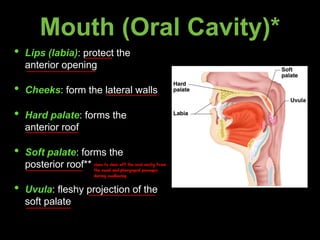
Mouth (Oral Cavity)*
•Lips (labia): protect the
anterior opening
• Cheeks: form the lateral walls
• Hard palate: forms the
anterior roof
• Soft palate: forms the
posterior roof**
• Uvula: fleshy projection of the
soft palate
rises to close off the oral cavity from
the nasal and pharyngeal passages
during swallowing
5.
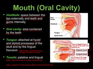
Mouth (Oral Cavity)
•Vestibule: space between the
lips externally and teeth and
gums internally
• Oral cavity: area contained
by the teeth
• Tongue: attached at hyoid
and styloid processes of the
skull and by the lingual
frenulum
• Tonsils: palatine and lingual
lingual frenulum - membrane that secures
the tongue to the floor of the mouth
papillae - found on the surface of the tongue; contain taste buds
masses of lymphoid tissue on
every side of the mouth at its
posterior end
cover the base of the tongue
posterior to the oral cavity
proper
part of the bodys defense system
amylase - component of the saliva; begins the digestion of starchy food in the mouth
X
L
6.
Processes of theMouth
• Mastication (chewing) of food
• Mixing masticated food with
saliva
• Initiation of swallowing by the
tongue
• Allowing for the sense of taste
form the bolus
- swallowing is voluntary in the upper part
- swallowing is involuntary in the lower part (peristalsis)
7.
Pharynx
• Nasopharynx:
not partof the
digestive system
• Oropharynx:
posterior to oral
cavity
• Laryngopharynx:
below the
oropharynx and
connected to the
esophagus
throat
8.
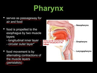
Pharynx
• serves aspassageway for
air and food
• food is propelled to the
esophagus by two muscle
layers
- longitudinal inner layer
- circular outer layer*
• food movement is by
alternating contractions of
the muscle layers
(peristalsis)
initiates wave-like contractions that
propel the food going down to the
esophagus
9.
Esophagus
• runs fromthe pharynx to
stomach through the
diaphragm
• conducts food by
peristalsis (slow rhythmic
squeezing)
• passageway for food only
gullet/food pipe
10 inch long
CARDIOESOPHAGEAL SPHINCTER/LOWER ESOPHAGEAL SPHINCTER/CARDIAC SPHINCTER
- thickening of the smooth muscle layer at the esophagus-stomach junction
- controls the food passage into the stomach
- prevents the stomach contents to flow back into the esophagus
10.
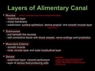
Layers of AlimentaryCanal
• Mucosa
- innermost layer
- moist membrane
- subdivision: surface epithelium, lamina propria* and smooth muscle layer
• Submucosa
- just beneath the mucosa
- soft connective tissue with blood vessels, nerve endings and lymphatics
• Muscularis Externa
- smooth muscle
- inner circular layer and outer longitudinal layer
• Serosa
- outermost layer: visceral peritoneum
- layer of serous fluid-producing cells
connective tissue
epithelium is mostly simple columnar; friction resisting stratified epithelium
PERITONEUM
- visceral - covers the external surfaces of most
abdominal organs including the intestinal tract
- parietal - lines the inside of the abdomen and pelvis
(peritoneal cavity)
11.
TUNICS
lines the lumen
foldof peritoneum w/c attaches the stomach, small intestine,
pancreas, spleen, and other organs to the posterior wall of the
abdomen
I
13.
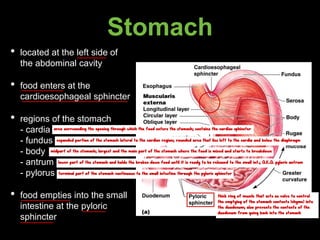
Stomach
• located atthe left side of
the abdominal cavity
• food enters at the
cardioesophageal sphincter
• regions of the stomach
- cardia
- fundus
- body
- antrum
- pylorus
• food empties into the small
intestine at the pyloric
sphincter
area surrounding the opening through which the food enters the stomach; contains the cardiac sphincter
expanded portion of the stomach lateral to the cardiac region; rounded area that lies left to the cardia and below the diaphragm
midpart of the stomach; largest and the main part of the stomach where the food is mixed and starts to breakdown
lower part of the stomach and holds the broken down food until it is ready to be released to the small int.; A.K.A. pyloric antrum
terminal part of the stomach continuous to the small intestine through the pyloric sphincter
thick ring of muscle that acts as valve to control
the emptying of the stomach contents (chyme) into
the duodenum; also prevents the contents of the
duodenum from going back into the stomach
14.
Stomach
• rugae: internalfolds of the
mucosa
• external regions:
- lesser curvature
- greater curvature
• layers of the peritoneum
attached to the stomach
- lesser omentum: attaches
the liver to the lesser
curvature
- greater omentum: attaches
the greater curvature to the
posterior body wall
- contains fat to insulate,
cushion and protect
abdominal organs
concave medial
surface of the
stomach
convex lateral surface
TWO MESENTERIES - omenta
15.
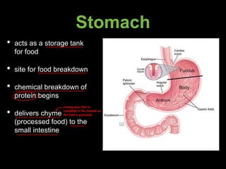
Stomach
• acts asa storage tank
for food
• site for food breakdown
• chemical breakdown of
protein begins
• delivers chyme
(processed food) to the
small intestine
creamy mass that is
resembled in the stomach as
the food is processed
W
16.
Specialized Mucosa ofthe Stomach
• simple columnar epithelium
• mucous neck cells: produce sticky alkaline mucus
• gastric glands: secrete gastric juice
• chief cells: produce protein-digesting ezymes (pepsinogen)
• parietal cells: produce hydrochloric acid
• endocrine cells: produce gastrin
converted into pepsin by the hydrochloric acid;
digest protein into polypeptide chains
17.
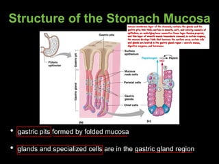
Structure of theStomach Mucosa
• gastric pits formed by folded mucosa
• glands and specialized cells are in the gastric gland region
mucous membrane layer of the stomach; contains the glands and the
gastric pits; 1mm thick; surface is smooth, soft, and velvety; consists of
epithelium, an underlying loose connective tissue layer (lamina propria),
and thin layer of smooth muscle (muscularis mucosa); in certain regions,
the mucosa develops folds that increase the surface area; certain cells
and glands are located in the gastric gland region - secrete mucus,
digestive enzymes, and hormones
18.
Small Intestine
• thebody’s major digestive
organ
• site of nutrient absorption
into the blood
• muscular tube extending
from the pyloric sphincter to
the ileocecal valve
• suspended from the
posterior abdominal wall by
the mesentery
5 meters
convoluted (coiled) tubule
valve between the ileum and cecum
19.
Subdivisions of SmallIntestine
• duodenum: attached to
the stomach; curves
around the head of the
pancreas
• jejunum: attaches
anteriorly to the
duodenum
• ileum: extends from
jejunum to large intestine
10inch long
8ft long
12ft long
joins the large intestine at the ileocecal valve
20.
Chemical Digestion inthe Small Intestine
• source of enzymes that are mixed with chyme
- intestinal cells
- pancreas
• bile enters from the gall bladder
brush border enzymes - enzymes bound to the microvilli of the columnar epithelial
cells and more important enzymes produced by the pancreas inducted into the
duodenum via the pancreatic duct - complete the chemical breakdown process in
the small int.
formed in the liver; enters the duodenum via the bile duct
21.
Villi of theSmall Intestine
• fingerlike structures formed
by the mucosa
• provide the small intestine
with more surface area
Microvilli of the Small Intestine
• small projections of the
plasma membrane
• found on absorptive cells
22.
Structures Involved inAbsorption of Nutrients
• absorptive cells
• blood capillaries
• lacteals (specialized lymphatic
capillaries)
Folds of the Small Intestine
• called circular folds or plicae circulares
• deep folds of the mucosa and submucosa
• do not disappear when filled with food
• the submucosa has Peyer’s patches
(collections of lymphatic tissue)
increase along the length of the small intestine
23.
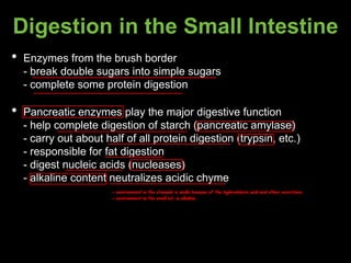
Digestion in theSmall Intestine
• Enzymes from the brush border
- break double sugars into simple sugars
- complete some protein digestion
• Pancreatic enzymes play the major digestive function
- help complete digestion of starch (pancreatic amylase)
- carry out about half of all protein digestion (trypsin, etc.)
- responsible for fat digestion
- digest nucleic acids (nucleases)
- alkaline content neutralizes acidic chyme
- environment in the stomach is acidic because of the hydrochloric acid and other secretions
- environment in the small int. is alkaline
24.
Absorption and Propulsionin the Small Intestine
• Water is absorbed along the length of the small intestine
• End products of digestion
- most substances are absorbed by active transport through
cell membranes
- lipids are absorbed by diffusion
• Substances are transported to the liver by the hepatic portal
vein or lymph
• Peristalsis is the major means of moving food
• Segmental movements
- mix chyme with digestive juices
- aid in propelling food
25.
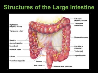
Large Intestine
• largerin diameter, but
shorter than the small
intestine
• frames the internal abdomen
• functions:
- absorption of water
- elimination of indigestible
food from the body as feces
- no participation in digestion
of food
- goblet cells produce mucus
to act as lubricant
26.
Structures of theLarge Intestine
• cecum: saclike first part of
the large intestine
• appendix: accumulation of
lymphatic tissue that
sometimes becomes
inflamed; hangs from the
cecum
• colon: ascending, transverse,
descending and sigmoidal
• rectum
• anus: external body opening
appendicitis
appendectomy
largest part
Food Breakdown andAbsorption
in the Large Intestine
• no digestive enzymes are produced
• resident bacteria digest remaining nutrients
- produce some vitamin K and B
- release gases
• water and vitamins K and B are absorbed
• remaining materials are eliminated via feces
29.
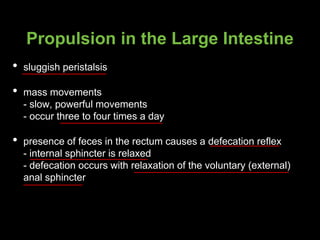
Propulsion in theLarge Intestine
• sluggish peristalsis
• mass movements
- slow, powerful movements
- occur three to four times a day
• presence of feces in the rectum causes a defecation reflex
- internal sphincter is relaxed
- defecation occurs with relaxation of the voluntary (external)
anal sphincter
Salivary Glands andSaliva
• saliva-producing glands
• parotid glands
• submandibular glands
• sublingual glands
• saliva
- mixture of mucus and serous
fluids*
- helps to form a bolus
- contains salivary amylase to
begun starch digestion**
- dissolves chemicals so they can
be tasted
for lubrication of food in the oral cavity
in front and behind the ears; secretes saliva with ptyalin to the oral
cavity through the parotid duct/Stensen’s duct
mumps - common childhood disease which is characterized by the inflammation of the parotid gland (parotitis)
below the mandible; secretes saliva through the Wharton’s duct
below the tongue; secretes saliva through the Bartholin’s and Rivinus ducts
empty secretions into the floor of the mouth through the tiny ducts
makes chewing and swallowing easier
mucus - noun
mucous - adjective
32.
Teeth
for mastication offood
20; fully formed by age 2
32; replace the deciduous teeth between ages 6-12
16 in the upper and 16 in the lower
33.
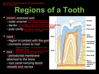
Regions of aTooth
• crown: exposed part
- outer enamel
- dentin
- pulp cavity
• neck
- region in contact with the gum
- connects crown to root
• root
- periodontal membrane
attached to the bone
- root canal carrying blood
vessels and nerves
TEETH - one of the hardest substances in the human body; also play an
important role in speech
hardest whiter, outer part of the
tooth; made up of calcium phosphate
layer underlying the enamel; hard tissue that contains microscopic tubes;
can cause sensitivity or pain when the outer enamel is damaged
softer living inner structure of teeth; blood
vessels and nerves run through it
PERIDONTAL LIGAMENT - tissue that holds the teeth
tightly against the jaw
CEMENTUM - connective tissue that binds the roots of
the teeth firmly to the gums and the jaw bone
34.
Pancreas
• produces awide spectrum of
digestive enzymes that
break down categories of
food
• enzymes are secreted into
the duodenum
• alkaline fluid introduced with
enzymes neutralizes acidic
chyme
• endocrine products of
pancreas
- insulin
- glucagon
has both endocrine and exocrine functions
alpha cells
beta cells increase protein and fat synthesis and slow down the breakdown of glycogen, protein, and fat
PANCREATIC AMYLASE - digests starch and
glycerol into maltose
PANCREATIC LIPASE - digests fat into fatty acids
and glycerol
TRYPSINOGEN - converted into trypsin by
enterokinase and digests the proteins into
polypeptides, peptones, and proteoses
CHEMOTRYPSINOGEN - activated to chemotrypsin
by trypsin and digests polypeptides to amino acids
35.
Liver
• located inghe right upper
quadrant of the abdomen
• divided into lobes
separated by a connective
tissue septum, the
falciform ligament
• receives blood from 2
sources: hepatic artery
and hepatic portal vein*
• blood exits the liver
through the hepatic veins
which empty into IVC
brings O2 rich blood to the liver and supplies liver cells with O2
carries O2 poor but rich in absorbed nutrients and other substances from the digestive tract to
the liver
liver cells process nutrients and detoxify harmful substances from the blood
36.
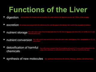
Functions of theLiver
• digestion
• excretion
• nutrient storage
• nutrient conversion
• detoxification of harmful
chemicals
• synthesis of new molecules
bile neutralizes the stomach acid and emulsifies fat which facilitates fat digestion; secretes abt. 700mL of bile everyday
bile contains excretory products like cholesterol, fats, and bile pigments like bilirubin that results from hemoglobin breakdown
liver cells remove sugar from the blood and store it in the form of glycogen; also stores fat, vit. A, B12, D, E, K, and
minerals such as iron
liver cells convert some nutrients into others like amino acids = lipids/glucose; fats = phospholipids; vit. D = its
active form
liver cells remove ammonia from the circulation and convert it to urea which is eliminated in the
urine; other substances are detoxified and secreted in the bile or excreted in the urine
liver synthesizes blood proteins like albumin, fibrinogen, globulins, and clotting factors
37.
Gall Bladder
• sacfound in hollow fossa
of the liver
• stores bile from the liver
by way of the cystic duct
• bile is introduced into the
duodenum in the
presence of fatty food
• gallstones can cause
blockages
emulsify the fat
cholecystectomy - removal of the gallbladder
cholecystitis - inflammation of the gallbladder
38.
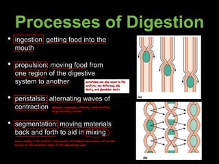
Processes of Digestion
•ingestion: getting food into the
mouth
• propulsion: moving food from
one region of the digestive
system to another
• peristalsis: alternating waves of
contraction
• segmentation: moving materials
back and forth to aid in mixing
pharynx, esophagus, stomach, small intestine,
large intestine, rectum
peristalsis can also occur in the
ureters, vas deferens, bile
ducts, and glandular ducts
occurs mainly in the small int. and consists of localized contractions of circular
muscles of the muscularis layer of the alimentary canal
39.
Processes of Digestion
•absorption
- end products of absorption
are absorbed in the blood or
lymph
- food must enter mucosal
cells and then into blood or
lymph capillaries
• defecation
- elimination of indigestible
substances as feces
40.
Control of DigestiveActivity
• mostly controlled by reflexes via the parasympathetic division
• chemical and mechanical receptors are located in organ walls
that trigger reflexes
• stimuli include stretch of the organ, pH of the contents, presence
of breakdown products
• reflexes include activation or inhibition of glandular secretions
and smooth muscle activity
rest and digest/feed and breathe