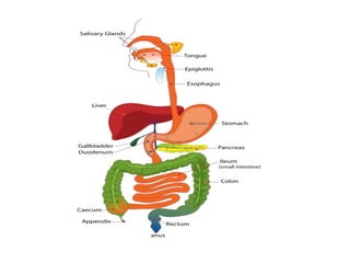
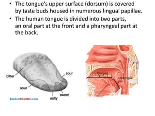
The digestive system breaks down food and absorbs nutrients. It includes the mouth, esophagus, stomach, small intestine, large intestine, liver, pancreas and gallbladder. The mouth chews food, the stomach acids break it down and the small intestine further breaks it down and absorbs nutrients using enzymes from the pancreas and bile from the liver. The large intestine absorbs water and passes waste to the rectum to be eliminated. Accessory organs like the liver, pancreas and gallbladder produce enzymes and bile to aid digestion.
An overview of the GIT with detailed study of the organs, along with their anatomy and physiology. It will find u easier to go through this complex function within our body.
The digestive system is made up of the digestive tract and other organs that help the body break down and absorb food. It is a long, twisting tube that starts at the mouth and goes through the oesophagus, stomach, small intestine, large intestine and anus.
The digestive system is made up of the gastrointestinal tract—also called the GI tract or digestive tract—and the liver, pancreas, and gallbladder. ... The hollow organs that make up the GI tract are the mouth, esophagus, stomach, small intestine, large intestine, and anus.
Anatomy and Physiology of Digestive system.
Different Digestive process for absorption of food in GIT.
Different parts GIT Tract where food move from Mouth to the anus.
Grade 7 chapter1 lesson1 - the digestive systemLermaPendon1
Lesson 2 The Digestive system
-Functions of Digestive system
-Digestion
-Types of Digestion
-Enzymes
-The role of Enzymes in Digestion
-Organs of Digestive system
-The Mouth
-the Esophagus
-Peristalsis
-The Stomach
-Chyme
-the Small Intestine
-Villi
-the Large intestine
-Bacteria and Digestion
-the Digestive system and Homeostasis
-Common Ailments and Disorders of the Digestive system
1 GNM Anatomy Unit - 7 GIT by thirumurugan.pptxthiru murugan
By:M. Thiru murugan
Unit - 7
Structure and functions of the alimentary tract & is accessory organs.
The process of digestion, absorption & metabolism of food constituents
Digestive system
Also known as gastrointestinal tract, digestive tract, digestional tract, GI tract, GIT, gut, or alimentary canal.
Consist of alimentary tract and Accessory organs
It involve in Ingestion, Digestion, Absorption & Excretion
It is started from mouth and ended at anal canal
Parts of Digestive System
Accessory organs:
Teeth
Tongue
Salivary glands
Liver
Gallbladder
Pancreas
Diagram
Functions of Digestive System
Ingestion: taking of food into the GIT. i.e. eating & drinking.
Propulsion: mixes & moves the contents along the GIT
Digestion: breakdown of food
Absorption: this is the process by which digested food substances pass into the blood for circulation.
Elimination: undigested food substances removed from the GIT as feces by the process of defecation.
Mouth:
First part of GIT
The mouth is an oval-shaped cavity inside the skull.
Parts:
Lips (Labia), vestibule, mouth cavity, gums, teeth, hard & soft palate, tongue and salivary glands. The mouth is also known as the oral cavity or the buccal cavity.
Function of mouth:
Mastication (chewing) of food
Mixing masticated food with saliva
Initiation of swallowing by the tongue
Allowing for the sense of taste
Tongue or Lingua:
The tongue is a muscular organ in the mouth.
Small parts called papillae give the tongue its rough structure.
Many taste buds present on the surfaces of the papillae
Taste buds are sensory organs that are found on your tongue and allow you to experience tastes that are sweet, salty, sour, and bitter
Function of tongue:
Mastication
Deglutition
Speech
Taste
Oral cleaning
Teeth:
The teeth are the hardest white in oral cavity.
There are 20 temporary or deciduous teeth(10 in each Jaw 4 incisors, 2 canines & 4 molars)
The permanent or secondary teeth 32 teeth(16 in each jaw - 4 incisors, 2 canines, 4 premolars & 6 molars )
Function of teeth:
Chewing food
Cutting and grinding food
speech
Structure of tooth:
Crown: part protrude from gums or gingiva
Neck: in between crown & root
Root: bottom part of the tooth; may be single, double, or triple root embedded in bone
Enamel: The hardest, white outer part of the tooth.
Dentin: A layer underlying the enamel.
Pulp: The softer, living inner structure of teeth. Blood vessels and nerves run through the pulp of the teeth.
Cementum: A layer that attach the roots of the teeth to the gums and jawbone.
Periodontal ligament: Tissue that helps hold the teeth tightly in jaw.
Salivary glands:
It secreting saliva
There are 3 types
Parotid - each side of the face
Submandibular - below the mandibular
Sublingual – below the tongue
All have ducts to supply saliva in mouth
Functions: chewing and swallowing, Lubricating effect, dissolves food, taste food, Cleaning mouth, fight against pathogenic microorganisms
Pharynx:
Muscular tube situated behind th
Title: Sense of Taste
Presenter: Dr. Faiza, Assistant Professor of Physiology
Qualifications:
MBBS (Best Graduate, AIMC Lahore)
FCPS Physiology
ICMT, CHPE, DHPE (STMU)
MPH (GC University, Faisalabad)
MBA (Virtual University of Pakistan)
Learning Objectives:
Describe the structure and function of taste buds.
Describe the relationship between the taste threshold and taste index of common substances.
Explain the chemical basis and signal transduction of taste perception for each type of primary taste sensation.
Recognize different abnormalities of taste perception and their causes.
Key Topics:
Significance of Taste Sensation:
Differentiation between pleasant and harmful food
Influence on behavior
Selection of food based on metabolic needs
Receptors of Taste:
Taste buds on the tongue
Influence of sense of smell, texture of food, and pain stimulation (e.g., by pepper)
Primary and Secondary Taste Sensations:
Primary taste sensations: Sweet, Sour, Salty, Bitter, Umami
Chemical basis and signal transduction mechanisms for each taste
Taste Threshold and Index:
Taste threshold values for Sweet (sucrose), Salty (NaCl), Sour (HCl), and Bitter (Quinine)
Taste index relationship: Inversely proportional to taste threshold
Taste Blindness:
Inability to taste certain substances, particularly thiourea compounds
Example: Phenylthiocarbamide
Structure and Function of Taste Buds:
Composition: Epithelial cells, Sustentacular/Supporting cells, Taste cells, Basal cells
Features: Taste pores, Taste hairs/microvilli, and Taste nerve fibers
Location of Taste Buds:
Found in papillae of the tongue (Fungiform, Circumvallate, Foliate)
Also present on the palate, tonsillar pillars, epiglottis, and proximal esophagus
Mechanism of Taste Stimulation:
Interaction of taste substances with receptors on microvilli
Signal transduction pathways for Umami, Sweet, Bitter, Sour, and Salty tastes
Taste Sensitivity and Adaptation:
Decrease in sensitivity with age
Rapid adaptation of taste sensation
Role of Saliva in Taste:
Dissolution of tastants to reach receptors
Washing away the stimulus
Taste Preferences and Aversions:
Mechanisms behind taste preference and aversion
Influence of receptors and neural pathways
Impact of Sensory Nerve Damage:
Degeneration of taste buds if the sensory nerve fiber is cut
Abnormalities of Taste Detection:
Conditions: Ageusia, Hypogeusia, Dysgeusia (parageusia)
Causes: Nerve damage, neurological disorders, infections, poor oral hygiene, adverse drug effects, deficiencies, aging, tobacco use, altered neurotransmitter levels
Neurotransmitters and Taste Threshold:
Effects of serotonin (5-HT) and norepinephrine (NE) on taste sensitivity
Supertasters:
25% of the population with heightened sensitivity to taste, especially bitterness
Increased number of fungiform papillae
Lung Cancer: Artificial Intelligence, Synergetics, Complex System Analysis, S...Oleg Kshivets
RESULTS: Overall life span (LS) was 2252.1±1742.5 days and cumulative 5-year survival (5YS) reached 73.2%, 10 years – 64.8%, 20 years – 42.5%. 513 LCP lived more than 5 years (LS=3124.6±1525.6 days), 148 LCP – more than 10 years (LS=5054.4±1504.1 days).199 LCP died because of LC (LS=562.7±374.5 days). 5YS of LCP after bi/lobectomies was significantly superior in comparison with LCP after pneumonectomies (78.1% vs.63.7%, P=0.00001 by log-rank test). AT significantly improved 5YS (66.3% vs. 34.8%) (P=0.00000 by log-rank test) only for LCP with N1-2. Cox modeling displayed that 5YS of LCP significantly depended on: phase transition (PT) early-invasive LC in terms of synergetics, PT N0—N12, cell ratio factors (ratio between cancer cells- CC and blood cells subpopulations), G1-3, histology, glucose, AT, blood cell circuit, prothrombin index, heparin tolerance, recalcification time (P=0.000-0.038). Neural networks, genetic algorithm selection and bootstrap simulation revealed relationships between 5YS and PT early-invasive LC (rank=1), PT N0—N12 (rank=2), thrombocytes/CC (3), erythrocytes/CC (4), eosinophils/CC (5), healthy cells/CC (6), lymphocytes/CC (7), segmented neutrophils/CC (8), stick neutrophils/CC (9), monocytes/CC (10); leucocytes/CC (11). Correct prediction of 5YS was 100% by neural networks computing (area under ROC curve=1.0; error=0.0).
CONCLUSIONS: 5YS of LCP after radical procedures significantly depended on: 1) PT early-invasive cancer; 2) PT N0--N12; 3) cell ratio factors; 4) blood cell circuit; 5) biochemical factors; 6) hemostasis system; 7) AT; 8) LC characteristics; 9) LC cell dynamics; 10) surgery type: lobectomy/pneumonectomy; 11) anthropometric data. Optimal diagnosis and treatment strategies for LC are: 1) screening and early detection of LC; 2) availability of experienced thoracic surgeons because of complexity of radical procedures; 3) aggressive en block surgery and adequate lymph node dissection for completeness; 4) precise prediction; 5) adjuvant chemoimmunoradiotherapy for LCP with unfavorable prognosis.
Title: Sense of Smell
Presenter: Dr. Faiza, Assistant Professor of Physiology
Qualifications:
MBBS (Best Graduate, AIMC Lahore)
FCPS Physiology
ICMT, CHPE, DHPE (STMU)
MPH (GC University, Faisalabad)
MBA (Virtual University of Pakistan)
Learning Objectives:
Describe the primary categories of smells and the concept of odor blindness.
Explain the structure and location of the olfactory membrane and mucosa, including the types and roles of cells involved in olfaction.
Describe the pathway and mechanisms of olfactory signal transmission from the olfactory receptors to the brain.
Illustrate the biochemical cascade triggered by odorant binding to olfactory receptors, including the role of G-proteins and second messengers in generating an action potential.
Identify different types of olfactory disorders such as anosmia, hyposmia, hyperosmia, and dysosmia, including their potential causes.
Key Topics:
Olfactory Genes:
3% of the human genome accounts for olfactory genes.
400 genes for odorant receptors.
Olfactory Membrane:
Located in the superior part of the nasal cavity.
Medially: Folds downward along the superior septum.
Laterally: Folds over the superior turbinate and upper surface of the middle turbinate.
Total surface area: 5-10 square centimeters.
Olfactory Mucosa:
Olfactory Cells: Bipolar nerve cells derived from the CNS (100 million), with 4-25 olfactory cilia per cell.
Sustentacular Cells: Produce mucus and maintain ionic and molecular environment.
Basal Cells: Replace worn-out olfactory cells with an average lifespan of 1-2 months.
Bowman’s Gland: Secretes mucus.
Stimulation of Olfactory Cells:
Odorant dissolves in mucus and attaches to receptors on olfactory cilia.
Involves a cascade effect through G-proteins and second messengers, leading to depolarization and action potential generation in the olfactory nerve.
Quality of a Good Odorant:
Small (3-20 Carbon atoms), volatile, water-soluble, and lipid-soluble.
Facilitated by odorant-binding proteins in mucus.
Membrane Potential and Action Potential:
Resting membrane potential: -55mV.
Action potential frequency in the olfactory nerve increases with odorant strength.
Adaptation Towards the Sense of Smell:
Rapid adaptation within the first second, with further slow adaptation.
Psychological adaptation greater than receptor adaptation, involving feedback inhibition from the central nervous system.
Primary Sensations of Smell:
Camphoraceous, Musky, Floral, Pepperminty, Ethereal, Pungent, Putrid.
Odor Detection Threshold:
Examples: Hydrogen sulfide (0.0005 ppm), Methyl-mercaptan (0.002 ppm).
Some toxic substances are odorless at lethal concentrations.
Characteristics of Smell:
Odor blindness for single substances due to lack of appropriate receptor protein.
Behavioral and emotional influences of smell.
Transmission of Olfactory Signals:
From olfactory cells to glomeruli in the olfactory bulb, involving lateral inhibition.
Primitive, less old, and new olfactory systems with different path
Prix Galien International 2024 Forum ProgramLevi Shapiro
June 20, 2024, Prix Galien International and Jerusalem Ethics Forum in ROME. Detailed agenda including panels:
- ADVANCES IN CARDIOLOGY: A NEW PARADIGM IS COMING
- WOMEN’S HEALTH: FERTILITY PRESERVATION
- WHAT’S NEW IN THE TREATMENT OF INFECTIOUS,
ONCOLOGICAL AND INFLAMMATORY SKIN DISEASES?
- ARTIFICIAL INTELLIGENCE AND ETHICS
- GENE THERAPY
- BEYOND BORDERS: GLOBAL INITIATIVES FOR DEMOCRATIZING LIFE SCIENCE TECHNOLOGIES AND PROMOTING ACCESS TO HEALTHCARE
- ETHICAL CHALLENGES IN LIFE SCIENCES
- Prix Galien International Awards Ceremony
Report Back from SGO 2024: What’s the Latest in Cervical Cancer?bkling
Are you curious about what’s new in cervical cancer research or unsure what the findings mean? Join Dr. Emily Ko, a gynecologic oncologist at Penn Medicine, to learn about the latest updates from the Society of Gynecologic Oncology (SGO) 2024 Annual Meeting on Women’s Cancer. Dr. Ko will discuss what the research presented at the conference means for you and answer your questions about the new developments.
TEST BANK for Operations Management, 14th Edition by William J. Stevenson, Ve...kevinkariuki227
TEST BANK for Operations Management, 14th Edition by William J. Stevenson, Verified Chapters 1 - 19, Complete Newest Version.pdf
Factory Supply Best Quality Pmk Oil CAS 28578–16–7 PMK Powder in Stockrebeccabio
Factory Supply Best Quality Pmk Oil CAS 28578–16–7 PMK Powder in Stock
Telegram: bmksupplier
signal: +85264872720
threema: TUD4A6YC
You can contact me on Telegram or Threema
Communicate promptly and reply
Free of customs clearance, Double Clearance 100% pass delivery to USA, Canada, Spain, Germany, Netherland, Poland, Italy, Sweden, UK, Czech Republic, Australia, Mexico, Russia, Ukraine, Kazakhstan.Door to door service
Hot Selling Organic intermediates
Ethanol (CH3CH2OH), or beverage alcohol, is a two-carbon alcohol
that is rapidly distributed in the body and brain. Ethanol alters many
neurochemical systems and has rewarding and addictive properties. It
is the oldest recreational drug and likely contributes to more morbidity,
mortality, and public health costs than all illicit drugs combined. The
5th edition of the Diagnostic and Statistical Manual of Mental Disorders
(DSM-5) integrates alcohol abuse and alcohol dependence into a single
disorder called alcohol use disorder (AUD), with mild, moderate,
and severe subclassifications (American Psychiatric Association, 2013).
In the DSM-5, all types of substance abuse and dependence have been
combined into a single substance use disorder (SUD) on a continuum
from mild to severe. A diagnosis of AUD requires that at least two of
the 11 DSM-5 behaviors be present within a 12-month period (mild
AUD: 2–3 criteria; moderate AUD: 4–5 criteria; severe AUD: 6–11 criteria).
The four main behavioral effects of AUD are impaired control over
drinking, negative social consequences, risky use, and altered physiological
effects (tolerance, withdrawal). This chapter presents an overview
of the prevalence and harmful consequences of AUD in the U.S.,
the systemic nature of the disease, neurocircuitry and stages of AUD,
comorbidities, fetal alcohol spectrum disorders, genetic risk factors, and
pharmacotherapies for AUD.
3. WHAT IS THE DIGESTIVE SYSTEM?
• The gastrointestinal tract (digestive
tract, digestional tract, GI tract, GIT, gut,
or alimentary canal) is an organ system within
humans and other animals which takes in
food, digests it and absorb energy and nutrients, and
expels the remaining waste as feces.
• The major organs of the digestive system:
• Mouth.
• Pharynx.
• Esophagus.
• Stomach.
• Small Intestine.
• Large Intestine.
• Rectum.
4. Accessory digestive organs:
liver
gallbladder
pancreas.
Salivary gland
Functions of GI tract
• Ingestion: taking of food into the alimentary tract.
i.e. eating & drinking.
• Propulsion:mixes & moves the contents along the
alimentary tract.
• Digestion:consist of:
• Mechanical breakdown of food e.g. mastication
(chewing)
• Chemical digestion of food into small molecules by
enzymes.
5. • Absorption:this is the process by which
digested food substances pass through the walls
of some organs of the alimentary canal into the
blood.
• Elimination:food substances that have been
eaten but cannot be digested & absorbed are
excreted from the alimentary canal as faeces by
the process of defaecation.
6. MOUTH
• The mouth is the first portion of the alimentary
canal that receives food and produces saliva.
• Relations:
• Anteriorly-lips
• Posteriorly-continue with the oropharynx
• Laterally-muscles of cheeks
• Superiorly-bony hard palate
• Inferiorly-muscular tongue & the soft tissues of the
floor of the mouth
7.
8. • The palate forms the roof of the mouth & is divided
into the anterior hard palate & posterior soft
palate.
• The uvula is a curved fold of muscle covered with
mucous membrane,hanging down from the middle.
TONGUE
• The tongue is a muscular organ in the mouth, that
manipulates food for mastication, and is used in
the act of swallowing.
• It is of importance in the digestive system and is
the primary organ of taste in the gustatory system.
9. • The tongue's upper surface (dorsum) is covered
by taste buds housed in numerous lingual papillae.
• The human tongue is divided into two parts,
an oral part at the front and a pharyngeal part at
the back.
12. SALIVARY GLANDS
• The salivary glands are exocrine glands that
produce saliva through a system of ducts.
• Humans have 3 paired major salivary glands:
• Parotid
• submandibular
• Sublingual
• as well hundreds of minor salivary glands.
• Parotid glands
• The two parotid glands are major salivary glands
wrapped around the mandibular ramus in humans.
• The largest of the salivary glands.
13.
14. • THE PHARYNX
• The pharynx is the part of the throat that is behind
the mouth and nasal cavity and above the esophagus
and the larynx, or the tubes going down to the
stomach and the lungs.
• The pharynx is the portion of the digestive tract that
receives the food from your mouth.
15. • THE ESOPHAGUS
• The esophagus is a muscular tube connecting
the throat (pharynx) with the stomach.
• The esophagus runs behind the windpipe
(trachea) and heart, and in front of the spine.
• Length :25 cm
• Diameter:2 cm
16.
17.
18. FUNCTIONS
• Formation of a bolus
• Swallowing
• Food is ingested through the mouth and when
swallowed passes first into the pharynx and then
into the esophagus.
• Reducing gastric reflux
• Constriction of the upper and lower esophageal
sphincters help to prevent reflux (backflow) of
gastric contents and acid into the esophagus,
protecting the esophageal mucosa.
19. STOMACH
• The stomach is a muscular organ located on
the left side of the upper abdomen. The
stomach receives food from the esophagus.
• As food reaches the end of the esophagus, it
enters the stomach through a muscular valve
called the lower esophageal sphincter.
20.
21.
22. FUNCTIONS
• Digestion
• The stomach releases proteases (protein-digesting
enzymes such as pepsin) and hydrochloric acid,
which kills or inhibits bacteria and provides the
acidic pH of 2 for the proteases to work.
• Food is churned by the stomach through muscular
contractions of the wall called peristalsis
• Absorption
• some absorption of certain small molecules
nevertheless does occur in the stomach
through its lining
23. • GASTRIC JUICE
• Gastric acid, gastric juice or stomach acid, is a
digestive fluid formed in the stomach and is
composed of hydrochloric acid (HCl), potassium
chloride (KCl) and sodium chloride (NaCl).
• The acid plays a key role in digestion of proteins, by
activating digestive enzymes, and making ingested
proteins unravel so that digestive enzymes break
down the long chains of amino acids.
24. • Gastric Secretory Cells
• Chief cells: secrete pepsinogen (an inactive
enzyme).
• Parietal cells: secrete hydrochloric and (HCl)
and "intrinsic factor" (which helps absorption of
vitamin B12 in the intestines).
• Mucous cells: secrete mucus and alkaline
substances to help neutralize HCl in the gastric
juice .
• G cells: secrete a hormone called gastrin , which
stimulates the parietal cells and overall gastric.
25.
26.
27. THE PANCREAS
• The pancreas is a glandular organ in the digestive
system and endocrine system.
• It is located in the abdominal cavity behind the
stomach.
• It is an endocrine gland producing several
important hormones, including insulin, glucagon,
somatostatin, and pancreatic polypeptide, all of
which circulate in the blood.
• Length: 15 cm or 6 inch
28. • FUNCTION
• The pancreas is involved in blood sugar control and
metabolism within the body.
• Four main types of cells are involved in the regulation of
blood glucose levels.
• Each type of cell secretes a different type of hormone:
• α alpha cells secrete glucagon (increase glucose in blood)
• β beta cells secrete insulin (decrease glucose in blood)
• δ delta cells secrete somatostatin (regulates/stops α and β
cells) and
• γ (gamma) cells, secrete pancreatic polypeptide.
29. • THE LIVER
• The liver‘s main function: detoxifies various
metabolites, synthesizes proteins, and produces
biochemicals necessary for digestion
30. • STRUCTURE
• The liver is a reddish-brown wedge-shaped
organ with four lobes of unequal size and
shape.
• weighs 1.44–1.66 kg
• width -15 cm.
• It is both the heaviest internal organ and the
largest gland in the human body.
31. • FUNCTIONS
• Synthesis
• The liver plays a major role in carbohydrate, protein,
amino acid, and lipid metabolism.
• Breakdown
• The liver is responsible for the breakdown of insulin
and other hormones.
32. • The liver breaks down bilirubin to produce bile
• Other
• The liver stores a multitude of substances,
including glucose (in the form of glycogen)
• vitamin A vitamin D vitamin B12
• vitamin K, iron, and copper.
• The liver produces albumin, the most abundant
protein in blood serum.
33. • Contains phagocytes to destroy damaged
erythrocytes and foreign substances, using
phagocytosis .
34. • THE GALLBLADDER
• The gallbladder is a small hollow organ where
bile is stored and concentrated before it is
released into the small intestine.
• In humans, the pear-shaped gallbladder lies
beneath the liver.
35.
36.
37. • STRUCTURE
• sits in a shallow depression below the right lobe of the
liver, that is grey-blue in life.
• Length-7 to 10 cm or 2.8 to 3.9 inches
• Diameter -4 cm or 1.6 inch
• The gallbladder has a capacity of about 50 millilitres
• The gallbladder is shaped like a pear, with its tip
opening into the cystic duct.
• gallbladder is divided into three sections:
• the fundus,
• The body,
• and the neck.
38. • FUNCTIONS
• The main purpose of the gallbladder is to
store bile, also called gall, needed for the
digestion of fats in food.
• bile flows through small vessels into the larger
hepatic ducts and ultimately though the cystic
duct into the gallbladder, where it is stored.
39.
40.
41. • PARTS
• The small intestine is divided into three structural
parts:
• (I)The duodenum
• (II)The jejunum
• (III)The ileum
• The duodenum is a short structure ranging from
20 cm to 25 cm in length, and shaped like a "C".
• The jejunum is the midsection of the small
intestine, connecting the duodenum to the ileum.
It is about 2.5 m long.
42. • The ileum is the final section of the small
intestine. It is about 3 m long.
• FUNCTIONS
• Digestion
• The small intestine is where most chemical
digestion takes place.
• Many of the digestive enzymes that act in the
small intestine are secreted by the pancreas and
liver and enter the small intestine via the
pancreatic duct.
• Digestion of proteins & carbohydrate
43.
44. • Absorption
• Digested food is now able to pass into the blood
vessels in the wall of the intestine through
either diffusion or active transport.
• The small intestine is the site where most of
the nutrients from ingested food are absorbed.
• Immunological
• The small intestine supports the body's immune
system.
• The presence of gut flora appears to contribute
positively to the host's immune system.
45. THE LARGE INTESTINE
• The large intestine, also known as the large
bowel or colon, is the last part of the
gastrointestinal tract and of the digestive
system.
• Water is absorbed here and the remaining
waste material is stored as feces before being
removed by defecation.
46.
47. • STRUCTURE
• The length of male colon is 166 cm .
• female colon 155 cm
• The colon consists of five sections:
• the cecum
• ascending colon,
• the transverse colon,
• the descending colon,
• the sigmoid colon and the rectum.
48. • The average inner diameter of sections of the
colon in centimeters
• cecum 8.7cm
• ascending colon 6.6cm
• transverse colon 5.8cm
• descending/sigmoid colon 6.3cm
• and rectum near rectal/sigmoid junction
5.7cm
49. • The cecum
• The cecum is the first section of the colon and
involved in the digestion, while the appendix is a
structure of the colon, not involved in digestion.
• The function of the appendix is uncertain.
• Containing Ileocecal valve
• The ileocecal valve is a sphincter muscle valve that
separates the small intestine and the large
intestine.
• Its critical function is to limit the reflux of colonic
contents into the ileum.
50. • The ascending colon
• It is connected to the small intestine by a
section of bowel called the cecum.
• The unwanted waste material is moved
upwards toward the transverse colon by the
action of peristalsis.
• Peristalsis is a radially symmetrical contraction
and relaxation of muscles that propagates in a
wave down a tube
•
51. • Transverse colon
• The transverse colon is the part of the colon from the hepatic
flexure to the splenic flexure.
• Descending colon
• The descending colon is the part of the colon from the splenic
flexure to the beginning of the sigmoid colon,descending colon is
also called the distal gut.
• One function of the descending colon in the digestive system is to
store feces that will be emptied into the rectum.
52. • Sigmoid colon
• The sigmoid colon is the part of the large intestine after the descending
colon and before the rectum.
• The name sigmoid means S-shaped .
• The walls of the sigmoid colon are muscular, and contract to increase the
pressure inside the colon, causing the stool to move into the rectum.
• Rectum
• The rectum is the last section of the large intestine. It holds the
formed feces awaiting elimination via defecation.
53. • the anus
• The anus is the external opening of the
rectum.
• Its function is to control the expulsion of feces.
• Two sphincters control the exit of feces from the body
during an act of defecation.
• These are the internal anal sphincter and the external
anal sphincter, which are circular muscles that
normally maintain constriction of the orifice and which
relaxes as required by normal physiological functioning.
54. • FUNCTIONS
• The large intestine absorbs water and any remaining
absorbable nutrients from the food before sending the
indigestible matter to the rectum.
• The colon absorbs vitamins that are created by the colonic
bacteria, such as vitamin K.
• Gut flora
• The large intestine houses over 700 species of bacteria that
perform a variety of functions.
• The large intestine absorbs some of the products formed
by the bacteria inhabiting this region.
55. PHYSIOLOGY OF DIGESTION
• The mouth is the beginning of the digestive tract.
• Chewing breaks the food into pieces that are more
easily digested, while saliva mixes with food to
begin the process of breaking it down into a form
your body can absorb and use.
• From pharynx food travels to the esophagus
or swallowing tube.
• By means of a series of contractions, called
peristalsis, the esophagus delivers food to
the stomach.
• The lower esophageal sphincter keep food
from passing backwards into the esophagus.
56. • The stomach secretes acid and powerful enzymes that
continue the process of breaking down the food.
• When it leaves the stomach, food is the
consistency of a liquid or paste.
• From there the food moves to the small intestine.
• The small intestine continues the process of breaking
down food by using enzymes released by the pancreas and
bile from the liver.
• Bile is a compound that aids in the digestion of fat.
57. • Peristalsis is also at work in this organ, moving food through
and mixing it up with digestive secretions.
• The duodenum is largely responsible for continuing the
process of breaking down food, with the jejunum and ileum
being mainly responsible for the absorption of nutrients into
the bloodstream.
• pancreas secretes enzymes into the small intestine.
• These enzymes break down protein, fat, and
carbohydrates from the food we eat.
58. • Feces, or waste left over from the digestive
process, is passed through the colon by means
of peristalsis, first in a liquid state and
ultimately in solid form as the water is
removed from the feces.
• Feces are stored in the sigmoid colon until a
"mass movement" empties it into the rectum
once or twice a day.