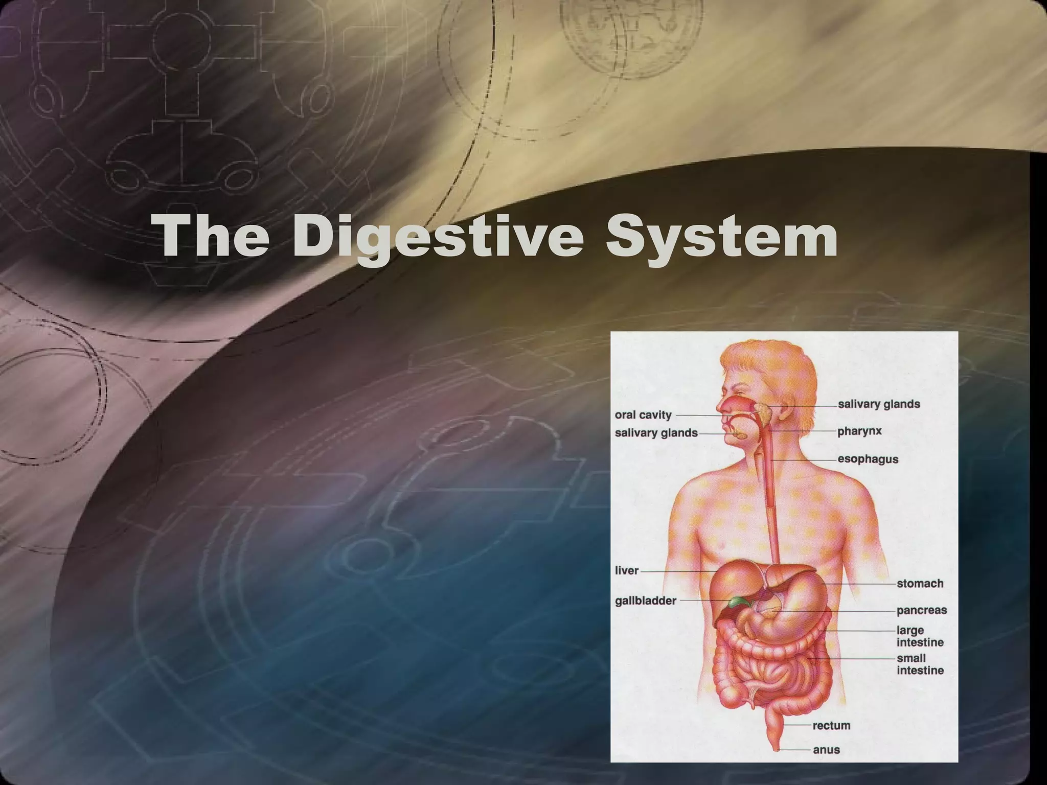
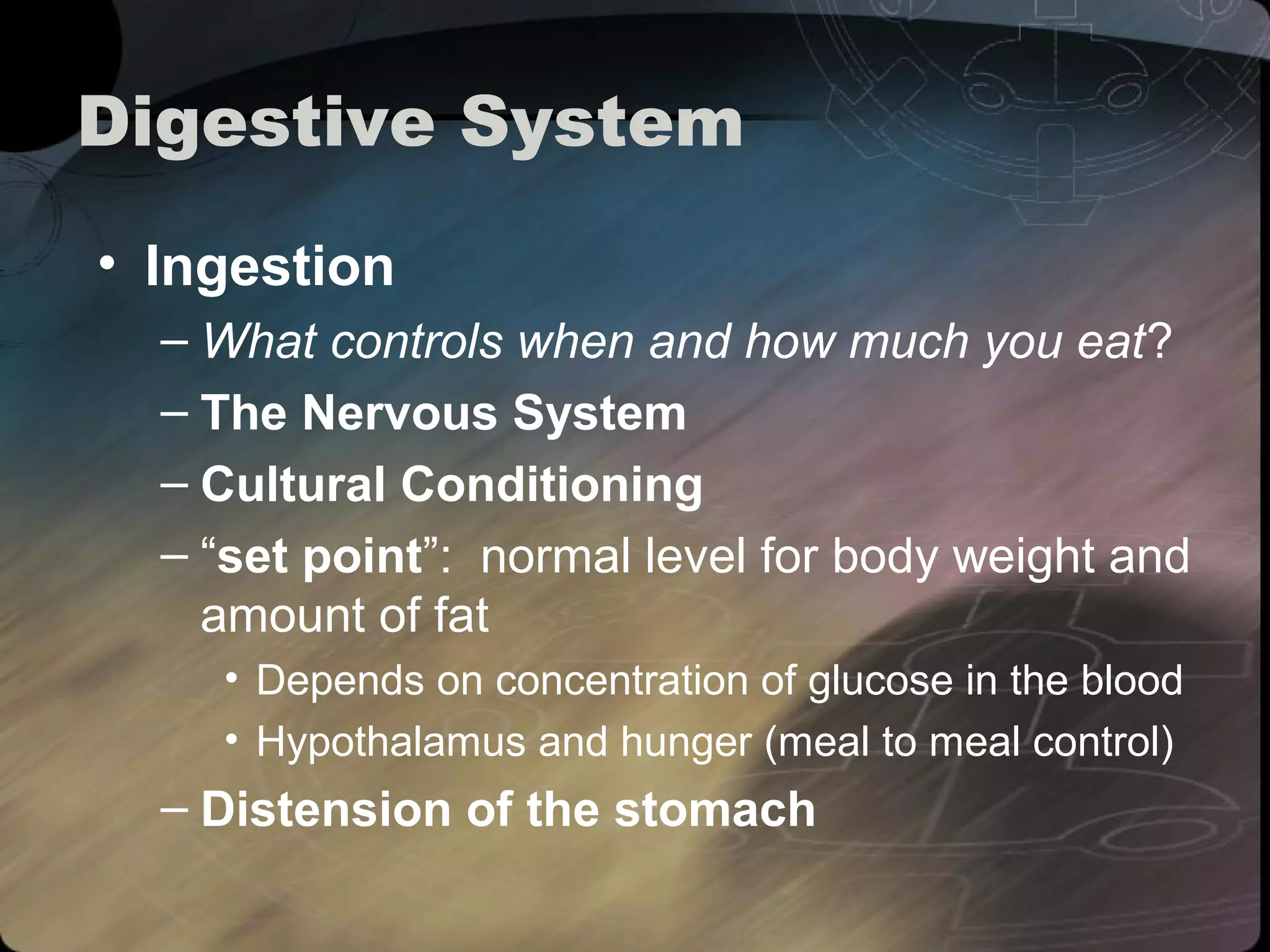
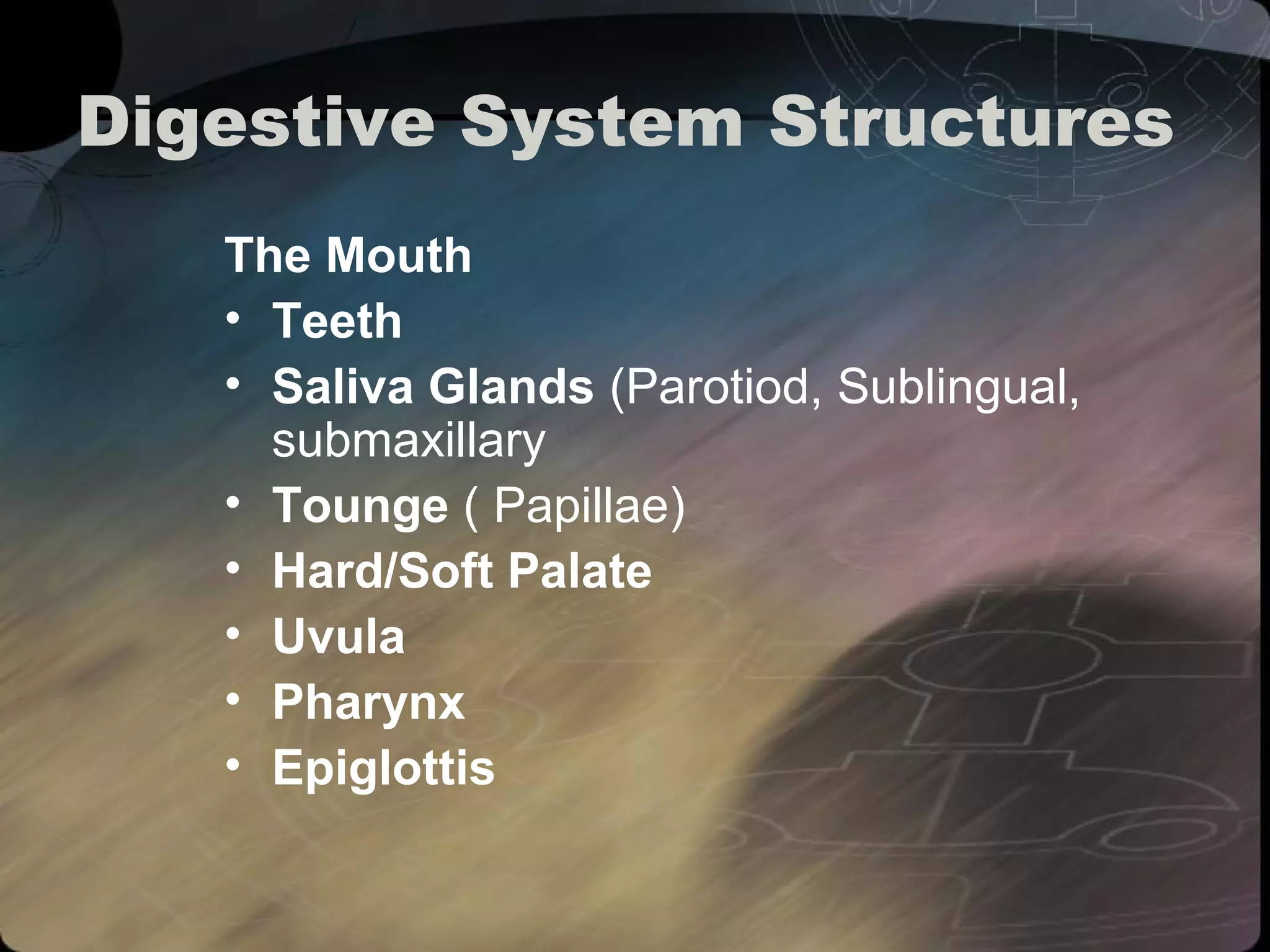
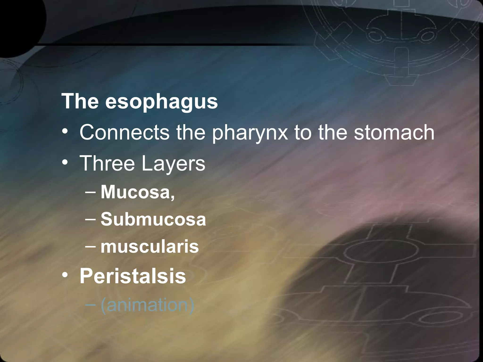
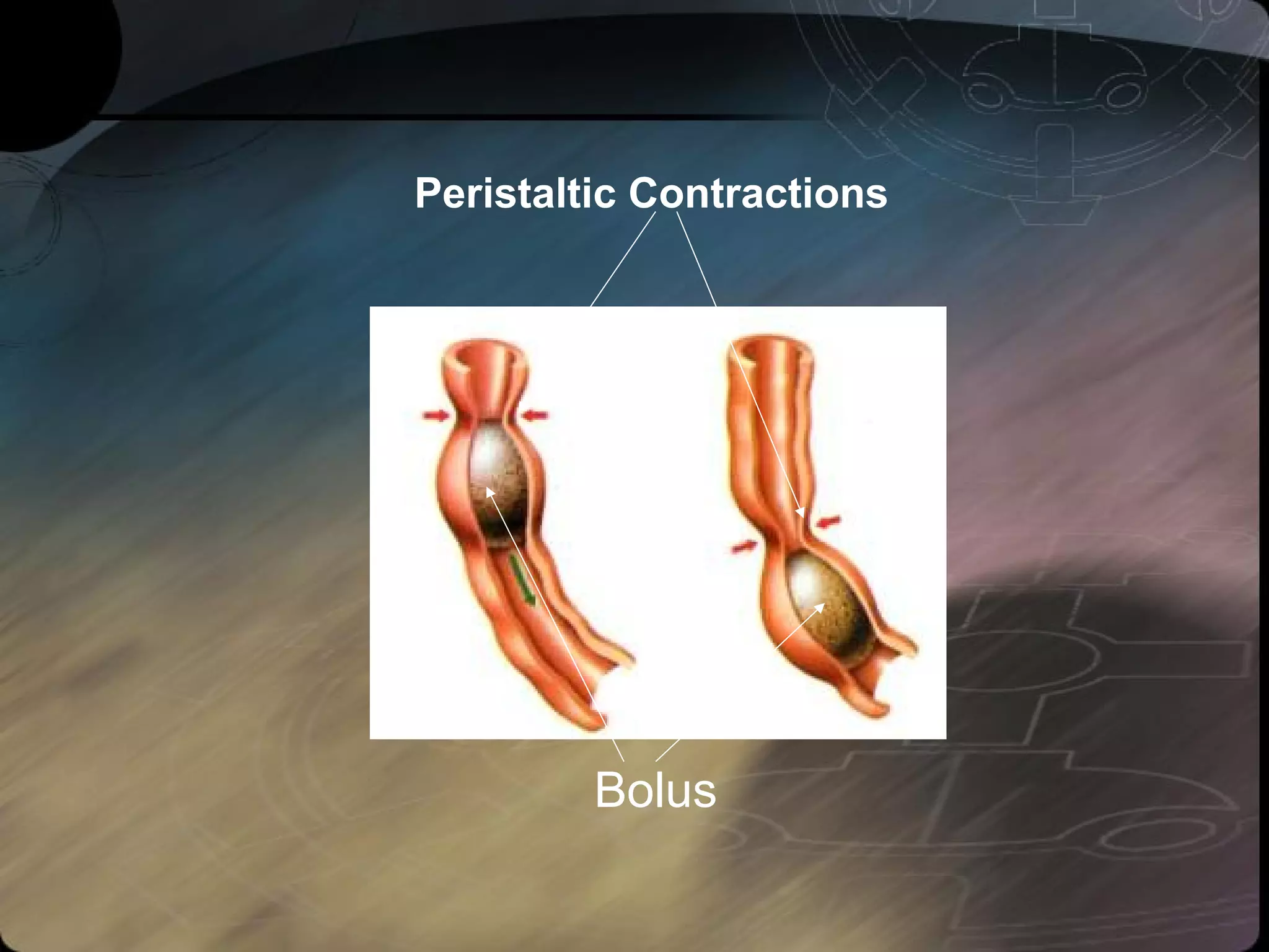
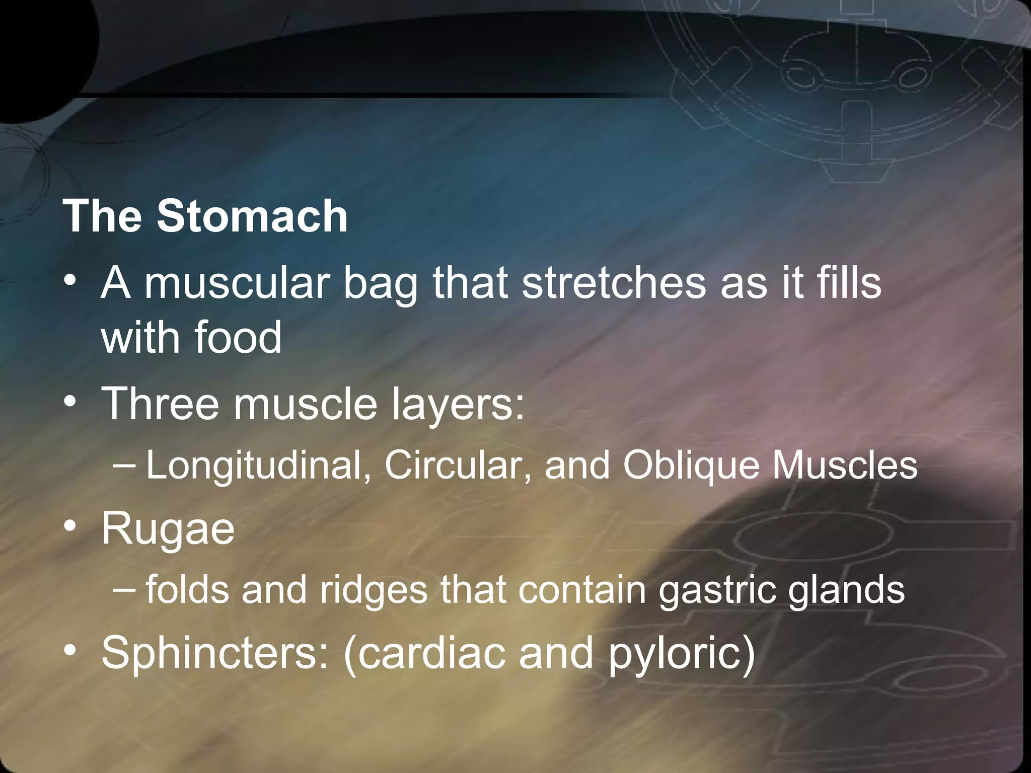
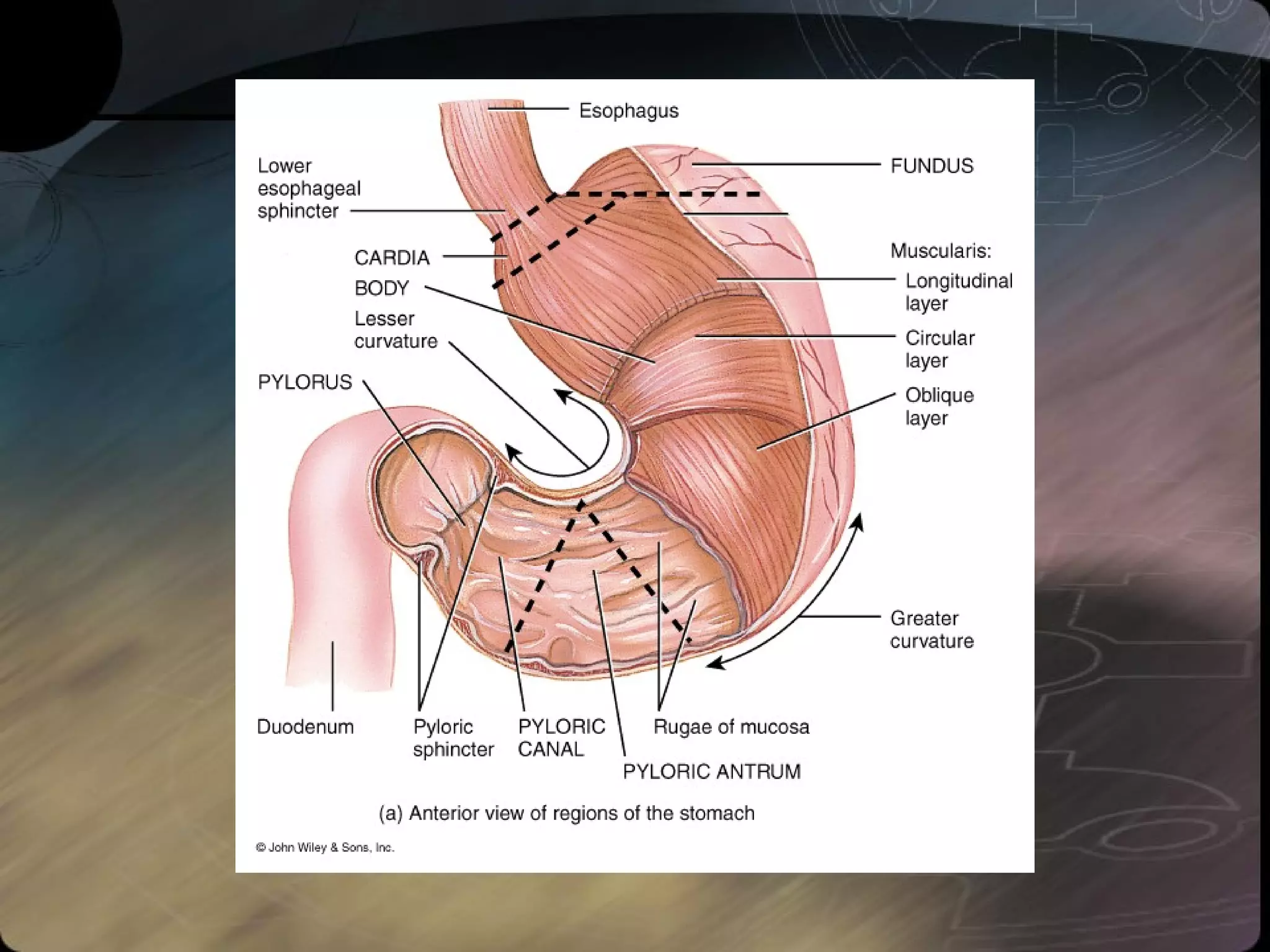
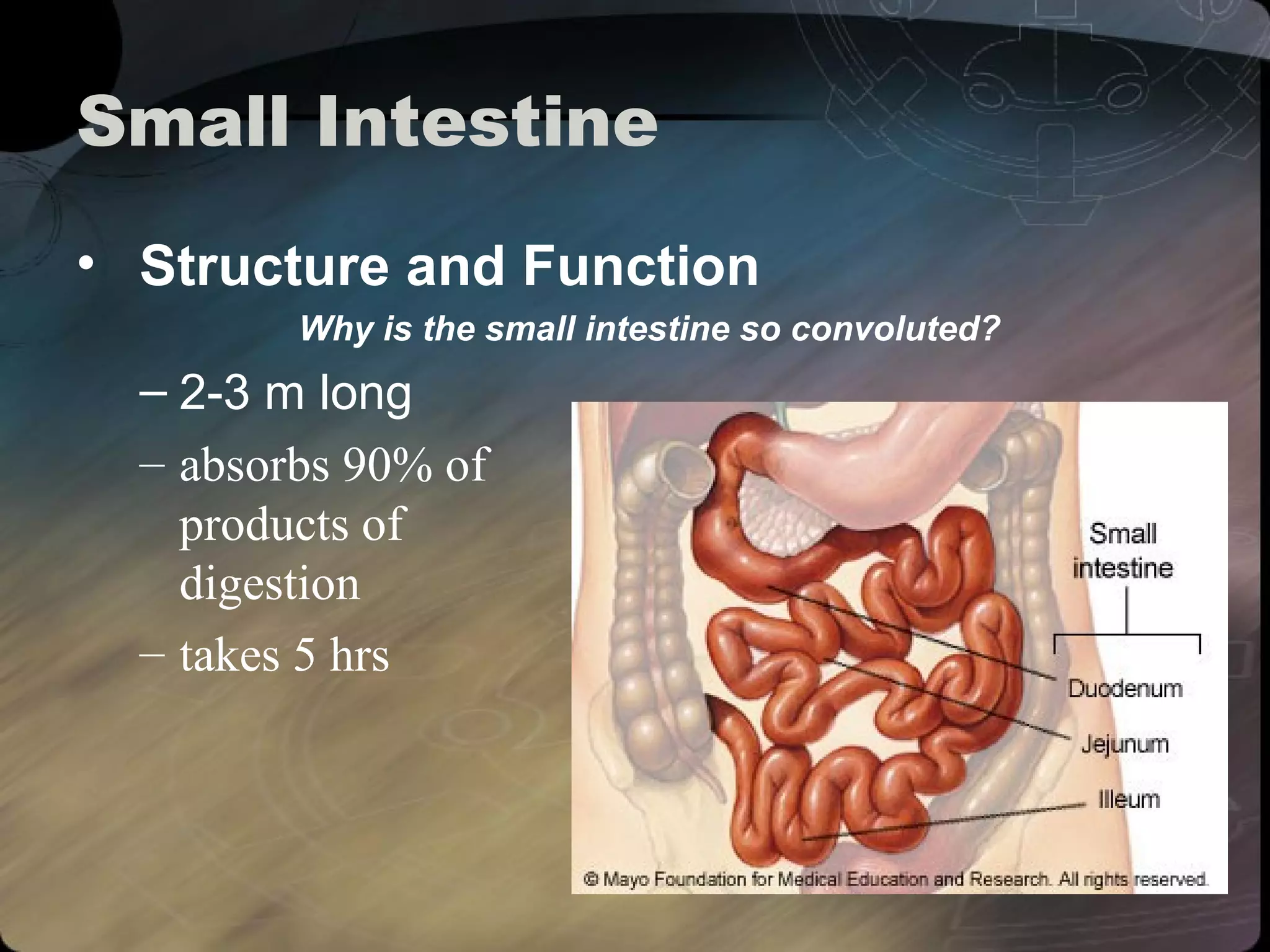
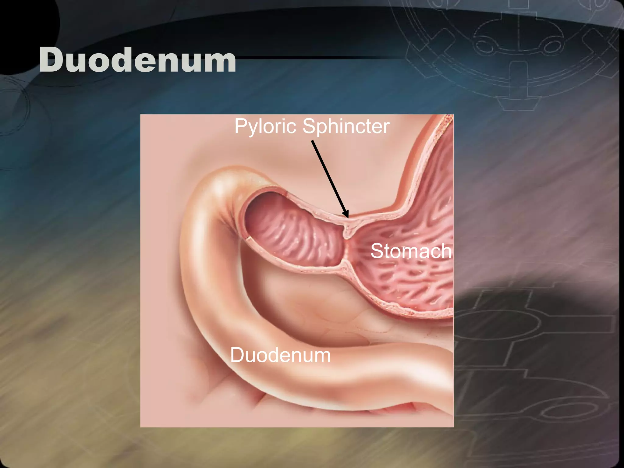
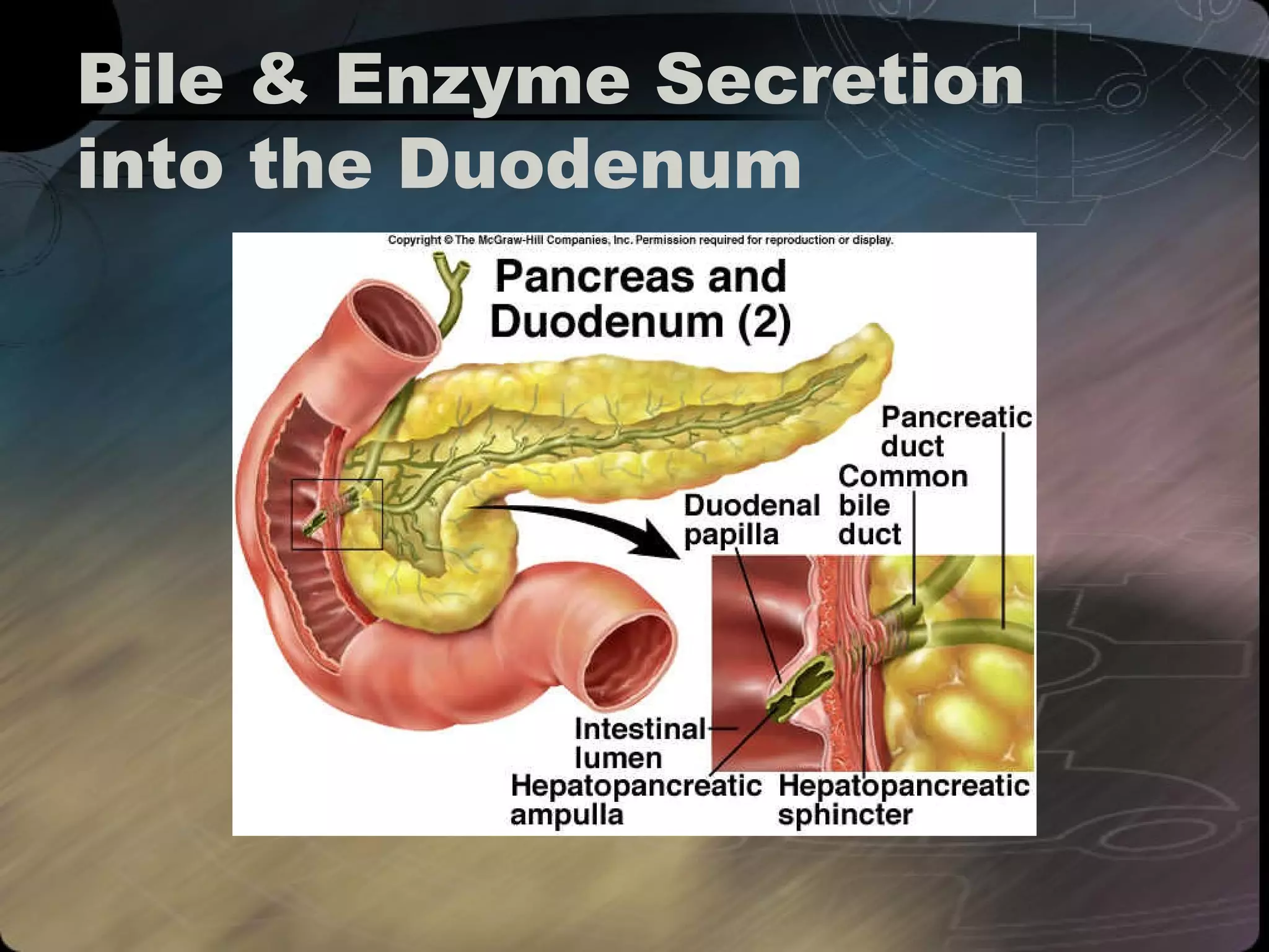
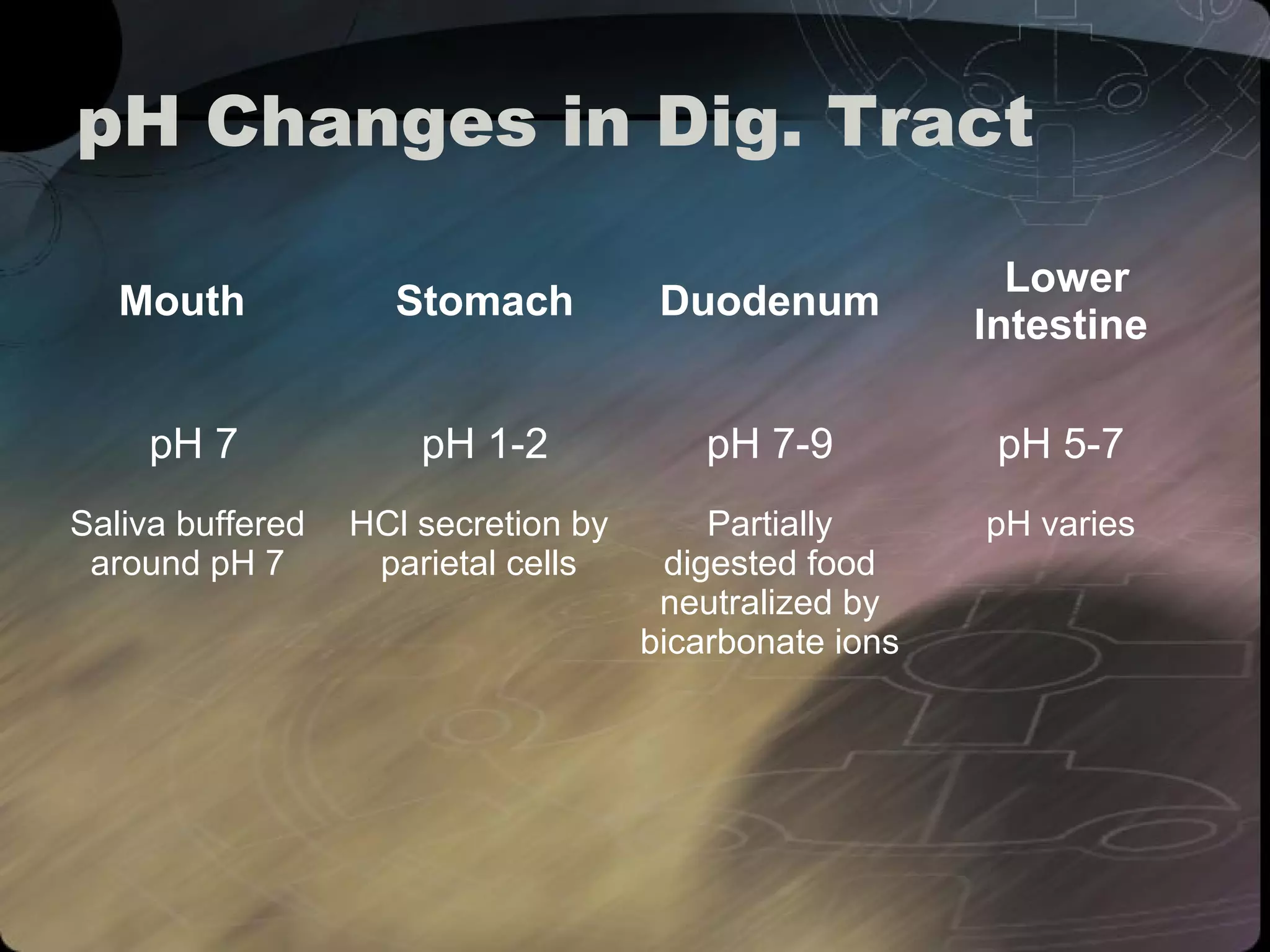
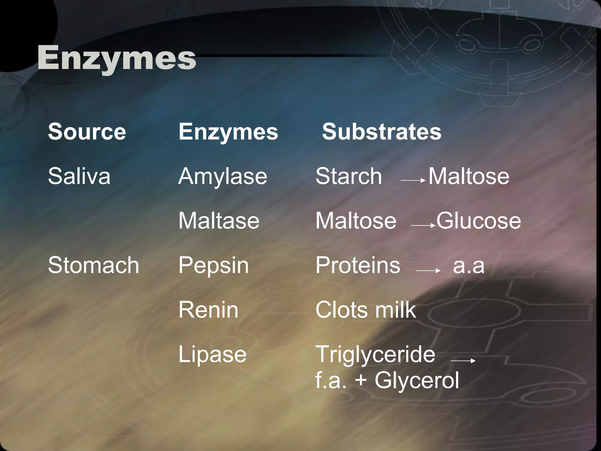
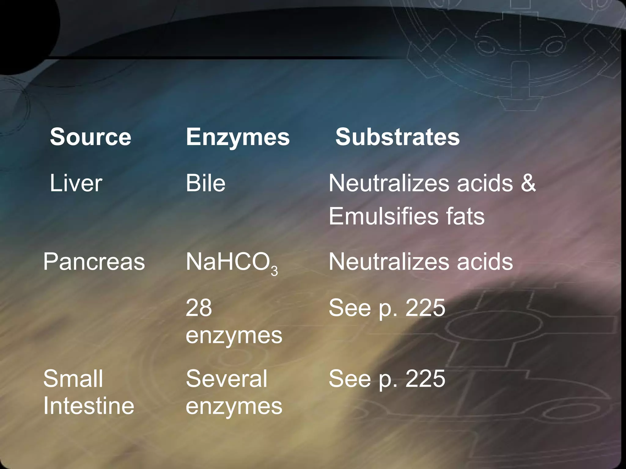
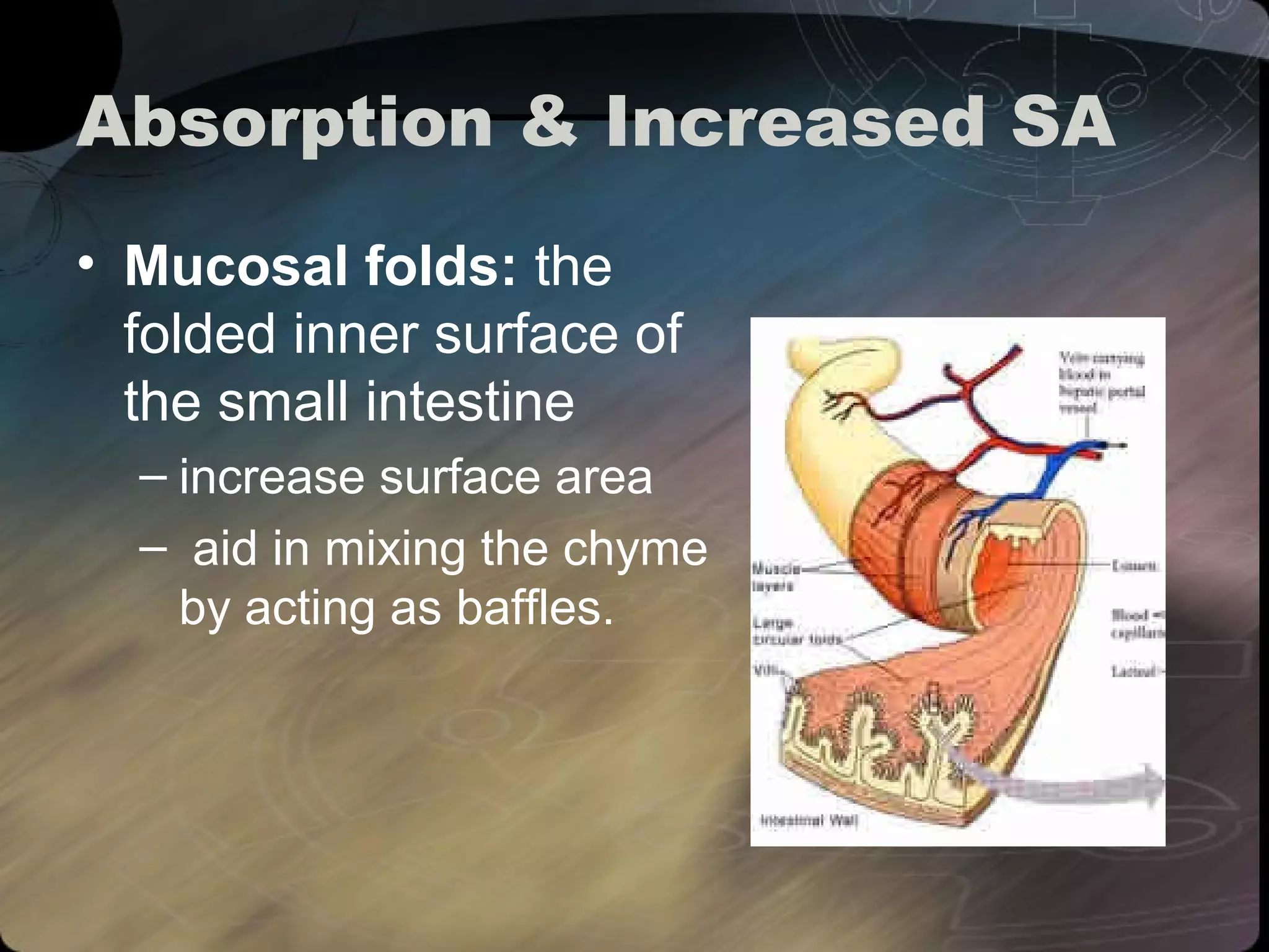
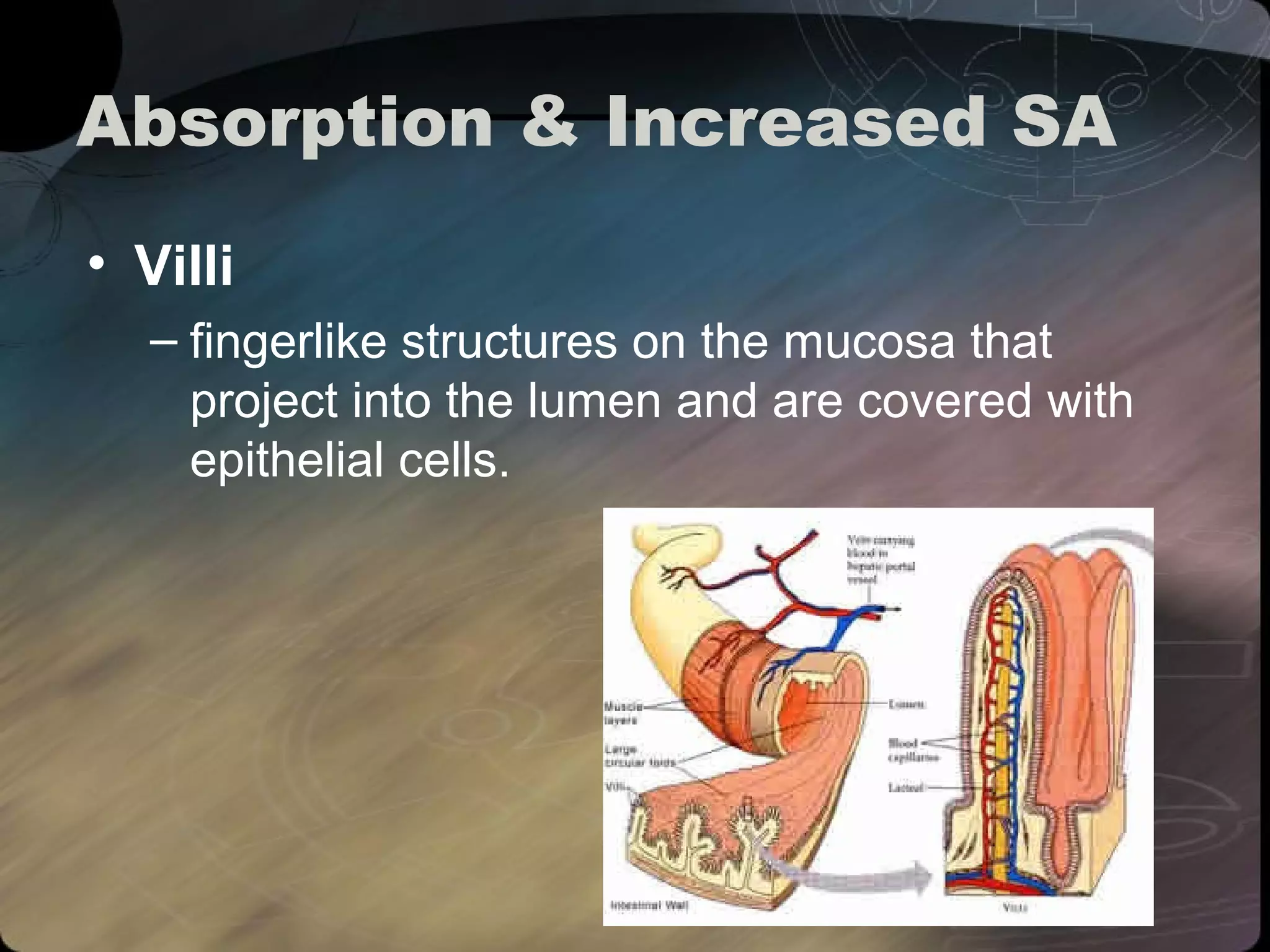
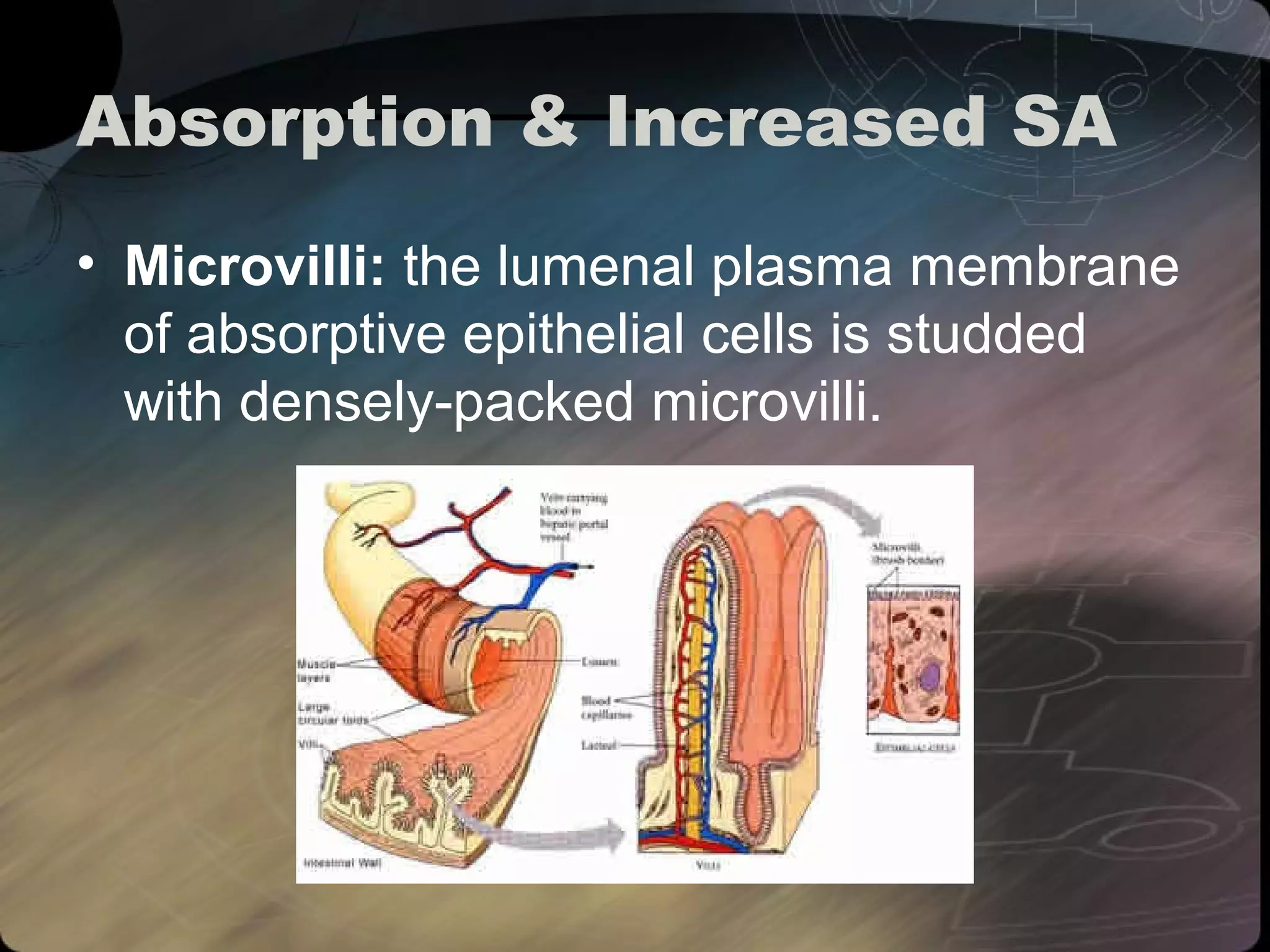
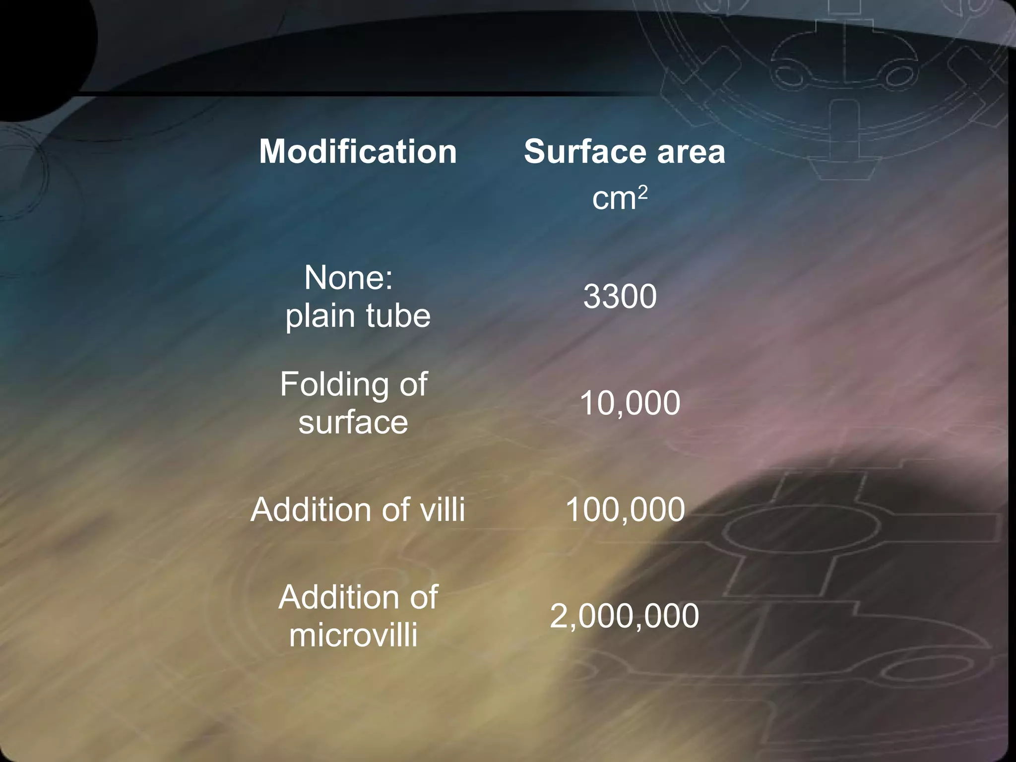
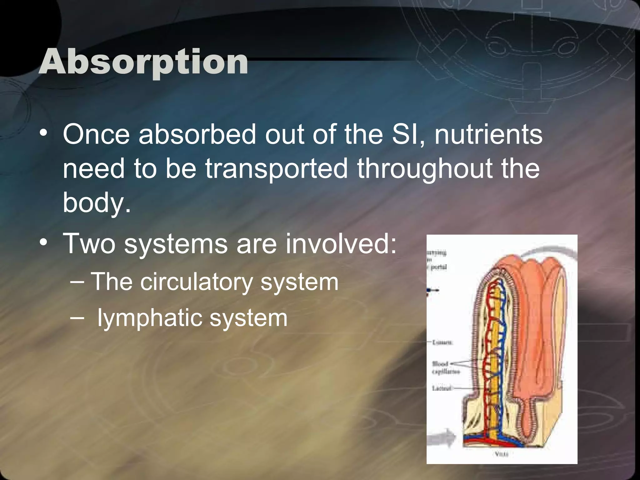
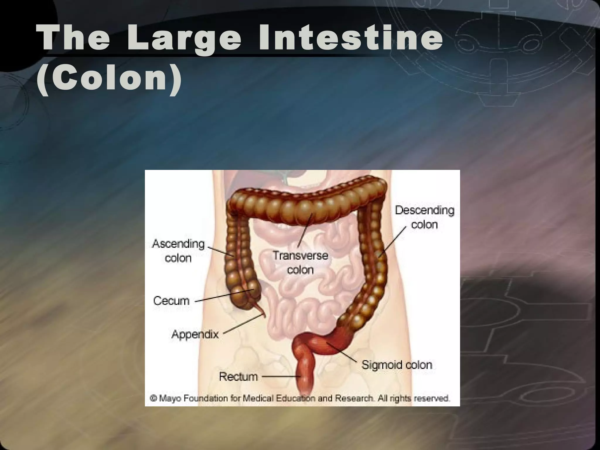
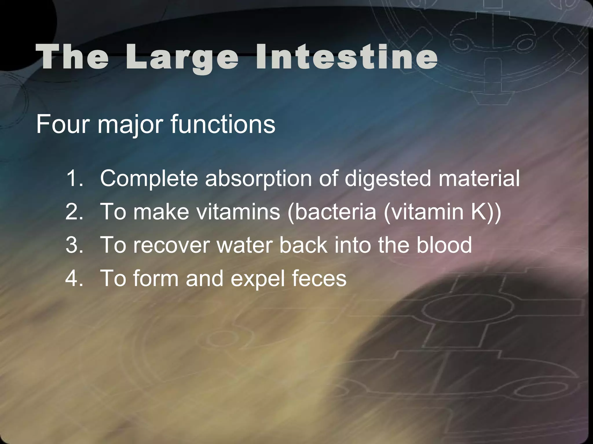
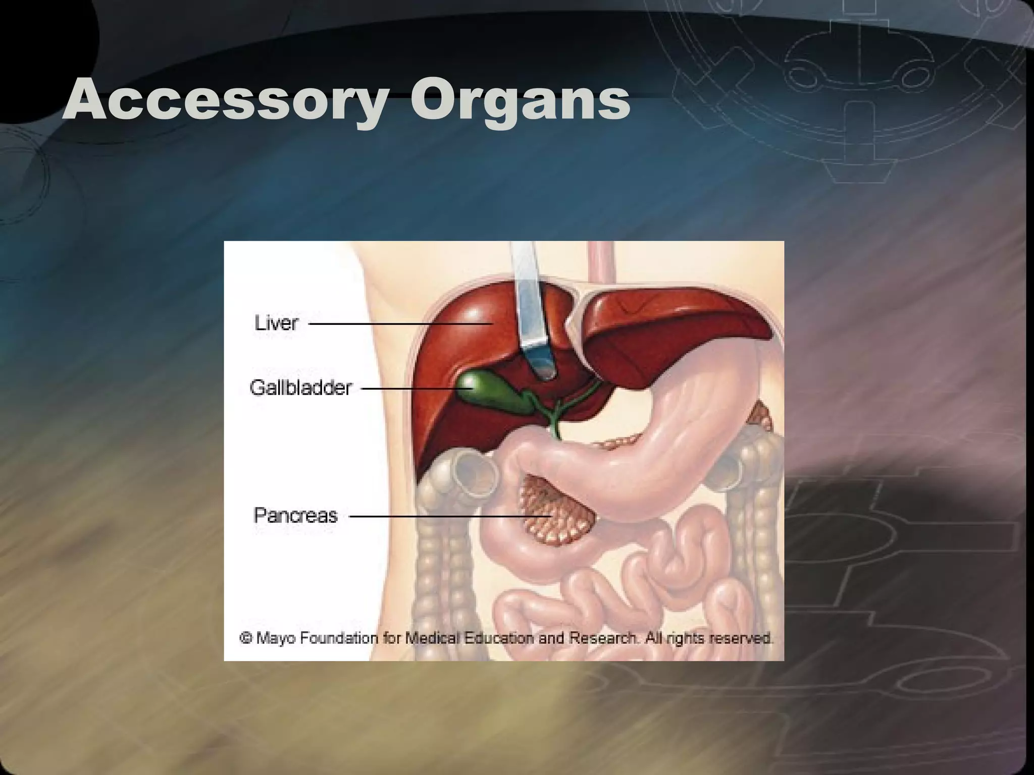
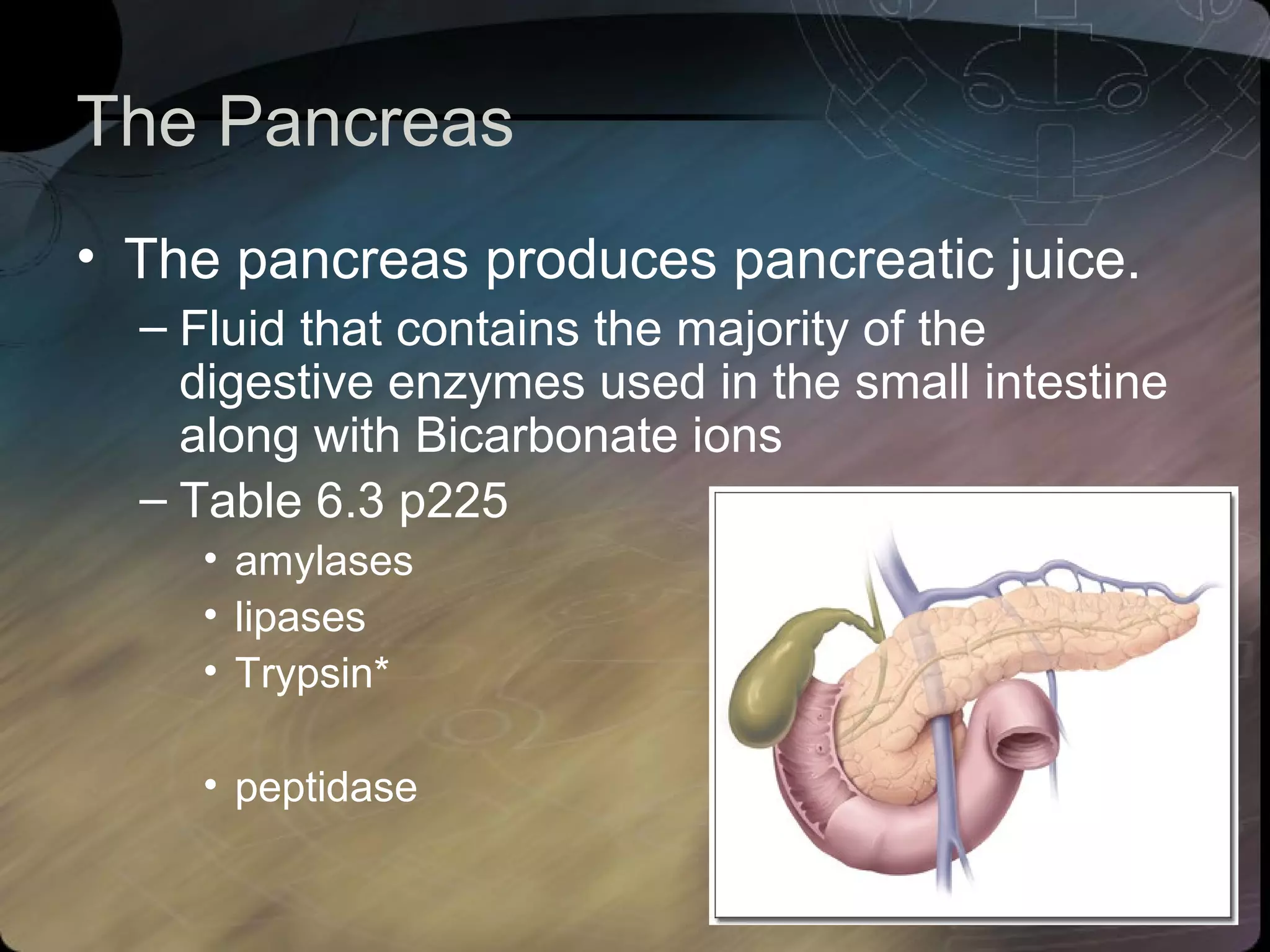
The Digestive System
The Digestive System 
• Alimentation 
– Ingestion 
– Digestion 
– Egestion
Digestive System 
• Ingestion 
– What controls when and how much you eat? 
– The Nervous System 
– Cultural Conditioning 
– “set point”: normal level for body weight and 
amount of fat 
• Depends on concentration of glucose in the blood 
• Hypothalamus and hunger (meal to meal control) 
– Distension of the stomach
Digestive System Structures 
The Mouth 
• Teeth 
• Saliva Glands (Parotiod, Sublingual, 
submaxillary 
• Tounge ( Papillae) 
• Hard/Soft Palate 
• Uvula 
• Pharynx 
• Epiglottis
The esophagus 
• Connects the pharynx to the stomach 
• Three Layers 
– Mucosa, 
– Submucosa 
– muscularis 
• Peristalsis 
– (animation)
Peristaltic Contractions 
Bolus
The Stomach 
• A muscular bag that stretches as it fills 
with food 
• Three muscle layers: 
– Longitudinal, Circular, and Oblique Muscles 
• Rugae 
– folds and ridges that contain gastric glands 
• Sphincters: (cardiac and pyloric)
Digestive system 
Small Intestine to Large Intestine
Small Intestine 
• Structure and Function 
Why is the small intestine so convoluted? 
– 2-3 m long 
– absorbs 90% of 
products of 
digestion 
– takes 5 hrs
Small Intestine 
• Duodenum: 
– approx. 20 cm long 
– receives digestive enzymes from pancreas and 
liver/gall bladder 
• Jejenum 
– approx. 1.7m long 
– most absorption occurs in Duodenum and Jejenum 
• Ileum 
– approx. 1.1m long
Duodenum 
Pyloric Sphincter 
Stomach 
Duodenum
Bile & Enzyme Secretion 
into the Duodenum
Chemical Digestion 
Enzymes
pH Changes in Dig. Tract 
Mouth Stomach Duodenum Lower 
Intestine 
pH 7 pH 1-2 pH 7-9 pH 5-7 
Saliva buffered 
around pH 7 
HCl secretion by 
parietal cells 
Partially 
digested food 
neutralized by 
bicarbonate ions 
pH varies
Enzymes 
Source Enzymes Substrates 
Saliva Amylase Starch Maltose 
Maltase Maltose Glucose 
Stomach Pepsin Proteins a.a 
Renin Clots milk 
Lipase Triglyceride 
f.a. + Glycerol
Source Enzymes Substrates 
Liver Bile Neutralizes acids & 
Emulsifies fats 
Pancreas NaHCO3 Neutralizes acids 
28 
enzymes 
See p. 225 
Small 
Intestine 
Several 
enzymes 
See p. 225
Absorption & Increased SA 
• Mucosal folds: the 
folded inner surface of 
the small intestine 
– increase surface area 
– aid in mixing the chyme 
by acting as baffles.
Absorption & Increased SA 
• Villi 
– fingerlike structures on the mucosa that 
project into the lumen and are covered with 
epithelial cells.
Absorption & Increased SA 
• Microvilli: the lumenal plasma membrane 
of absorptive epithelial cells is studded 
with densely-packed microvilli.
Modification Surface area 
cm2 
None: 
plain tube 3300 
Folding of 
surface 10,000 
Addition of villi 100,000 
Addition of 
microvilli 2,000,000
Absorption 
• Peristaltic contractions in the intestinal 
walls move the chyme through the SI and 
increases the contact between the 
digested nutrients and absorptive surfaces 
• Movement of sugars (glucose) and amino 
acids into villi is accomplished through 
active transport
Absorption 
• Once absorbed out of the SI, nutrients 
need to be transported throughout the 
body. 
• Two systems are involved: 
– The circulatory system 
– lymphatic system
Absorption 
• These transport structures are located in 
close proximity to absorptive surfaces 
– Capillary Network 
• within the villi, absorbed glucose and amino acids 
enter directly into the blood of the capillaries that 
will then be transported to the liver before being 
distributed throughout the body.
Absorption 
• Lacteal: 
– vessel in the villi that takes 
absorbed fats and 
transports them to larger 
lymphatic vessels that lead 
to the liver
Enzymes secreted by S.I. 
• carbohydrases 
(list 3) 
• enterokinase* 
• peptidases
Absorption
The Large Intestine 
(Colon)
The Large Intestine 
Four major functions 
1. Complete absorption of digested material 
2. To make vitamins (bacteria (vitamin K)) 
3. To recover water back into the blood 
4. To form and expel feces
Accessory Organs
Accessory Organs 
• Functions of the Liver 
– Production of bile (stored in the gallbladder) 
– The liver helps to maintain balance of 
nutrients in the body by regulating the 
concentration of nutrients in the blood before 
being sent out to the body.
Functions of the Liver 
• Interconversion of carbohydrates to fats and 
a.a.’s to carbohydrates or fats. 
• Glycogen 
– Low glucose levels in the blood cause the release of 
the hormone glucagon that stimulates the breakdown 
of glycogen into glucose. 
– When no glucose or glycogen is available, a.a.’s are 
converted into glucose in the liver. The process of 
deamination removes the amino groups from amino 
acids. Urea is formed and passed through the blood 
to the kidney for export from the body.
Functions of the Liver 
– Storage of fat soluble vitamins and iron 
– Synthesizes cholesterol and modifies lipids 
coming from fat-storage tissues 
– Responsible for break down of amino acids 
and converts ammonia to urea 
– Detoxifies poisonous chemical substances 
such as alcohol and drugs 
– Produces proteins found in the blood that fight 
against infection and clotting
The Pancreas 
• The pancreas produces pancreatic juice. 
– Fluid that contains the majority of the 
digestive enzymes used in the small intestine 
along with Bicarbonate ions 
– Table 6.3 p225 
• amylases 
• lipases 
• Trypsin* 
• peptidase

Digestive System

  • 1.
  • 2.
    The Digestive System • Alimentation – Ingestion – Digestion – Egestion
  • 3.
    Digestive System •Ingestion – What controls when and how much you eat? – The Nervous System – Cultural Conditioning – “set point”: normal level for body weight and amount of fat • Depends on concentration of glucose in the blood • Hypothalamus and hunger (meal to meal control) – Distension of the stomach
  • 4.
    Digestive System Structures The Mouth • Teeth • Saliva Glands (Parotiod, Sublingual, submaxillary • Tounge ( Papillae) • Hard/Soft Palate • Uvula • Pharynx • Epiglottis
  • 6.
    The esophagus •Connects the pharynx to the stomach • Three Layers – Mucosa, – Submucosa – muscularis • Peristalsis – (animation)
  • 7.
  • 8.
    The Stomach •A muscular bag that stretches as it fills with food • Three muscle layers: – Longitudinal, Circular, and Oblique Muscles • Rugae – folds and ridges that contain gastric glands • Sphincters: (cardiac and pyloric)
  • 10.
    Digestive system SmallIntestine to Large Intestine
  • 11.
    Small Intestine •Structure and Function Why is the small intestine so convoluted? – 2-3 m long – absorbs 90% of products of digestion – takes 5 hrs
  • 12.
    Small Intestine •Duodenum: – approx. 20 cm long – receives digestive enzymes from pancreas and liver/gall bladder • Jejenum – approx. 1.7m long – most absorption occurs in Duodenum and Jejenum • Ileum – approx. 1.1m long
  • 13.
    Duodenum Pyloric Sphincter Stomach Duodenum
  • 14.
    Bile & EnzymeSecretion into the Duodenum
  • 15.
  • 16.
    pH Changes inDig. Tract Mouth Stomach Duodenum Lower Intestine pH 7 pH 1-2 pH 7-9 pH 5-7 Saliva buffered around pH 7 HCl secretion by parietal cells Partially digested food neutralized by bicarbonate ions pH varies
  • 17.
    Enzymes Source EnzymesSubstrates Saliva Amylase Starch Maltose Maltase Maltose Glucose Stomach Pepsin Proteins a.a Renin Clots milk Lipase Triglyceride f.a. + Glycerol
  • 18.
    Source Enzymes Substrates Liver Bile Neutralizes acids & Emulsifies fats Pancreas NaHCO3 Neutralizes acids 28 enzymes See p. 225 Small Intestine Several enzymes See p. 225
  • 19.
    Absorption & IncreasedSA • Mucosal folds: the folded inner surface of the small intestine – increase surface area – aid in mixing the chyme by acting as baffles.
  • 20.
    Absorption & IncreasedSA • Villi – fingerlike structures on the mucosa that project into the lumen and are covered with epithelial cells.
  • 21.
    Absorption & IncreasedSA • Microvilli: the lumenal plasma membrane of absorptive epithelial cells is studded with densely-packed microvilli.
  • 22.
    Modification Surface area cm2 None: plain tube 3300 Folding of surface 10,000 Addition of villi 100,000 Addition of microvilli 2,000,000
  • 23.
    Absorption • Peristalticcontractions in the intestinal walls move the chyme through the SI and increases the contact between the digested nutrients and absorptive surfaces • Movement of sugars (glucose) and amino acids into villi is accomplished through active transport
  • 24.
    Absorption • Onceabsorbed out of the SI, nutrients need to be transported throughout the body. • Two systems are involved: – The circulatory system – lymphatic system
  • 25.
    Absorption • Thesetransport structures are located in close proximity to absorptive surfaces – Capillary Network • within the villi, absorbed glucose and amino acids enter directly into the blood of the capillaries that will then be transported to the liver before being distributed throughout the body.
  • 26.
    Absorption • Lacteal: – vessel in the villi that takes absorbed fats and transports them to larger lymphatic vessels that lead to the liver
  • 27.
    Enzymes secreted byS.I. • carbohydrases (list 3) • enterokinase* • peptidases
  • 28.
  • 29.
  • 30.
    The Large Intestine Four major functions 1. Complete absorption of digested material 2. To make vitamins (bacteria (vitamin K)) 3. To recover water back into the blood 4. To form and expel feces
  • 31.
  • 32.
    Accessory Organs •Functions of the Liver – Production of bile (stored in the gallbladder) – The liver helps to maintain balance of nutrients in the body by regulating the concentration of nutrients in the blood before being sent out to the body.
  • 33.
    Functions of theLiver • Interconversion of carbohydrates to fats and a.a.’s to carbohydrates or fats. • Glycogen – Low glucose levels in the blood cause the release of the hormone glucagon that stimulates the breakdown of glycogen into glucose. – When no glucose or glycogen is available, a.a.’s are converted into glucose in the liver. The process of deamination removes the amino groups from amino acids. Urea is formed and passed through the blood to the kidney for export from the body.
  • 34.
    Functions of theLiver – Storage of fat soluble vitamins and iron – Synthesizes cholesterol and modifies lipids coming from fat-storage tissues – Responsible for break down of amino acids and converts ammonia to urea – Detoxifies poisonous chemical substances such as alcohol and drugs – Produces proteins found in the blood that fight against infection and clotting
  • 35.
    The Pancreas •The pancreas produces pancreatic juice. – Fluid that contains the majority of the digestive enzymes used in the small intestine along with Bicarbonate ions – Table 6.3 p225 • amylases • lipases • Trypsin* • peptidase