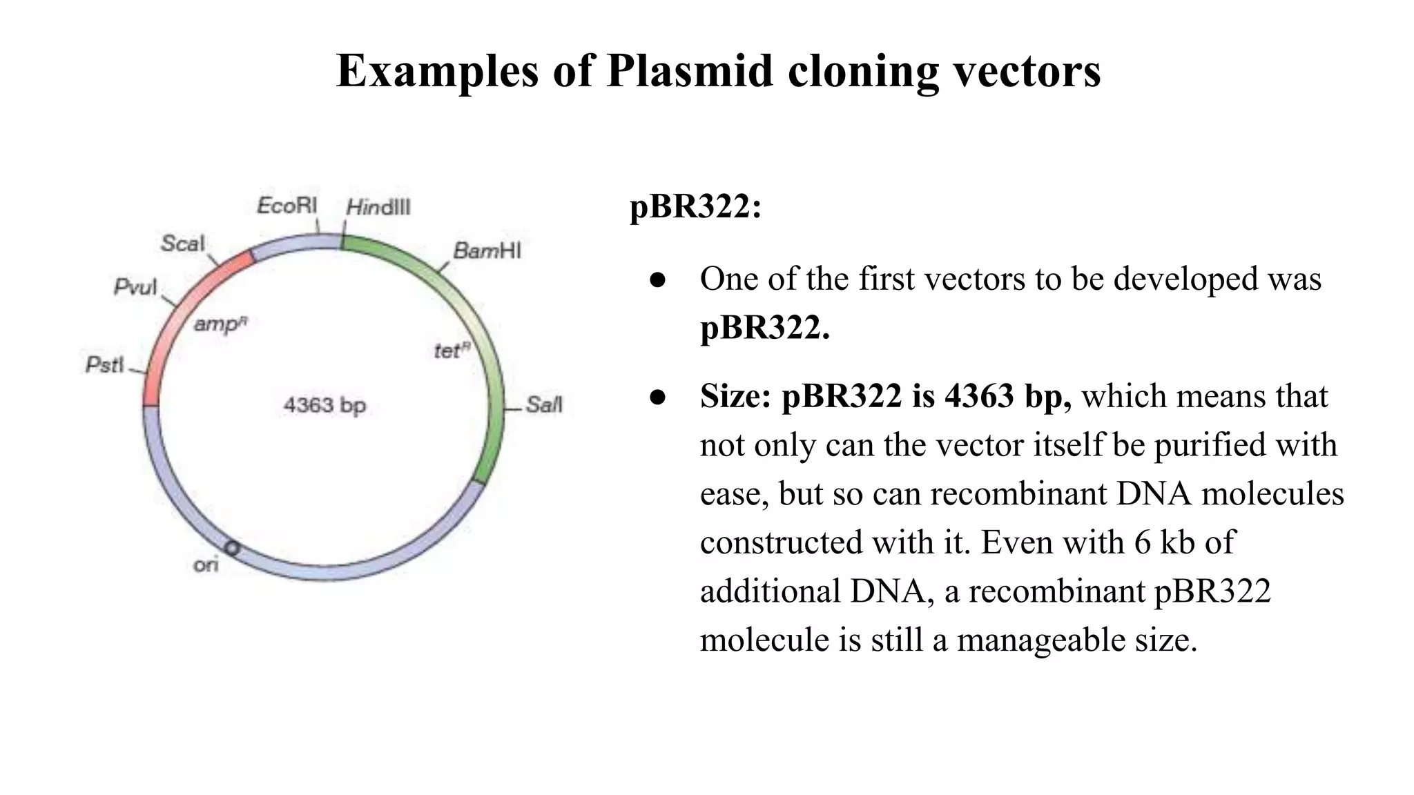
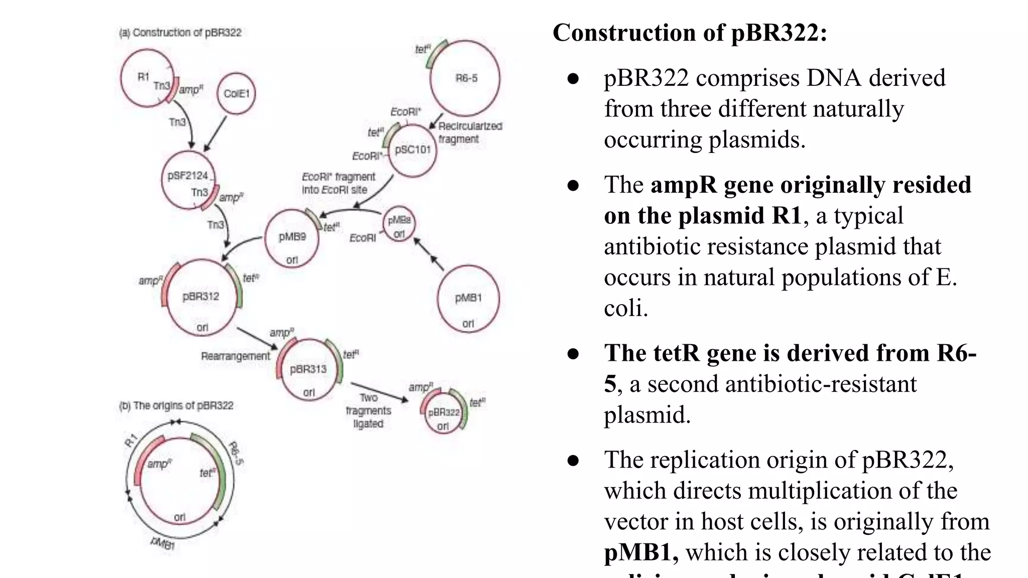
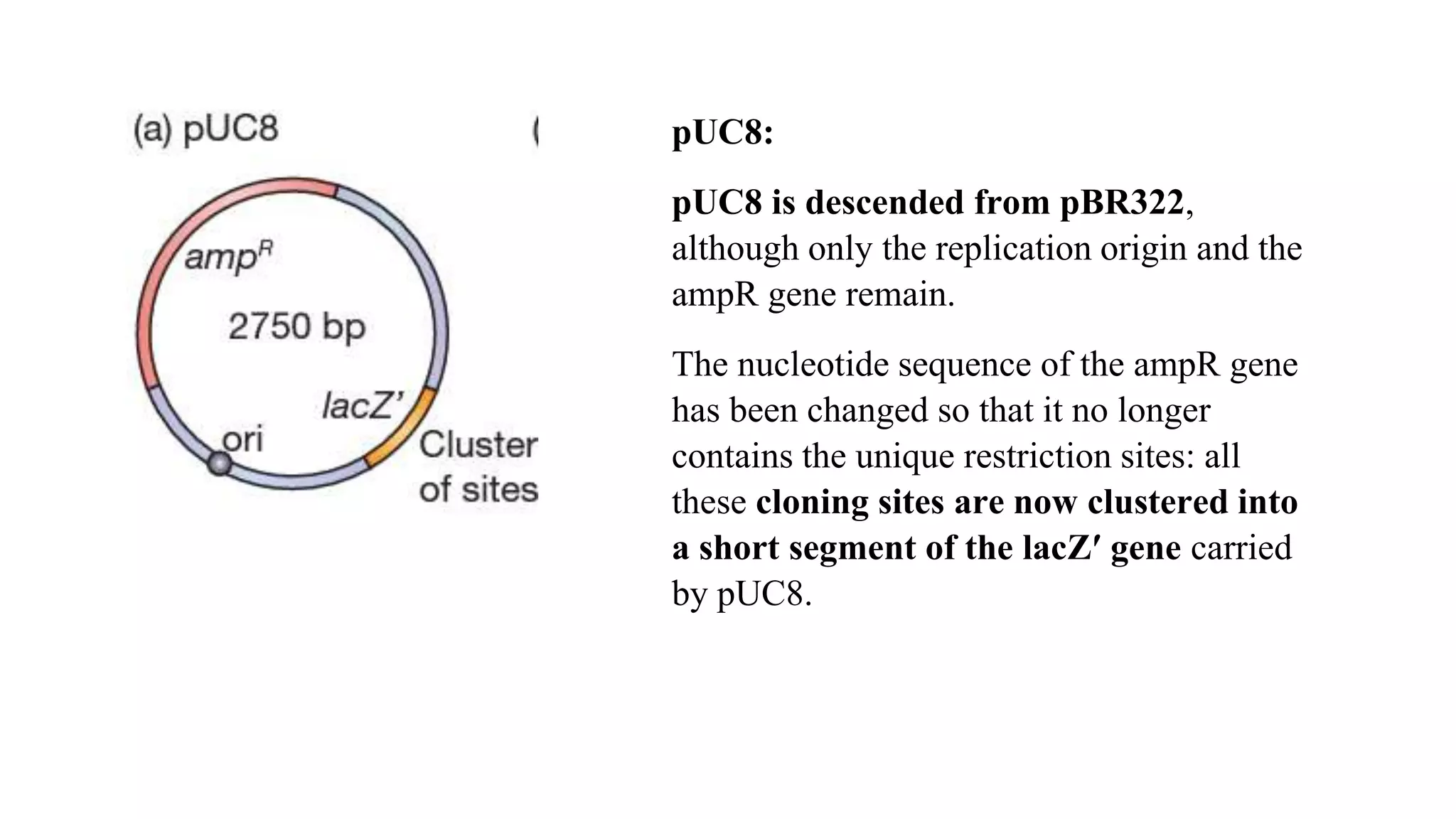
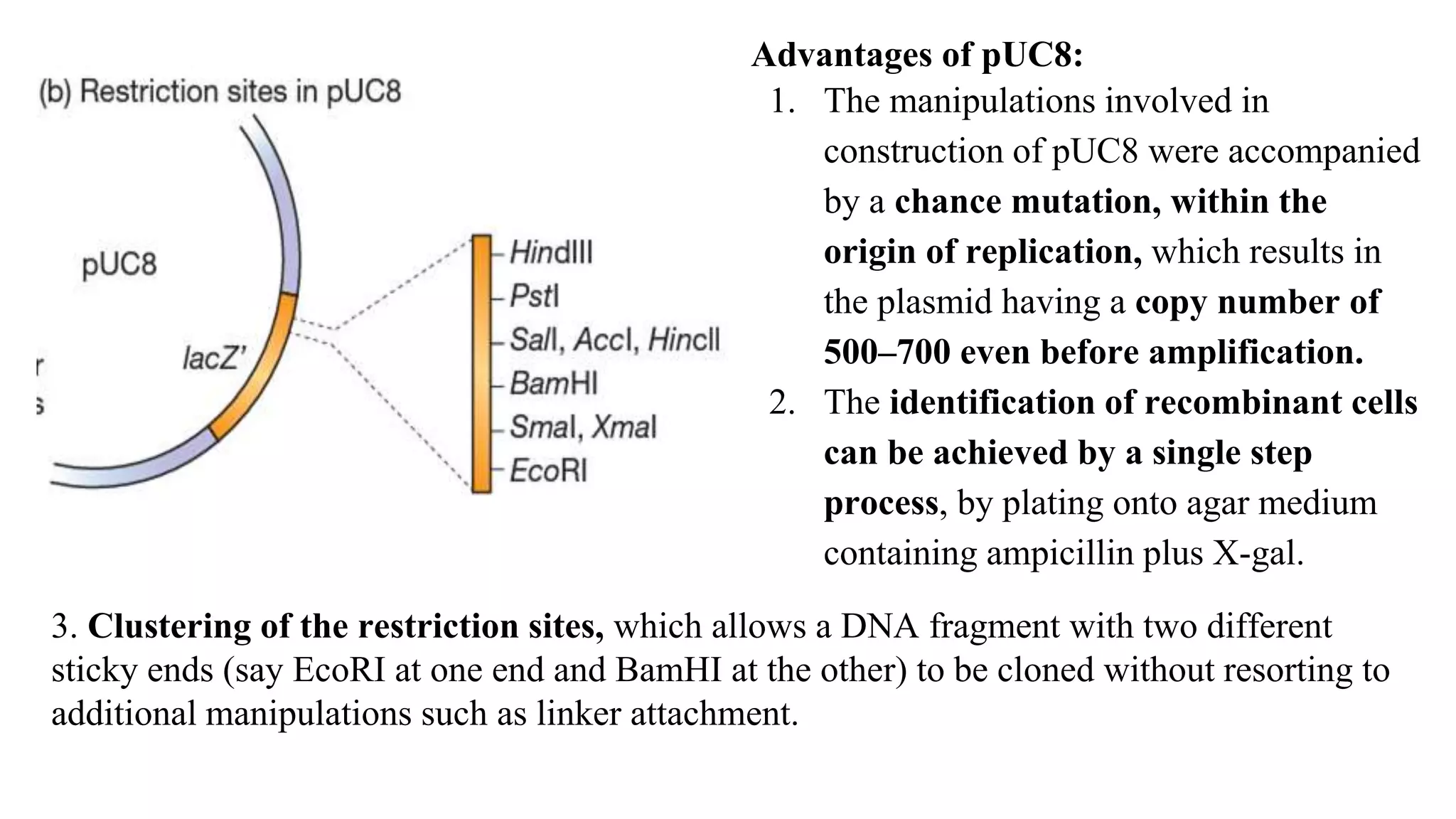
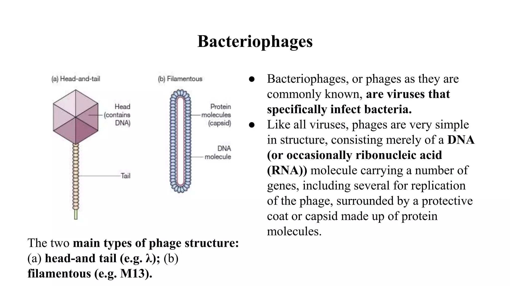
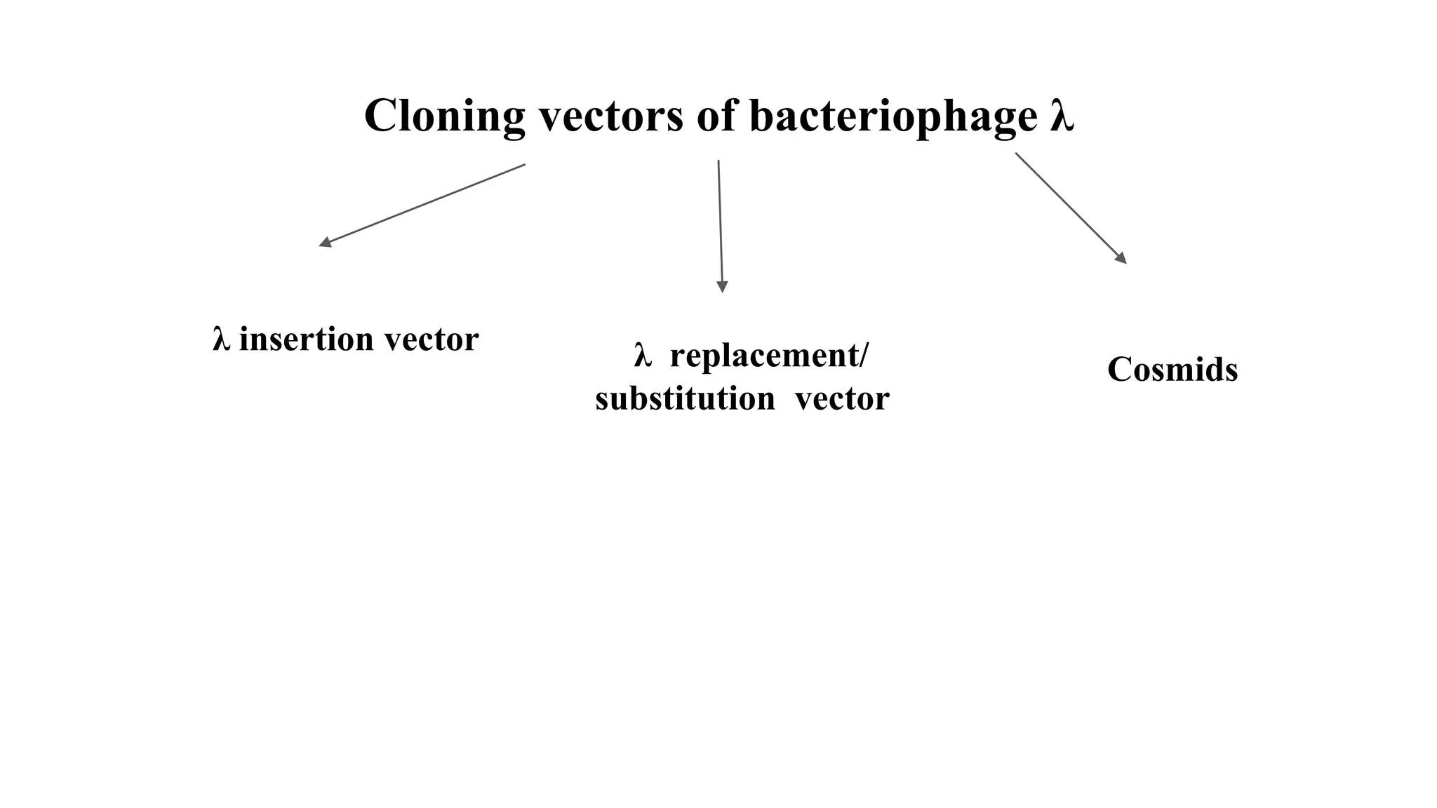
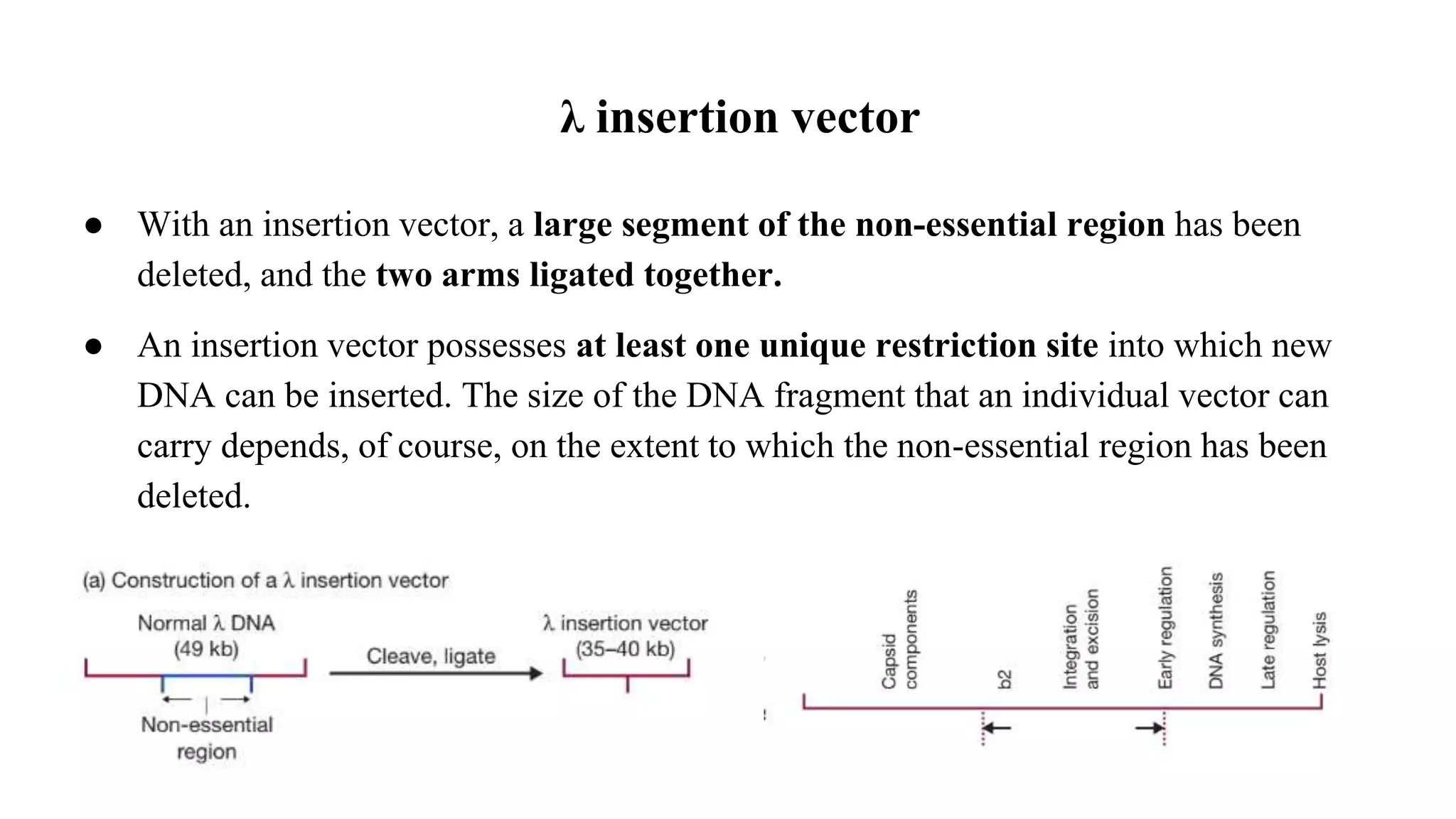
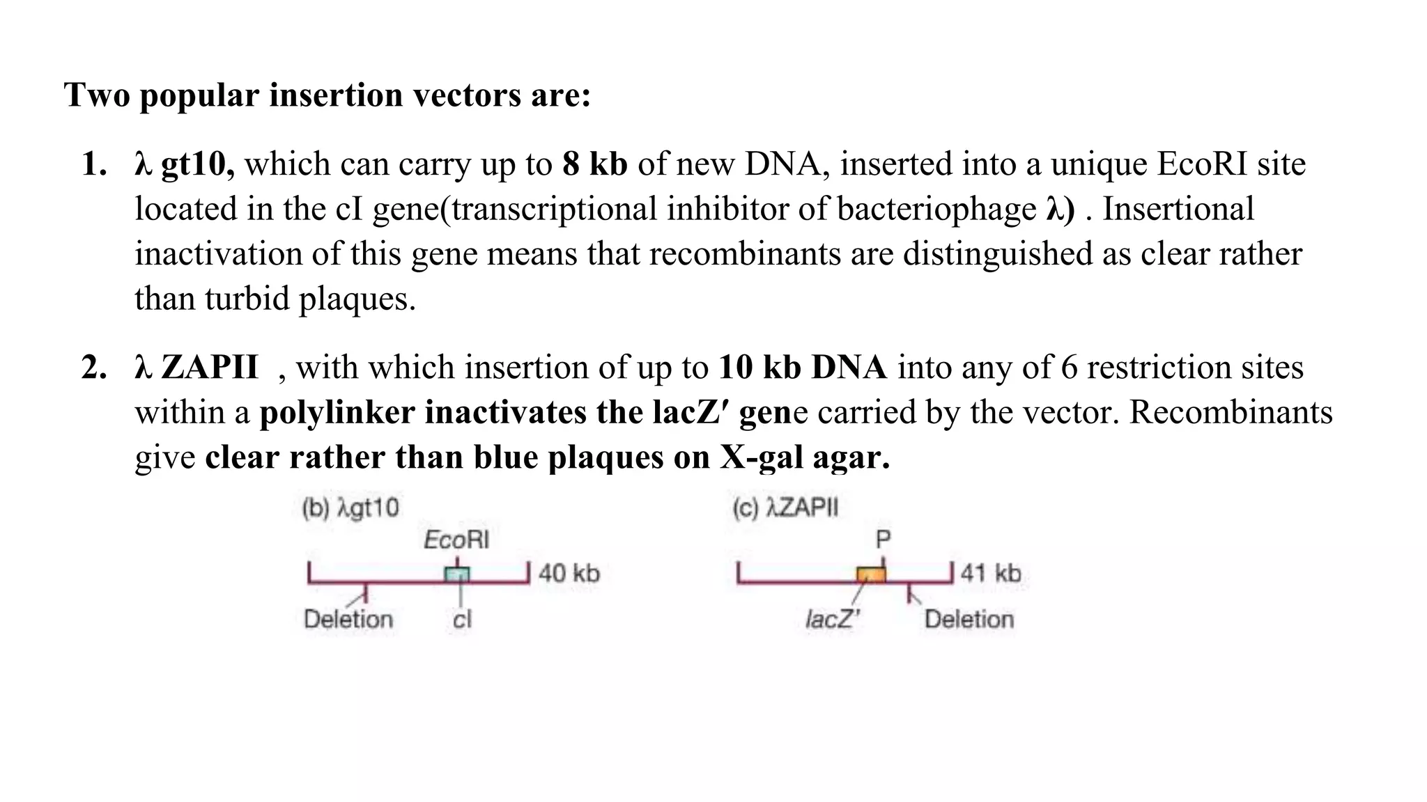
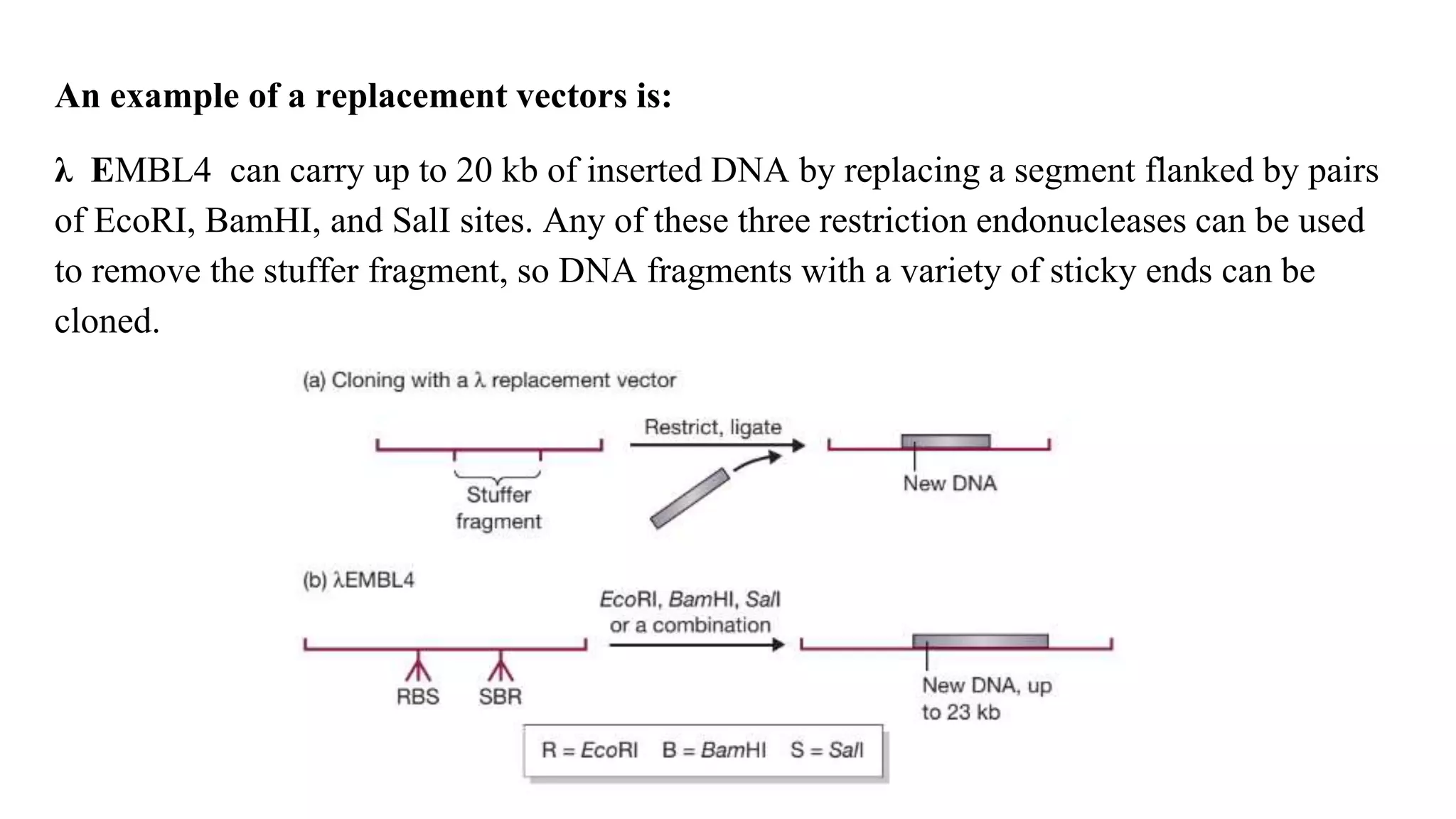
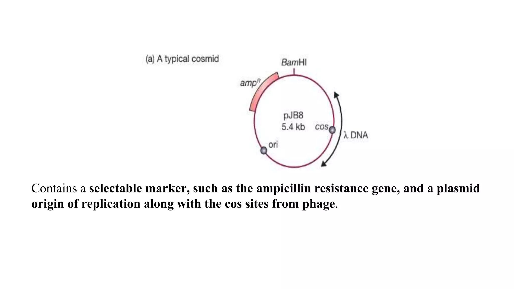
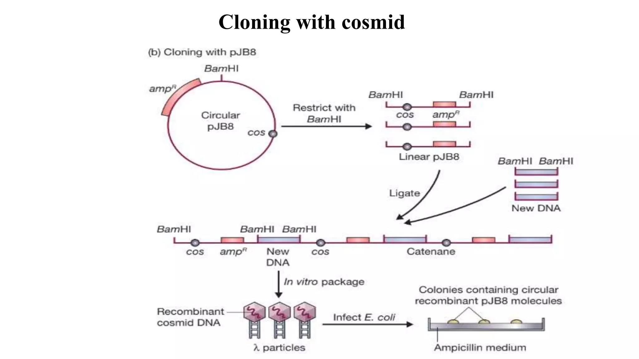
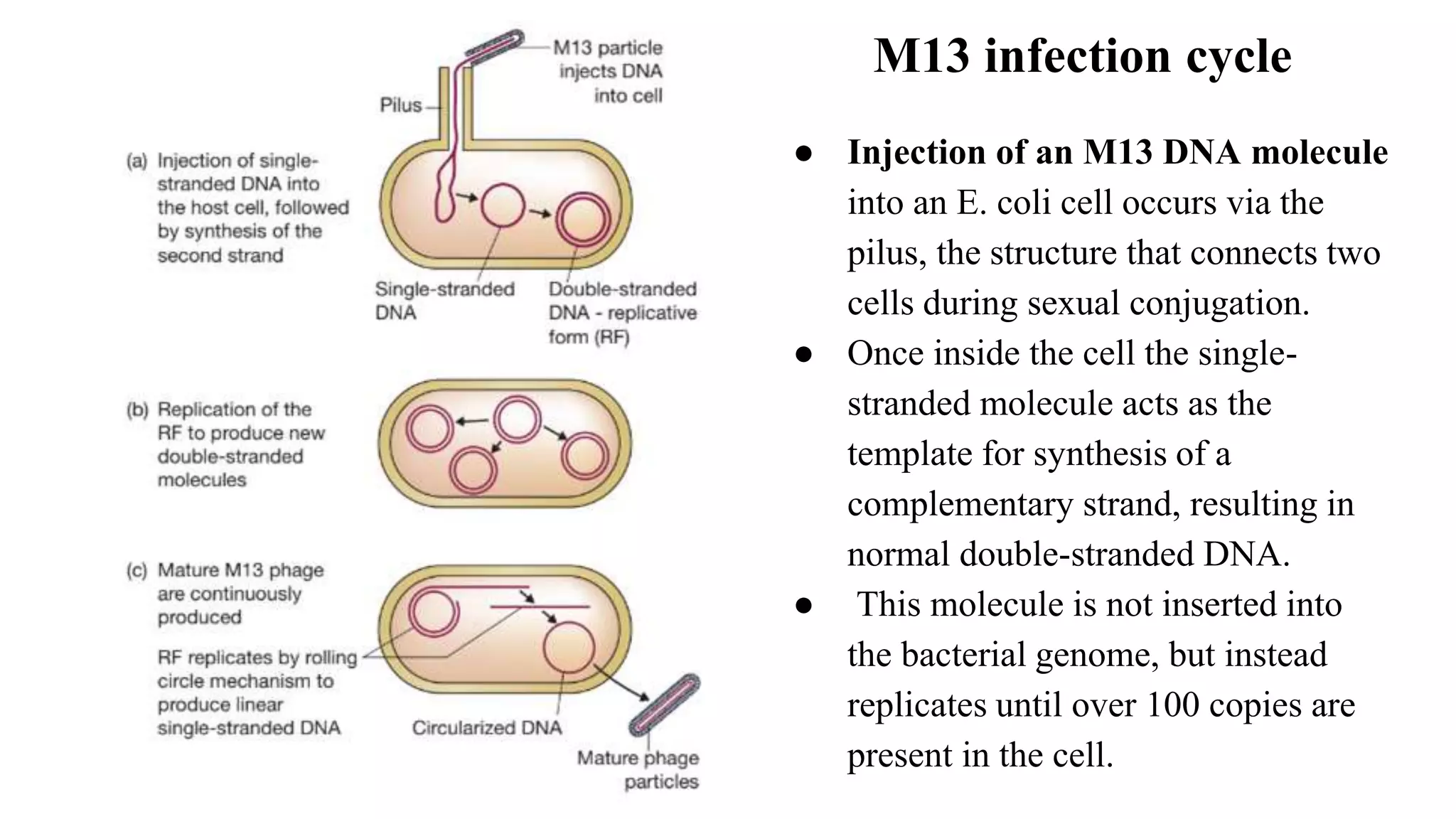
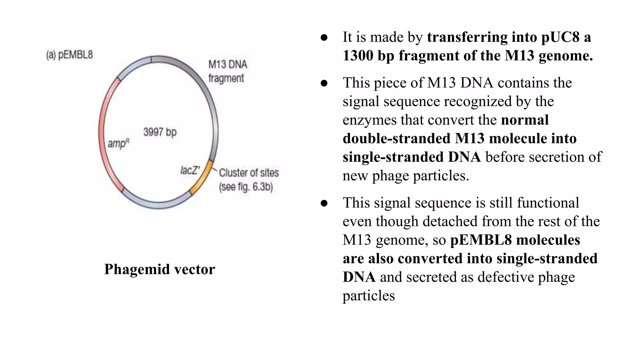
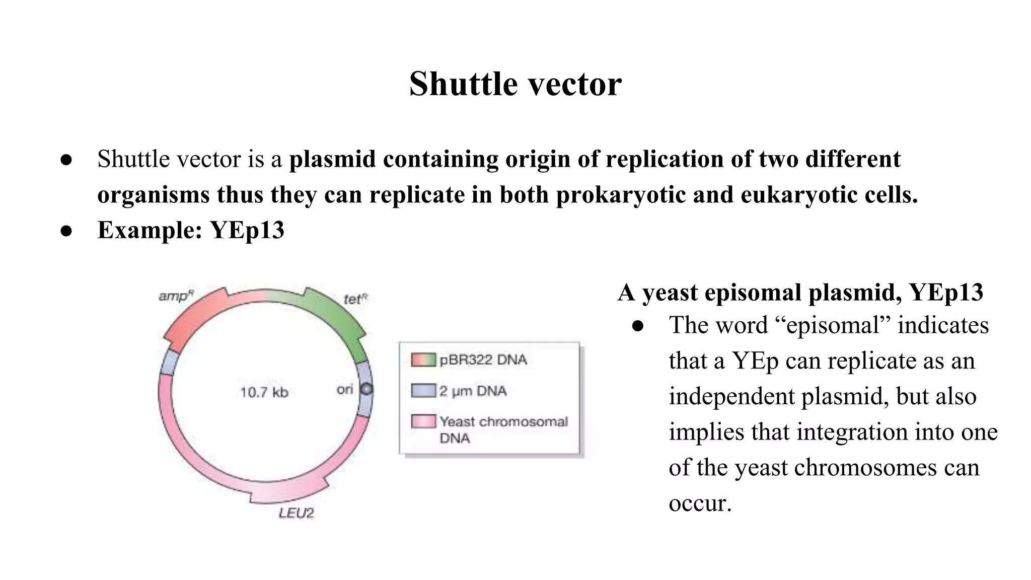
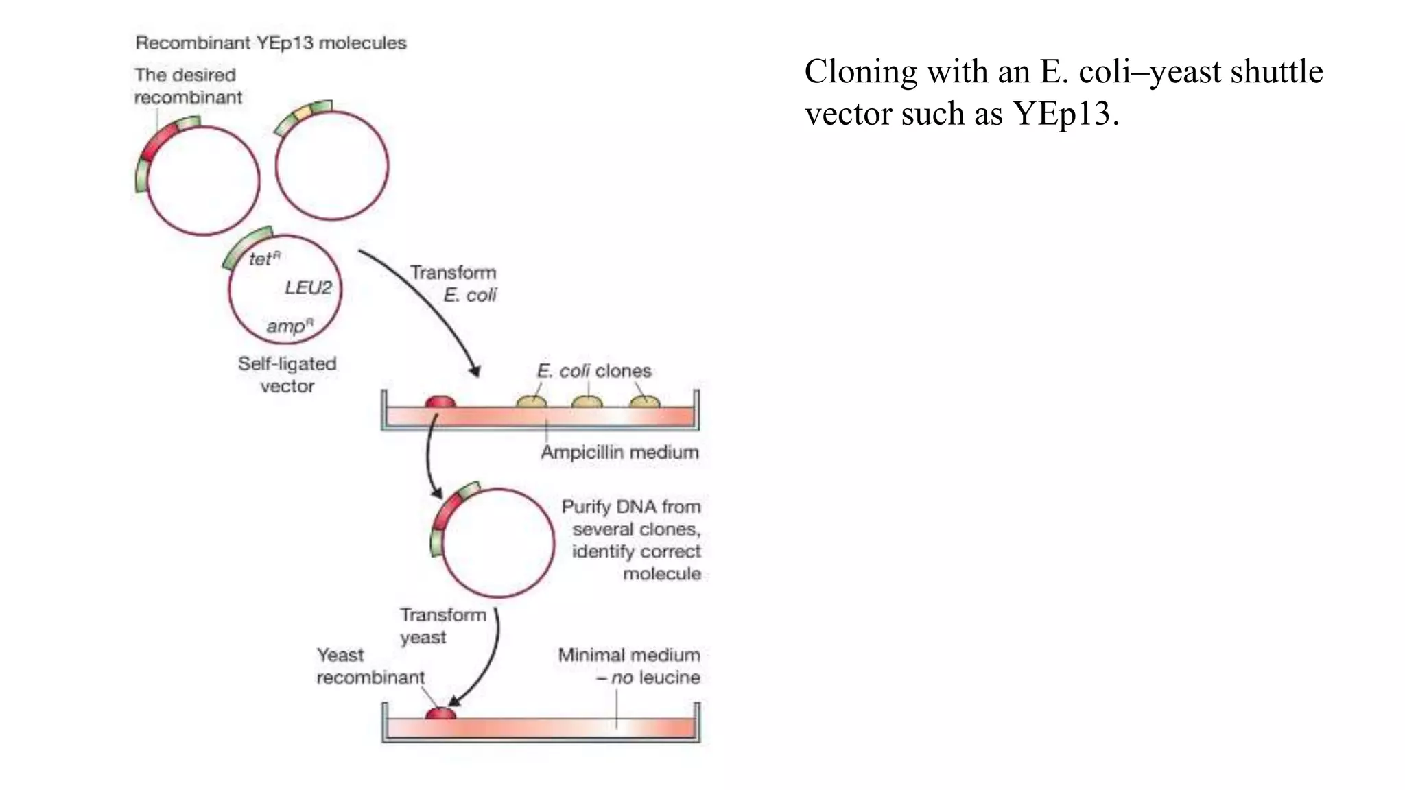
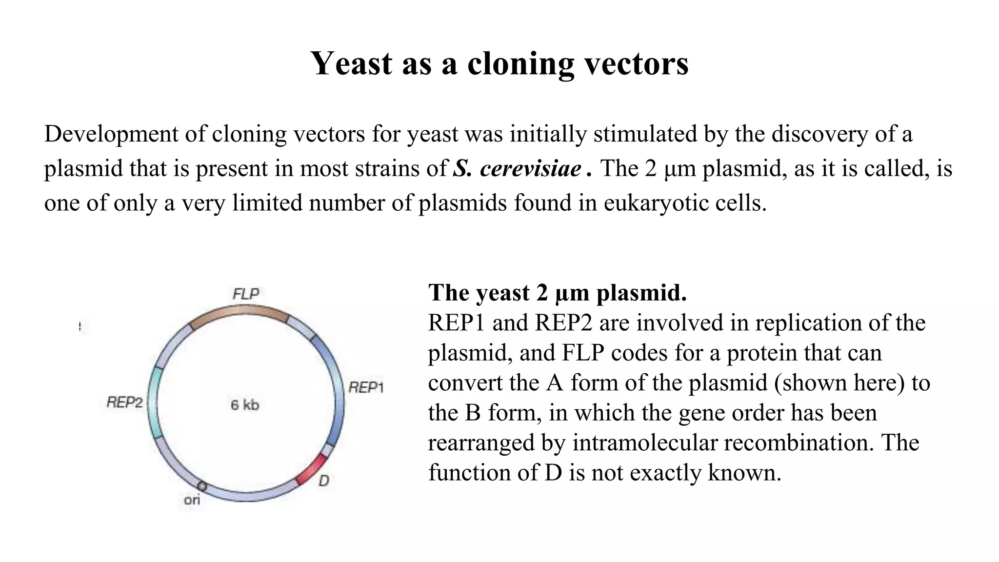
Different types of vectors
Module II
Genetic Engineering
Vector and criteria for ideal cloning vector
Cloning vector is a genome that can accept the target DNA and increase the number
of copies through its own autonomous replication. Cloning vectors provide a basic
backbone for the DNA insert to be reproduced.
Criteria for ideal cloning vectors:
1. It must be self-replicating inside host cell.
2. Insert Size: When it is desired that a particular piece of DNA be cloned, one
consideration is the size of the insert DNA. Most general cloning plasmids can carry
a DNA insert up to around 15 kb in size.
3. It must possess restriction site for Restriction Endonuclease enzymes
4. It must possess some marker gene such that it can be used for later identification of
recombinant cell.
5. It should have multiple cloning sites.
Plasmids
● Plasmids are circular molecules of DNA that lead an independent existence in the
bacterial cell.
● Plasmids almost always carry one or more genes, and often these genes are
responsible for a useful characteristic displayed by the host bacterium.
● For example, the ability to survive in normally toxic concentrations of antibiotics
such as chloramphenicol or ampicillin is often due to the presence in the bacterium of
a plasmid carrying antibiotic resistance genes. In the laboratory, antibiotic resistance
is often used as a selectable marker to ensure that bacteria in a culture contain a
particular plasmid.
Non integrative Plasmid: The smaller plasmids
make use of the host cell’s own DNA replicative
enzymes in order to make copies of themselves,
whereas some of the larger ones carry genes that
code for special enzymes that are specific for
plasmid replication.
Integrative plasmids or episomes: A few types
of plasmid are also able to replicate by inserting
themselves into the bacterial chromosome. These
may be stably maintained in this form through
numerous cell divisions, but always at some stage
exist as independent elements.
Types of plasmid based on mechanism of replication
Classification of plasmids
The most useful classification of naturally occurring plasmids is based on the main
characteristic coded by the plasmid genes. The five major types of plasmid according to
this classification are as follows:
1. Fertility or F plasmids: They carry only tra genes and have no characteristic
beyond the ability to promote conjugal transfer of plasmids. A well-known example
is the F plasmid of E. coli.
2. Resistance or R plasmids: They carry genes conferring on the host bacterium
resistance to one or more antibacterial agents, such as chloramphenicol, ampicillin,
and mercury. R plasmids are very important in clinical microbiology as their spread
through natural populations can have profound consequences in the treatment of
bacterial infections. An example is RP4, which is commonly found in
Pseudomonas, but also occurs in many other bacteria.
3. Col plasmids: They code for colicins, proteins that kill other bacteria. An example is
ColE1 of E. coli.
4. Degradative plasmids: They allow the host bacterium to metabolize unusual molecules
such as toluene and salicylic acid, an example being TOL of Pseudomonas putida.
5. Virulence plasmids: confer pathogenicity on the host bacterium; these include the Ti
plasmids of Agrobacterium tumefaciens, which induce crown gall disease on
dicotyledonous plants.
Size and copy number of plasmid
Size:
Plasmids range from about 1.0 kb for the smallest to over 250 kb for the largest plasmids,
so only a few are useful for cloning purposes.
The copy number:
It refers to the number of molecules of an individual plasmid that are normally found in a
single bacterial cell.Based on copy number there are two types of plasmids:
1. Stringent: low copy number of perhaps just one or two per cell. Specifically, large
plasmids are the stringent plasmids.
2. Relaxed: are present in multiple copies of 50 or more per cell.
Generally speaking, a useful cloning vector needs to be present in the cell in multiple
copies so that large quantities of the recombinant DNA molecule can be obtained.
Conjugation of plasmids
Plasmids fall into two groups: conjugative
and non-conjugative.
Conjugative plasmids are characterized by
the ability to promote sexual conjugation
between bacterial cells, a process that can
result in a conjugative plasmid spreading
from one cell to all the other cells in a
bacterial culture.
Fig: Plasmid transfer by conjugation between bacterial cells. The donor and recipient cells attach to each
other by a pilus, a hollow appendage present on the surface of the donor cell. A copy of the plasmid is
then passed to the recipient cell. Transfer is thought to occur through the pilus, but this has not been
proven and transfer by some other means (e.g. directly across the bacterial cell walls) remains a
possibility.
● Compatibility of plasmid refers to coexistence of different kinds of plasmids in a
single bacterial cell. For example: cells of E. coli have been known to contain up to
seven different plasmids at once.
● If two plasmids are incompatible then one or the other will be rapidly lost from
the cell.
Compatibility of plasmids
Cloning vectors based on E. coli plasmids
● The simplest cloning vectors, and the ones most widely used in gene cloning, are
those based on small bacterial plasmids.
● A large number of different plasmid vectors are available for use with E. coli.
● They combine ease of purification with desirable properties such as high
transformation efficiency, convenient selectable markers for transformants and
recombinants, and the ability to clone reasonably large (up to about 8 kb) pieces
of DNA.
● Most “routine” gene cloning experiments make use of one or other of these plasmid
vectors.
Examples of Plasmid cloning vectors
pBR322:
● One of the first vectors to be developed was
pBR322.
● Size: pBR322 is 4363 bp, which means that
not only can the vector itself be purified with
ease, but so can recombinant DNA molecules
constructed with it. Even with 6 kb of
additional DNA, a recombinant pBR322
molecule is still a manageable size.
● Selection Marker: pBR322 carries two sets of antibiotic resistance genes. Either
ampicillin or tetracycline resistance can be used as a selectable marker for cells
containing the plasmid, and each marker gene includes unique restriction sites that can be
used in cloning experiments. Insertion of new DNA into pBR322 that has been restricted
with PstI, PvuI, or ScaI inactivates the ampR gene, and insertion using any one of eight
restriction endonucleases (notably BamHI and HindIII) inactivates tetracycline
resistance. This great variety of restriction sites that can be used for insertional
inactivation means that pBR322 can be used to clone DNA fragments with any of several
kinds of sticky end.
● Copy number: pBR322 is that it has a reasonably high copy number. Generally there
are about 15 molecules present in a transformed E. coli cell, but this number can be
increased, up to 1000–3000, by plasmid amplification in the presence of a protein
synthesis inhibitor such as chloramphenicol. An E. coli culture therefore provides a good
yield of recombinant pBR322 molecules.
Construction of pBR322:
● pBR322 comprises DNA derived
from three different naturally
occurring plasmids.
● The ampR gene originally resided
on the plasmid R1, a typical
antibiotic resistance plasmid that
occurs in natural populations of E.
coli.
● The tetR gene is derived from R6-
5, a second antibiotic-resistant
plasmid.
● The replication origin of pBR322,
which directs multiplication of the
vector in host cells, is originally from
pMB1, which is closely related to the
pBR327:
pBR327, which was produced by removing a 1089 bp segment from pBR322. This
deletion left the ampR and tetR genes intact, but changed the replicative and
conjugative abilities of the resulting plasmid. As a result, pBR327 differs from pBR322
in two important ways:
1. pBR327 has a higher copy number than pBR322, being present at about 30–45
molecules per E. coli cell. This is not of great relevance as far as plasmid yield is
concerned, as both plasmids can be amplified to copy numbers greater than 1000.
However, the higher copy number of pBR327 in normal cells makes this vector more
suitable if the aim of the experiment is to study the function of the cloned
gene.More copies are of a cloned gene, the more likely it is that the effect of the
cloned gene on the host cell will be detectable. pBR327, with its high copy number,
is therefore a better choice than pBR322 for this kind of work.
● The deletion also destroys the conjugative ability of pBR322, making pBR327 a
non-conjugative plasmid that cannot direct its own transfer to other E. coli cells. This
is important for biological containment, averting the possibility of a recombinant
pBR327 molecule escaping from the test tube and colonizing bacteria in the gut
“Although pBR327, like pBR322, is no longer widely used, its properties have been
inherited by most of today’s modern plasmid vectors.”
pUC8:
pUC8 is descended from pBR322,
although only the replication origin and the
ampR gene remain.
The nucleotide sequence of the ampR gene
has been changed so that it no longer
contains the unique restriction sites: all
these cloning sites are now clustered into
a short segment of the lacZ′ gene carried
by pUC8.
Advantages of pUC8:
1. The manipulations involved in
construction of pUC8 were accompanied
by a chance mutation, within the
origin of replication, which results in
the plasmid having a copy number of
500–700 even before amplification.
2. The identification of recombinant cells
can be achieved by a single step
process, by plating onto agar medium
containing ampicillin plus X-gal.
3. Clustering of the restriction sites, which allows a DNA fragment with two different
sticky ends (say EcoRI at one end and BamHI at the other) to be cloned without resorting to
additional manipulations such as linker attachment.
Bacteriophages
● Bacteriophages, or phages as they are
commonly known, are viruses that
specifically infect bacteria.
● Like all viruses, phages are very simple
in structure, consisting merely of a DNA
(or occasionally ribonucleic acid
(RNA)) molecule carrying a number of
genes, including several for replication
of the phage, surrounded by a protective
coat or capsid made up of protein
molecules.
The two main types of phage structure:
(a) head-and tail (e.g. λ); (b)
filamentous (e.g. M13).
The Phage infection cycle
Lytic cycle Lysogenic cycle
● Release of the new phage
particles is associated with lysis
of the bacterial cell.
● Phage DNA replication is
immediately followed by
synthesis of capsid proteins, and
the phage DNA molecule is never
maintained in a stable condition in
the host cell.
● Retention of the phage DNA
molecule in the host
bacterium, possibly for many
thousands of cell divisions.
● The integrated form of the
phage DNA (called the
prophage) is quiescent. E.g.
Bacteriophage λ and M13
Lytic cycle Lysogenic cycle
Bacteriophage λ : Gene organization
● λ is a typical example of a head-and-tail phage. The DNA is contained in the
polyhedral head structure and the tail serves to attach the phage to the bacterial
surface and to inject the DNA into the cell.
● The λ DNA molecule is 49 kb in size and has been intensively studied by the
techniques of gene mapping and DNA sequencing.
● A feature of the λ genetic map is that genes related in terms of function are clustered
together in the genome. For example, all of the genes coding for components of the
capsid are grouped together in the left-hand third of the molecule, and genes controlling
integration of the prophage into the host genome are clustered in the middle of the
molecule.
● Clustering of related genes is profoundly important for controlling expression of the
λ genome, as it allows genes to be switched on and off as a group rather than
individually. Clustering is also important in the construction of λ -based cloning
vectors.
Fig: The λ genetic map, showing the positions of the important genes and the
functions of the gene clusters.
The linear and circular forms of λ DNA
● linear λ DNA has two free ends, and represents the DNA present in the phage head
structure. This linear molecule consists of two complementary strands of DNA,
base-paired according to the Watson–Crick rules (that is, double-stranded DNA).
However, at either end of the molecule is a short 12-nucleotide stretch in which
the DNA is single-stranded. The two single strands are complementary, and so can
base pair with one another to form a circular, completely double-stranded molecule.
● Complementary single strands are often referred to as “sticky” ends or cohesive
ends, because base pairing between them can “stick” together the two ends of a DNA
molecule (or the ends of two different DNA molecules). The λ cohesive ends are
called the cos sites and they play two distinct roles during the λ infection cycle.
Role of Cos Sites
1. They allow the linear DNA molecule that is injected into the cell to be circularized,
which is a necessary prerequisite for insertion into the bacterial genome.
2. After the prophage has excised from the host genome. At this stage a large number of
new λ DNA molecules are produced by the rolling circle mechanism of
replication in which a continuous DNA strand is “rolled off” the template
molecule.The result is a catenane consisting of a series of linear λ genomes joined
together at the cos sites.
3. Act as recognition sequences for an endonuclease that cleaves the catenane at the
cos sites, producing individual λ genomes.
Cloning vectors of bacteriophage λ
λ insertion vector λ replacement/
substitution vector
Cosmids
λ insertion vector
● With an insertion vector, a large segment of the non-essential region has been
deleted, and the two arms ligated together.
● An insertion vector possesses at least one unique restriction site into which new
DNA can be inserted. The size of the DNA fragment that an individual vector can
carry depends, of course, on the extent to which the non-essential region has been
deleted.
Two popular insertion vectors are:
1. λ gt10, which can carry up to 8 kb of new DNA, inserted into a unique EcoRI site
located in the cI gene(transcriptional inhibitor of bacteriophage λ) . Insertional
inactivation of this gene means that recombinants are distinguished as clear rather
than turbid plaques.
2. λ ZAPII , with which insertion of up to 10 kb DNA into any of 6 restriction sites
within a polylinker inactivates the lacZ′ gene carried by the vector. Recombinants
give clear rather than blue plaques on X-gal agar.
λ replacement/ substitution vector
● A λ replacement vector has two recognition sites for the restriction endonuclease
used for cloning. These sites flank a segment of DNA that is replaced by the DNA to
be cloned.
● Often the replaceable fragment (or “stuffer fragment” in cloning jargon) carries
additional restriction sites that can be used to cut it up into small pieces, so that its
own re-insertion during a cloning experiment is very unlikely. Replacement vectors
are generally designed to carry larger pieces of DNA than insertion vectors can
handle.
An example of a replacement vectors is:
λ EMBL4 can carry up to 20 kb of inserted DNA by replacing a segment flanked by pairs
of EcoRI, BamHI, and SalI sites. Any of these three restriction endonucleases can be used
to remove the stuffer fragment, so DNA fragments with a variety of sticky ends can be
cloned.
Different strategies for
cloning with a λ vector.
(a) Using the circular form
of λ as a plasmid.
(b) Using left and right
arms of the λ genome, plus
in vitro packaging, to
achieve a greater number of
recombinant plaques.
Cosmids
● Most sophisticated type of λ based vector is the cosmid.
● Cosmids are hybrids between a phage DNA molecule and a bacterial plasmid. A
cosmid is basically a plasmid that carries a cos site.
● Their design centers on the fact that the enzymes that package the λ DNA molecule
into the phage protein coat need only the cos sites in order to function.
● The in vitro packaging reaction works not only with λ genomes, but also with any
molecule that carries cos sites separated by 37–52 kb of DNA.
Contains a selectable marker, such as the ampicillin resistance gene, and a plasmid
origin of replication along with the cos sites from phage.
Cloning with cosmid
A cloning experiment with a cosmid is carried out as follows:
1. The cosmid is opened at its unique restriction site and new DNA fragments inserted.
These fragments are usually produced by partial digestion with a restriction
endonuclease, as total digestion almost invariably results in fragments that are too small
to be cloned with a cosmid.
2. Ligation is carried out so that catenanes are formed. Providing the inserted DNA is the
right size, in vitro packaging cleaves the cos sites and places the recombinant cosmids
in mature phage particles. These λ phage are then used to infect an E. coli culture, though
of course plaques are not formed.
3. Instead, infected cells are plated onto a selective medium and antibiotic-resistant
colonies are grown. All colonies are recombinants, as non-recombinant linear cosmids
are too small to be packaged into λ heads.
M13—a filamentous phage
● M13 is an example of a filamentous phage.
● The M13 DNA molecule is much smaller than the λ genome, being only 6407
nucleotides in length.
● It is circular and is unusual in that it consists entirely of single-stranded DNA.
● The smaller size of the M13 DNA molecule means that it has room for fewer
genes than the λ genome.
● M13 follows a simpler infection cycle than λ, and does not need genes for
insertion into the host genome.
M13 infection cycle
● Injection of an M13 DNA molecule
into an E. coli cell occurs via the
pilus, the structure that connects two
cells during sexual conjugation.
● Once inside the cell the single-
stranded molecule acts as the
template for synthesis of a
complementary strand, resulting in
normal double-stranded DNA.
● This molecule is not inserted into
the bacterial genome, but instead
replicates until over 100 copies are
present in the cell.
● When the bacterium divides, each daughter cell receives copies of the phage genome,
which continues to replicate, thereby maintaining its overall numbers per cell.
● New phage particles are continuously assembled and released, about 1000 new
phages being produced during each generation of an infected cell.
M13 as a Vector
● The genome is less than 10 kb in size, well within the range desirable for a potential
vector.
● In addition, the double-stranded replicative form (RF) of the M13 genome behaves
very much like a plasmid, and can be treated as such for experimental purposes.
● It is easily prepared from a culture of infected E. coli cells and can be reintroduced
by transfection.
● Most importantly, genes cloned with an M13-based vector can be obtained in the
form of single-stranded DNA.
● Single-stranded versions of cloned genes are useful for several techniques, notably
DNA sequencing and in vitro mutagenesis.
● Cloning in an M13 vector is an easy and reliable way of obtaining single-stranded
DNA for this type of work. M13 vectors are also used in phage display, a technique
for identifying pairs of genes whose protein products interact with one another.
Phagemid- hybrid plasmid and M13 vector
● M13 vectors are very useful for the production of single-stranded versions of cloned
genes but there is a limit to the size of DNA fragment that can be cloned with an M13
vector, with 1500 bp generally being looked on as the maximum capacity, though
fragments up to 3 kb have occasionally been cloned.
● To get around this problem a number of hybrid vectors (“phagemids”) have been
developed by combining a part of the M13 genome with plasmid DNA
● An example is provided by pEMBL8
● It is made by transferring into pUC8 a
1300 bp fragment of the M13 genome.
● This piece of M13 DNA contains the
signal sequence recognized by the
enzymes that convert the normal
double-stranded M13 molecule into
single-stranded DNA before secretion of
new phage particles.
● This signal sequence is still functional
even though detached from the rest of the
M13 genome, so pEMBL8 molecules
are also converted into single-stranded
DNA and secreted as defective phage
particles
Phagemid vector
● All that is necessary is that the E. coli cells used as hosts for a pEMBL8 cloning
experiment are subsequently infected with normal M13 to act as a helper phage,
providing the necessary replicative enzymes and phage coat proteins.
● pEMBL8, being derived from pUC8, has the polylinker cloning sites within the
lacZ′ gene, so recombinant plaques can be identified in the standard way on
agar containing X-gal. With pEMBL8, single-stranded versions of cloned DNA
fragments up to 10 kb in length can be obtained, greatly extending the range of the
M13 cloning system.
Shuttle vector
● Shuttle vector is a plasmid containing origin of replication of two different
organisms thus they can replicate in both prokaryotic and eukaryotic cells.
● Example: YEp13
A yeast episomal plasmid, YEp13
● The word “episomal” indicates
that a YEp can replicate as an
independent plasmid, but also
implies that integration into one
of the yeast chromosomes can
occur.
Cloning with an E. coli–yeast shuttle
vector such as YEp13.
Yeast as a cloning vectors
Development of cloning vectors for yeast was initially stimulated by the discovery of a
plasmid that is present in most strains of S. cerevisiae . The 2 μm plasmid, as it is called, is
one of only a very limited number of plasmids found in eukaryotic cells.
The yeast 2 µm plasmid.
REP1 and REP2 are involved in replication of the
plasmid, and FLP codes for a protein that can
convert the A form of the plasmid (shown here) to
the B form, in which the gene order has been
rearranged by intramolecular recombination. The
function of D is not exactly known.
● The 2 μm plasmid is an excellent basis for a cloning vector.
● It is 6 kb in size, which is ideal for a vector, and exists in the yeast cell at a copy
number of between 70 and 200.
● Replication makes use of a plasmid origin, several enzymes provided by the host cell,
and the proteins coded by the REP1 and REP2 genes carried by the plasmid.

Different types of vectors.pptx

  • 1.
    Different types ofvectors Module II Genetic Engineering
  • 2.
    Vector and criteriafor ideal cloning vector Cloning vector is a genome that can accept the target DNA and increase the number of copies through its own autonomous replication. Cloning vectors provide a basic backbone for the DNA insert to be reproduced. Criteria for ideal cloning vectors: 1. It must be self-replicating inside host cell. 2. Insert Size: When it is desired that a particular piece of DNA be cloned, one consideration is the size of the insert DNA. Most general cloning plasmids can carry a DNA insert up to around 15 kb in size. 3. It must possess restriction site for Restriction Endonuclease enzymes 4. It must possess some marker gene such that it can be used for later identification of recombinant cell. 5. It should have multiple cloning sites.
  • 3.
    Plasmids ● Plasmids arecircular molecules of DNA that lead an independent existence in the bacterial cell. ● Plasmids almost always carry one or more genes, and often these genes are responsible for a useful characteristic displayed by the host bacterium. ● For example, the ability to survive in normally toxic concentrations of antibiotics such as chloramphenicol or ampicillin is often due to the presence in the bacterium of a plasmid carrying antibiotic resistance genes. In the laboratory, antibiotic resistance is often used as a selectable marker to ensure that bacteria in a culture contain a particular plasmid.
  • 4.
    Non integrative Plasmid:The smaller plasmids make use of the host cell’s own DNA replicative enzymes in order to make copies of themselves, whereas some of the larger ones carry genes that code for special enzymes that are specific for plasmid replication. Integrative plasmids or episomes: A few types of plasmid are also able to replicate by inserting themselves into the bacterial chromosome. These may be stably maintained in this form through numerous cell divisions, but always at some stage exist as independent elements. Types of plasmid based on mechanism of replication
  • 5.
    Classification of plasmids Themost useful classification of naturally occurring plasmids is based on the main characteristic coded by the plasmid genes. The five major types of plasmid according to this classification are as follows: 1. Fertility or F plasmids: They carry only tra genes and have no characteristic beyond the ability to promote conjugal transfer of plasmids. A well-known example is the F plasmid of E. coli. 2. Resistance or R plasmids: They carry genes conferring on the host bacterium resistance to one or more antibacterial agents, such as chloramphenicol, ampicillin, and mercury. R plasmids are very important in clinical microbiology as their spread through natural populations can have profound consequences in the treatment of bacterial infections. An example is RP4, which is commonly found in Pseudomonas, but also occurs in many other bacteria.
  • 6.
    3. Col plasmids:They code for colicins, proteins that kill other bacteria. An example is ColE1 of E. coli. 4. Degradative plasmids: They allow the host bacterium to metabolize unusual molecules such as toluene and salicylic acid, an example being TOL of Pseudomonas putida. 5. Virulence plasmids: confer pathogenicity on the host bacterium; these include the Ti plasmids of Agrobacterium tumefaciens, which induce crown gall disease on dicotyledonous plants.
  • 7.
    Size and copynumber of plasmid Size: Plasmids range from about 1.0 kb for the smallest to over 250 kb for the largest plasmids, so only a few are useful for cloning purposes. The copy number: It refers to the number of molecules of an individual plasmid that are normally found in a single bacterial cell.Based on copy number there are two types of plasmids: 1. Stringent: low copy number of perhaps just one or two per cell. Specifically, large plasmids are the stringent plasmids. 2. Relaxed: are present in multiple copies of 50 or more per cell. Generally speaking, a useful cloning vector needs to be present in the cell in multiple copies so that large quantities of the recombinant DNA molecule can be obtained.
  • 8.
    Conjugation of plasmids Plasmidsfall into two groups: conjugative and non-conjugative. Conjugative plasmids are characterized by the ability to promote sexual conjugation between bacterial cells, a process that can result in a conjugative plasmid spreading from one cell to all the other cells in a bacterial culture. Fig: Plasmid transfer by conjugation between bacterial cells. The donor and recipient cells attach to each other by a pilus, a hollow appendage present on the surface of the donor cell. A copy of the plasmid is then passed to the recipient cell. Transfer is thought to occur through the pilus, but this has not been proven and transfer by some other means (e.g. directly across the bacterial cell walls) remains a possibility.
  • 9.
    ● Compatibility ofplasmid refers to coexistence of different kinds of plasmids in a single bacterial cell. For example: cells of E. coli have been known to contain up to seven different plasmids at once. ● If two plasmids are incompatible then one or the other will be rapidly lost from the cell. Compatibility of plasmids
  • 10.
    Cloning vectors basedon E. coli plasmids ● The simplest cloning vectors, and the ones most widely used in gene cloning, are those based on small bacterial plasmids. ● A large number of different plasmid vectors are available for use with E. coli. ● They combine ease of purification with desirable properties such as high transformation efficiency, convenient selectable markers for transformants and recombinants, and the ability to clone reasonably large (up to about 8 kb) pieces of DNA. ● Most “routine” gene cloning experiments make use of one or other of these plasmid vectors.
  • 11.
    Examples of Plasmidcloning vectors pBR322: ● One of the first vectors to be developed was pBR322. ● Size: pBR322 is 4363 bp, which means that not only can the vector itself be purified with ease, but so can recombinant DNA molecules constructed with it. Even with 6 kb of additional DNA, a recombinant pBR322 molecule is still a manageable size.
  • 12.
    ● Selection Marker:pBR322 carries two sets of antibiotic resistance genes. Either ampicillin or tetracycline resistance can be used as a selectable marker for cells containing the plasmid, and each marker gene includes unique restriction sites that can be used in cloning experiments. Insertion of new DNA into pBR322 that has been restricted with PstI, PvuI, or ScaI inactivates the ampR gene, and insertion using any one of eight restriction endonucleases (notably BamHI and HindIII) inactivates tetracycline resistance. This great variety of restriction sites that can be used for insertional inactivation means that pBR322 can be used to clone DNA fragments with any of several kinds of sticky end. ● Copy number: pBR322 is that it has a reasonably high copy number. Generally there are about 15 molecules present in a transformed E. coli cell, but this number can be increased, up to 1000–3000, by plasmid amplification in the presence of a protein synthesis inhibitor such as chloramphenicol. An E. coli culture therefore provides a good yield of recombinant pBR322 molecules.
  • 13.
    Construction of pBR322: ●pBR322 comprises DNA derived from three different naturally occurring plasmids. ● The ampR gene originally resided on the plasmid R1, a typical antibiotic resistance plasmid that occurs in natural populations of E. coli. ● The tetR gene is derived from R6- 5, a second antibiotic-resistant plasmid. ● The replication origin of pBR322, which directs multiplication of the vector in host cells, is originally from pMB1, which is closely related to the
  • 14.
    pBR327: pBR327, which wasproduced by removing a 1089 bp segment from pBR322. This deletion left the ampR and tetR genes intact, but changed the replicative and conjugative abilities of the resulting plasmid. As a result, pBR327 differs from pBR322 in two important ways: 1. pBR327 has a higher copy number than pBR322, being present at about 30–45 molecules per E. coli cell. This is not of great relevance as far as plasmid yield is concerned, as both plasmids can be amplified to copy numbers greater than 1000. However, the higher copy number of pBR327 in normal cells makes this vector more suitable if the aim of the experiment is to study the function of the cloned gene.More copies are of a cloned gene, the more likely it is that the effect of the cloned gene on the host cell will be detectable. pBR327, with its high copy number, is therefore a better choice than pBR322 for this kind of work.
  • 15.
    ● The deletionalso destroys the conjugative ability of pBR322, making pBR327 a non-conjugative plasmid that cannot direct its own transfer to other E. coli cells. This is important for biological containment, averting the possibility of a recombinant pBR327 molecule escaping from the test tube and colonizing bacteria in the gut “Although pBR327, like pBR322, is no longer widely used, its properties have been inherited by most of today’s modern plasmid vectors.”
  • 16.
    pUC8: pUC8 is descendedfrom pBR322, although only the replication origin and the ampR gene remain. The nucleotide sequence of the ampR gene has been changed so that it no longer contains the unique restriction sites: all these cloning sites are now clustered into a short segment of the lacZ′ gene carried by pUC8.
  • 17.
    Advantages of pUC8: 1.The manipulations involved in construction of pUC8 were accompanied by a chance mutation, within the origin of replication, which results in the plasmid having a copy number of 500–700 even before amplification. 2. The identification of recombinant cells can be achieved by a single step process, by plating onto agar medium containing ampicillin plus X-gal. 3. Clustering of the restriction sites, which allows a DNA fragment with two different sticky ends (say EcoRI at one end and BamHI at the other) to be cloned without resorting to additional manipulations such as linker attachment.
  • 18.
    Bacteriophages ● Bacteriophages, orphages as they are commonly known, are viruses that specifically infect bacteria. ● Like all viruses, phages are very simple in structure, consisting merely of a DNA (or occasionally ribonucleic acid (RNA)) molecule carrying a number of genes, including several for replication of the phage, surrounded by a protective coat or capsid made up of protein molecules. The two main types of phage structure: (a) head-and tail (e.g. λ); (b) filamentous (e.g. M13).
  • 19.
    The Phage infectioncycle Lytic cycle Lysogenic cycle ● Release of the new phage particles is associated with lysis of the bacterial cell. ● Phage DNA replication is immediately followed by synthesis of capsid proteins, and the phage DNA molecule is never maintained in a stable condition in the host cell. ● Retention of the phage DNA molecule in the host bacterium, possibly for many thousands of cell divisions. ● The integrated form of the phage DNA (called the prophage) is quiescent. E.g. Bacteriophage λ and M13
  • 20.
  • 21.
    Bacteriophage λ :Gene organization ● λ is a typical example of a head-and-tail phage. The DNA is contained in the polyhedral head structure and the tail serves to attach the phage to the bacterial surface and to inject the DNA into the cell. ● The λ DNA molecule is 49 kb in size and has been intensively studied by the techniques of gene mapping and DNA sequencing. ● A feature of the λ genetic map is that genes related in terms of function are clustered together in the genome. For example, all of the genes coding for components of the capsid are grouped together in the left-hand third of the molecule, and genes controlling integration of the prophage into the host genome are clustered in the middle of the molecule.
  • 22.
    ● Clustering ofrelated genes is profoundly important for controlling expression of the λ genome, as it allows genes to be switched on and off as a group rather than individually. Clustering is also important in the construction of λ -based cloning vectors. Fig: The λ genetic map, showing the positions of the important genes and the functions of the gene clusters.
  • 23.
    The linear andcircular forms of λ DNA
  • 24.
    ● linear λDNA has two free ends, and represents the DNA present in the phage head structure. This linear molecule consists of two complementary strands of DNA, base-paired according to the Watson–Crick rules (that is, double-stranded DNA). However, at either end of the molecule is a short 12-nucleotide stretch in which the DNA is single-stranded. The two single strands are complementary, and so can base pair with one another to form a circular, completely double-stranded molecule. ● Complementary single strands are often referred to as “sticky” ends or cohesive ends, because base pairing between them can “stick” together the two ends of a DNA molecule (or the ends of two different DNA molecules). The λ cohesive ends are called the cos sites and they play two distinct roles during the λ infection cycle.
  • 25.
    Role of CosSites 1. They allow the linear DNA molecule that is injected into the cell to be circularized, which is a necessary prerequisite for insertion into the bacterial genome. 2. After the prophage has excised from the host genome. At this stage a large number of new λ DNA molecules are produced by the rolling circle mechanism of replication in which a continuous DNA strand is “rolled off” the template molecule.The result is a catenane consisting of a series of linear λ genomes joined together at the cos sites. 3. Act as recognition sequences for an endonuclease that cleaves the catenane at the cos sites, producing individual λ genomes.
  • 27.
    Cloning vectors ofbacteriophage λ λ insertion vector λ replacement/ substitution vector Cosmids
  • 28.
    λ insertion vector ●With an insertion vector, a large segment of the non-essential region has been deleted, and the two arms ligated together. ● An insertion vector possesses at least one unique restriction site into which new DNA can be inserted. The size of the DNA fragment that an individual vector can carry depends, of course, on the extent to which the non-essential region has been deleted.
  • 29.
    Two popular insertionvectors are: 1. λ gt10, which can carry up to 8 kb of new DNA, inserted into a unique EcoRI site located in the cI gene(transcriptional inhibitor of bacteriophage λ) . Insertional inactivation of this gene means that recombinants are distinguished as clear rather than turbid plaques. 2. λ ZAPII , with which insertion of up to 10 kb DNA into any of 6 restriction sites within a polylinker inactivates the lacZ′ gene carried by the vector. Recombinants give clear rather than blue plaques on X-gal agar.
  • 30.
    λ replacement/ substitutionvector ● A λ replacement vector has two recognition sites for the restriction endonuclease used for cloning. These sites flank a segment of DNA that is replaced by the DNA to be cloned. ● Often the replaceable fragment (or “stuffer fragment” in cloning jargon) carries additional restriction sites that can be used to cut it up into small pieces, so that its own re-insertion during a cloning experiment is very unlikely. Replacement vectors are generally designed to carry larger pieces of DNA than insertion vectors can handle.
  • 31.
    An example ofa replacement vectors is: λ EMBL4 can carry up to 20 kb of inserted DNA by replacing a segment flanked by pairs of EcoRI, BamHI, and SalI sites. Any of these three restriction endonucleases can be used to remove the stuffer fragment, so DNA fragments with a variety of sticky ends can be cloned.
  • 32.
    Different strategies for cloningwith a λ vector. (a) Using the circular form of λ as a plasmid. (b) Using left and right arms of the λ genome, plus in vitro packaging, to achieve a greater number of recombinant plaques.
  • 33.
    Cosmids ● Most sophisticatedtype of λ based vector is the cosmid. ● Cosmids are hybrids between a phage DNA molecule and a bacterial plasmid. A cosmid is basically a plasmid that carries a cos site. ● Their design centers on the fact that the enzymes that package the λ DNA molecule into the phage protein coat need only the cos sites in order to function. ● The in vitro packaging reaction works not only with λ genomes, but also with any molecule that carries cos sites separated by 37–52 kb of DNA.
  • 34.
    Contains a selectablemarker, such as the ampicillin resistance gene, and a plasmid origin of replication along with the cos sites from phage.
  • 35.
  • 36.
    A cloning experimentwith a cosmid is carried out as follows: 1. The cosmid is opened at its unique restriction site and new DNA fragments inserted. These fragments are usually produced by partial digestion with a restriction endonuclease, as total digestion almost invariably results in fragments that are too small to be cloned with a cosmid. 2. Ligation is carried out so that catenanes are formed. Providing the inserted DNA is the right size, in vitro packaging cleaves the cos sites and places the recombinant cosmids in mature phage particles. These λ phage are then used to infect an E. coli culture, though of course plaques are not formed. 3. Instead, infected cells are plated onto a selective medium and antibiotic-resistant colonies are grown. All colonies are recombinants, as non-recombinant linear cosmids are too small to be packaged into λ heads.
  • 37.
    M13—a filamentous phage ●M13 is an example of a filamentous phage. ● The M13 DNA molecule is much smaller than the λ genome, being only 6407 nucleotides in length. ● It is circular and is unusual in that it consists entirely of single-stranded DNA. ● The smaller size of the M13 DNA molecule means that it has room for fewer genes than the λ genome. ● M13 follows a simpler infection cycle than λ, and does not need genes for insertion into the host genome.
  • 38.
    M13 infection cycle ●Injection of an M13 DNA molecule into an E. coli cell occurs via the pilus, the structure that connects two cells during sexual conjugation. ● Once inside the cell the single- stranded molecule acts as the template for synthesis of a complementary strand, resulting in normal double-stranded DNA. ● This molecule is not inserted into the bacterial genome, but instead replicates until over 100 copies are present in the cell.
  • 39.
    ● When thebacterium divides, each daughter cell receives copies of the phage genome, which continues to replicate, thereby maintaining its overall numbers per cell. ● New phage particles are continuously assembled and released, about 1000 new phages being produced during each generation of an infected cell.
  • 40.
    M13 as aVector ● The genome is less than 10 kb in size, well within the range desirable for a potential vector. ● In addition, the double-stranded replicative form (RF) of the M13 genome behaves very much like a plasmid, and can be treated as such for experimental purposes. ● It is easily prepared from a culture of infected E. coli cells and can be reintroduced by transfection. ● Most importantly, genes cloned with an M13-based vector can be obtained in the form of single-stranded DNA. ● Single-stranded versions of cloned genes are useful for several techniques, notably DNA sequencing and in vitro mutagenesis. ● Cloning in an M13 vector is an easy and reliable way of obtaining single-stranded DNA for this type of work. M13 vectors are also used in phage display, a technique for identifying pairs of genes whose protein products interact with one another.
  • 41.
    Phagemid- hybrid plasmidand M13 vector ● M13 vectors are very useful for the production of single-stranded versions of cloned genes but there is a limit to the size of DNA fragment that can be cloned with an M13 vector, with 1500 bp generally being looked on as the maximum capacity, though fragments up to 3 kb have occasionally been cloned. ● To get around this problem a number of hybrid vectors (“phagemids”) have been developed by combining a part of the M13 genome with plasmid DNA ● An example is provided by pEMBL8
  • 42.
    ● It ismade by transferring into pUC8 a 1300 bp fragment of the M13 genome. ● This piece of M13 DNA contains the signal sequence recognized by the enzymes that convert the normal double-stranded M13 molecule into single-stranded DNA before secretion of new phage particles. ● This signal sequence is still functional even though detached from the rest of the M13 genome, so pEMBL8 molecules are also converted into single-stranded DNA and secreted as defective phage particles Phagemid vector
  • 44.
    ● All thatis necessary is that the E. coli cells used as hosts for a pEMBL8 cloning experiment are subsequently infected with normal M13 to act as a helper phage, providing the necessary replicative enzymes and phage coat proteins. ● pEMBL8, being derived from pUC8, has the polylinker cloning sites within the lacZ′ gene, so recombinant plaques can be identified in the standard way on agar containing X-gal. With pEMBL8, single-stranded versions of cloned DNA fragments up to 10 kb in length can be obtained, greatly extending the range of the M13 cloning system.
  • 45.
    Shuttle vector ● Shuttlevector is a plasmid containing origin of replication of two different organisms thus they can replicate in both prokaryotic and eukaryotic cells. ● Example: YEp13 A yeast episomal plasmid, YEp13 ● The word “episomal” indicates that a YEp can replicate as an independent plasmid, but also implies that integration into one of the yeast chromosomes can occur.
  • 46.
    Cloning with anE. coli–yeast shuttle vector such as YEp13.
  • 47.
    Yeast as acloning vectors Development of cloning vectors for yeast was initially stimulated by the discovery of a plasmid that is present in most strains of S. cerevisiae . The 2 μm plasmid, as it is called, is one of only a very limited number of plasmids found in eukaryotic cells. The yeast 2 µm plasmid. REP1 and REP2 are involved in replication of the plasmid, and FLP codes for a protein that can convert the A form of the plasmid (shown here) to the B form, in which the gene order has been rearranged by intramolecular recombination. The function of D is not exactly known.
  • 48.
    ● The 2μm plasmid is an excellent basis for a cloning vector. ● It is 6 kb in size, which is ideal for a vector, and exists in the yeast cell at a copy number of between 70 and 200. ● Replication makes use of a plasmid origin, several enzymes provided by the host cell, and the proteins coded by the REP1 and REP2 genes carried by the plasmid.