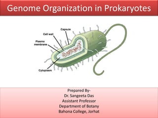
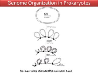
Prokaryotic genomes are typically organized as single circular chromosomes that are condensed into a nucleoid region within the cell. DNA supercoiling, which involves the over- or under-winding of DNA strands, facilitates compaction of prokaryotic genomes and enables DNA metabolism. Topoisomerases regulate DNA supercoiling by introducing temporary breaks in DNA strands, allowing strands to pass through one another and relieve torsional stress that builds during processes like transcription and replication. The two major types of topoisomerase are types I and II, which introduce single-strand or double-strand breaks, respectively, in regulating supercoiling levels.
DNA topology studies the geometric properties and spatial relationships of DNA that are unaffected by changes in shape or size. It includes phenomena like supercoiling, knots, and catenanes that involve the linking and twisting of the two DNA strands. DNA topology is characterized by parameters like the linking number, which represents the number of times the two strands are twisted around each other. Enzymes called topoisomerases regulate DNA topology by introducing temporary breaks in the DNA strands to allow strand passage and control supercoiling levels.
Genome organization in prokaryotes(molecular biology)IndrajaDoradla
1. In prokaryotes, the genome is located in an irregularly shaped region within the cell called the nucleoid, which is not surrounded by a membrane like the eukaryotic nucleus.
2. The prokaryotic genome is generally a circular piece of DNA that can exist in multiple copies and ranges in length but is at least a few million base pairs. It is packaged into the nucleoid through supercoiling facilitated by nucleoid-associated proteins.
3. DNA supercoiling allows for very long strands of DNA to be tightly packaged into a prokaryotic cell. This involves the introduction of plectonemic supercoils that twist the DNA into loops and wind it around nucle
1. The document discusses models of homologous recombination including the Holliday model and the double-strand break repair model. It describes the key steps and proteins involved in each model.
2. Recombination involves the breakage and rejoining of DNA. In eukaryotes, the MRN/X complex processes DNA breaks. The Rad51 and Rad54 proteins then facilitate strand invasion and D-loop formation during homologous pairing.
3. Homologous recombination proteins from bacteria and eukaryotes catalyze different steps of the process. In E. coli, RecBCD introduces breaks and generates single strands for RecA to perform strand exchange, while RuvAB and Ruv
This document discusses the phagemid vector pBluescript, which can be used in either the positive or negative orientation. pBluescript is a phagemid vector that can be used to clone DNA fragments for sequencing, mutagenesis, protein expression or other molecular biology experiments. References for further information about pBluescript are provided.
Bacteriophage vectors
Bacteriophage
WHY BACTERIOPHAGE AS A VECTOR?
M13 phage
Genome of m13 phage
Life cycle and dna replication of m13
CONSTRUCTION M13 AS PHAGE VECTOR
M13 MP 2 vector
M13MP7 VECTOR
Selection of recombinants
Lambda replacement vectors
LAMBDA EMBL 4 VECTOR
P1 PHAGE
GENOME OF P1 PHAGE
P1 PHAGE AS VECTOR
P1 phage vector system
Site-directed mutagenesis is a technique used to introduce specific changes to the DNA sequence of a gene by altering the nucleotide sequence. It allows researchers to study the impact of mutations by changing individual bases, deleting bases, or inserting new bases. There are different methods of site-directed mutagenesis including oligonucleotide-based methods and PCR-based methods. Site-directed mutagenesis has applications in research, production of desired proteins, and development of engineered proteins for commercial uses like detergents.
This document discusses DNA sequencing methods. It describes the Maxam-Gilbert sequencing method developed in 1976-1977 which uses chemical modification and cleavage of DNA at specific bases, followed by electrophoresis to separate fragments by size. It also mentions the popular Sanger sequencing method. The procedure for Maxam-Gilbert sequencing involves labeling DNA, cleaving it with chemicals, running the fragments on a gel, and analyzing the results to deduce the DNA sequence. Advantages include no premature termination and ability to sequence stretches not possible with enzymatic methods, while disadvantages include use of radioactivity and toxic chemicals.
Modified M13 vectors have a large number of cloning sites which allow for insertion of foreign DNA. These vectors are derived from the M13 bacteriophage and are commonly used for DNA sequencing, mapping and mutagenesis experiments in molecular biology research. The document appears to be a seminar topic submission about using the M13 phage for biotechnology applications.
This document discusses the organization of genetic material in prokaryotic and eukaryotic cells. It begins by defining key terms like genome and describing the overall structure of genetic material. It then contrasts prokaryotic and eukaryotic cells, noting things like prokaryotes having circular DNA without introns while eukaryotes have linear chromosomes and mRNA splicing. The document also discusses specific genetic elements like plasmids, viruses, and organelles. It provides details on their size, structure and content. Finally, the sizes of some viral, bacterial, and eukaryotic genomes are compared.
DNA footprinting is a technique used to identify protein binding regions on DNA. It involves treating DNA with nucleases like DNase I, which will degrade the DNA except for regions bound by proteins. These protected regions, called footprints, can identify transcription factor binding sites that regulate gene expression. The technique was originally developed in 1978 to study the binding specificity of the lac repressor protein, and it provides information on DNA-protein interactions and transcriptional regulation.
molecular biology phage vector, full lifecycle and all necessary information regarding lambda phage, it contain 2 types that is insertion and replacement.
A DNA library is a collection of DNA fragments that have been cloned into vectors. DNA libraries allow researchers to isolate and study specific DNA fragments of interest. To create a genomic library, DNA is extracted from an organism, cut into fragments, inserted into vectors, and introduced into host bacteria to generate clones containing all the organism's DNA sequences. This library can then be screened to identify and study particular genes. DNA libraries provide an efficient way to store, isolate, and analyze DNA sequences.
This document summarizes homologous recombination in eukaryotes and bacteria. In eukaryotes, homologous recombination repairs double-strand DNA breaks through either the double-strand break repair (DSBR) pathway or synthesis-dependent strand annealing (SDSA) pathway. The DSBR pathway forms double Holliday junctions that are resolved to result in crossover or non-crossover products. In bacteria, the RecBCD pathway repairs double-strand breaks and the RecF pathway repairs single-strand gaps. Both pathways involve strand invasion and branch migration to facilitate homologous recombination.
Lectut btn-202-ppt-l4. bacteriophage lambda and m13 vectors (1)Rishabh Jain
This document describes the bacteriophages λ and M13, which are commonly used as cloning vectors. λ phage is a temperate phage that infects E. coli and has a double-stranded linear DNA genome. Its genome is organized into regions that encode proteins for the phage head, tail, and lysogeny/lysis functions. M13 is a filamentous phage with a single-stranded circular genome. Both phages can be modified and used to insert and replicate foreign DNA fragments in E. coli for cloning purposes.
Yeast artificial chromosomes (YACs) are engineered DNA molecules that can clone and replicate large DNA sequences in yeast cells. YACs contain essential yeast elements like a centromere and telomeres that allow them to behave like natural yeast chromosomes. YACs can clone very large inserts of up to 10 megabases of foreign DNA, making them useful for generating whole genome libraries.
pUC vectors are plasmids derived from pBR322 that have a higher copy number of 500-600 copies per cell. They contain an ampicillin resistance gene for selection, as well as the lacZ' gene containing multiple cloning sites. When a gene of interest is inserted, it disrupts the lacZ' gene, allowing for blue-white screening on media containing IPTG and X-gal to identify recombinant colonies that appear white instead of blue. pUC vectors offer advantages over pBR322 such as high copy number and easy selection, though they cannot accommodate inserts larger than 15kb.
Chromosome walking jumping transposon tagging map based cloningPromila Sheoran
Chromosome walking, jumping, and transposon tagging are techniques used for gene mapping and cloning. Chromosome walking involves isolating overlapping DNA fragments in steps to characterize large chromosome regions. Chromosome jumping uses rare cutting enzymes to isolate larger DNA fragments spanning hundreds of kb. Transposon tagging involves inducing transposon insertion mutations, identifying the disrupted gene, and using the transposon as a tag to clone the gene. Map-based cloning localizes a gene of interest by identifying closely linked markers, screening libraries to find flanking markers, and identifying the gene between markers through complementation tests.
The document discusses three models of DNA replication:
1) Asymmetric replication - the leading and lagging strands are replicated differently due to the 5' to 3' directionality of DNA polymerase. The leading strand replicates continuously while the lagging strand replicates discontinuously in short Okazaki fragments.
2) D-loop model - replication in mitochondria where one strand is displaced to form a D-loop and replicates first before the other strand.
3) Rolling circle model - used by plasmids and viruses where one strand is nicked and displaced to be used as a template, forming multiple copies linked together in a concatemer.
Prokaryotic genomes are circular, double-stranded DNA contained within the nucleoid. They vary in length but are generally a few million base pairs. DNA supercoiling allows for tight packing of the genome.
Eukaryotic genomes are linear chromosomes associated with histone proteins within the nucleus. The DNA is wrapped around histone octamers to form nucleosomes, compacting the genome. Eukaryotic genomes are generally larger and contain more DNA than prokaryotic genomes.
Key differences between prokaryotic and eukaryotic genomes include genome size, number of chromosomes, ploidy level, association with histones, and method of compaction.
DNA ligase is an enzyme that catalyzes the formation of phosphodiester bonds between DNA fragments, joining two DNA strands together. It plays an important role in DNA replication by joining Okazaki fragments and filling in gaps, as well as in DNA repair and genetic engineering techniques like cloning. The most commonly used DNA ligase is from bacteriophage T4, which utilizes ATP as a cofactor and works efficiently at lower temperatures to ligate DNA strands with either sticky or blunt ends.
DNA repair mechanisms in prokaryotes involve direct repair, excision repair, and mismatch repair. Direct repair converts damaged nucleotides directly back to their original structure using enzymes like photolyase. Excision repair removes damaged sections of DNA through base excision repair which removes single damaged bases using glycosylases and AP endonucleases, or nucleotide excision repair which removes short oligonucleotides. Mismatch repair recognizes and fixes errors made during DNA replication by distinguishing the parental DNA strands and excising the newly synthesized strand containing mistakes.
Site-directed mutagenesis is a molecular biology technique used to make specific changes to DNA sequences. It involves using a primer containing the desired mutation in a PCR reaction to introduce the mutation into the gene of interest. There are different approaches for site-directed mutagenesis using PCR, including using a mutated primer in normal PCR or a primer extension method. The technique is used for applications like protein engineering to study the impact of sequence changes or insert restriction sites. However, it can be difficult to replicate the mutated DNA and screening mutations requires sequencing.
This document discusses bacteriophage T4, a virus that infects E. coli bacteria. It has a complex protein coat and large double-stranded DNA genome. T4 uses the host cell's machinery to replicate and kills the host cell. T4 plays a role in cholera and diphtheria by carrying toxin genes that allow the bacteria to cause disease. Bacteriophage may be useful for treating antibiotic-resistant bacteria or infections where antibiotics cannot reach. T4 is also used in recombinant DNA technology.
1. There are four main models of DNA replication: rolling circle replication, theta replication, bidirectional replication of linear DNA, and telomere replication.
2. Rolling circle replication involves nicking circular DNA and using one strand as a template to produce multiple copies of the original circular DNA.
3. Theta replication occurs in prokaryotes and involves unwinding circular DNA at an origin of replication and replicating bi-directionally to form a theta-shaped structure.
4. Bidirectional replication of linear DNA involves unwinding DNA at origins of replication and using leading and lagging strand synthesis to replicate in both directions until the ends of the linear genome are reached.
Prokaryotes are single-celled organisms that lack membrane-bound organelles. They have their DNA and other cellular components floating freely in the cytoplasm. They reproduce through binary fission. Eukaryotes are organisms with cells that contain membrane-bound organelles and a nucleus that holds their DNA. All eukaryotic cells share certain structures like the nucleus, plasma membrane, ribosomes, and cytoplasm. They also tend to have additional organelles not found in prokaryotes.
Prokaryotic genetic material differs from eukaryotes in several key ways:
1. Prokaryotes lack a membrane-bound nucleus and have their DNA located in the nucleoid. They typically have a single circular chromosome while eukaryotes have multiple linear chromosomes.
2. Prokaryotic genes are arranged in operons and expressed together, whereas eukaryotic genes each have their own promoter and are independently expressed.
3. DNA replication in prokaryotes is rapid and ongoing, starting from a single origin of replication site, while eukaryotes tightly regulate replication during the cell cycle.
This document provides information about molecular biology concepts including:
- The structure and function of eukaryotic cells including organelles like the nucleus, nucleolus, and mitochondria.
- The differences between prokaryotic and eukaryotic cells.
- DNA structure, replication, and the role of DNA and RNA in cells.
- Other genetic elements like viruses, viroids and prions.
This document contains the answers to an activity on biochemistry and cell organization submitted by a student named Maricris P. Nebiar. It defines several key terms related to cells and organelles. It also lists 5 differences between prokaryotic and eukaryotic cells, identifies which organelles contain DNA, are sites of energy production, and are surrounded by a double membrane. Finally, it summarizes the advantages of eukaryotic cells over prokaryotic cells and explains why a claim of discovering mitochondria in bacteria is unlikely.
Structure of prokariyotic and eukariyotic cells By KK Sahu SirKAUSHAL SAHU
This document provides an overview of the key differences between prokaryotic and eukaryotic cells. It discusses their origins, with prokaryotes originating 3.5 billion years ago and eukaryotes 1.5 billion years ago. The main structural differences are that prokaryotes lack membrane-bound organelles and have no nucleus, while eukaryotes have organelles like the nucleus enclosed in membranes. Some examples of large prokaryotes and reduced genome eukaryotes are also mentioned.
Analyzing the different organelles in eukaryotic & prokaryotic cells thro...Umair Raza
Analyzing the different organelles in Eukaryotic & Prokaryotic cells through their functions (List of organelles, structure, composition, diagram, charts, functions) by Engr. Umair Raza
Molecular different between Prokaryotic and eukaryotic cell By KK Sahu SirKAUSHAL SAHU
SYNOPSIS
Introduction to cell
Historical Aspects
Cell Diversity
Types Of Cell Diversity
Cell Diversity In Origin
Cell Diversity In size
Cell Diversity In Shape
Some Other Types
5) Differentiation And Specialisation Of Cell Diversity
6) Conclusion
7) References
What are the main functions of the components of prokaryotic vs. euk.pdfarihantstoneart
What are the main functions of the components of prokaryotic vs. eukaryotic cells? Could either
cell type survive without any of them? (Answer must be at least a page long, very detailed and in
paragraph form please)
Solution
Prokaryotes are unicellular living beings that need organelles or other interior film bound
structures . In this manner, they don\'t have a core, at the same time, rather, by and large have a
solitary chromosome: a bit of roundabout, twofold stranded DNA situated in a territory of the
phone called the nucleoid. Most prokaryotes have a cell divider outside the plasma film.
Like a prokaryotic cell, an eukaryotic cell has a plasma film, cytoplasm, and ribosomes.
Nonetheless, not at all like prokaryotic cells, eukaryotic cells have:
a film bound core
various film bound organelles (counting the endoplasmic reticulum, Golgi contraption,
chloroplasts, and mitochondria)
a few bar formed chromosomes
Since an eukaryotic cell\'s core is encompassed by a film, it is regularly said to have a \"genuine
core. \" Organelles (signifying \"little organ\") have specific cell parts, similarly as the organs of
your body have particular parts. They permit diverse capacities to be compartmentalized in
various territories of the cell.
The piece of the cell divider varies altogether between the areas Bacteria and Archaea, the two
spaces of life into which prokaryotes are separated . The structure of their cell dividers
additionally varies from the eukaryotic cell dividers found in plants (cellulose) or organisms and
bugs (chitin). The cell divider works as a defensive layer and is in charge of the life form\'s
shape. A few microorganisms have a container outside the cell divider. Different structures are
available in some prokaryotic species, yet not in others. For instance, the case found in a few
animal types empowers the living being to connect to surfaces, shields it from lack of hydration
and assault by phagocytic cells, and expands its imperviousness to our invulnerable reactions. A
few animal categories likewise have flagella utilized for velocity and pili utilized for connection
to surfaces. Plasmids, which comprise of additional chromosomal DNA, are likewise present in
numerous types of microorganisms and archaea.
Commonly, the core is the most conspicuous organelle in a cell. Eukaryotic cells have a genuine
core, which implies the cell\'s DNA is encompassed by a layer. Thusly, the core houses the cell\'s
DNA and coordinates the blend of proteins and ribosomes, the cell organelles in charge of
protein union. The atomic envelope is a twofold layer structure that constitutes the furthest bit of
the core. Both the internal and external layers of the atomic envelope are phospholipid bilayers.
The atomic envelope is punctuated with pores that control the section of particles, particles, and
RNA between the nucleoplasm and cytoplasm. The nucleoplasm is the semi-strong liquid inside
the core where we discover the chromatin and the nucleolus. M.
This document summarizes key aspects of genome organization in prokaryotes. It notes that prokaryotes like E. coli have a single circular chromosome composed of DNA that is compacted into a nucleoid structure. The DNA is highly condensed via supercoiling facilitated by enzymes like topoisomerases. Prokaryotic genomes are also typically smaller than eukaryotic ones and can contain extra DNA on plasmids that are exchanged between bacteria.
7_DNA organization in prokaryotes and eukaryotes.pptxAKHILRDONGA
DNA is the molecule that contains the genetic instructions used in the development and functioning of all known living organisms. In prokaryotes, DNA exists as a single circular chromosome located in the nucleoid region of the cell. Prokaryotic DNA is tightly packed using supercoiling, where the DNA winds further around itself. In eukaryotes, DNA is organized into linear chromosomes within the cell nucleus. Eukaryotic DNA is associated with histone proteins to form chromatin, which condenses into distinct chromosomes during cell division. The basic unit of hereditary information is the gene. Genes encode either functional products like proteins or regulatory elements that control gene expression.
All cells arise from preexisting cells, contain genetic material, and have a plasma membrane that encloses the cell. Eukaryotic cells are larger and more complex, compartmentalized with organelles like the nucleus and mitochondria that may have originated from engulfed prokaryotes. Plant cells have a cell wall and central vacuole while animal cells rely on an extracellular matrix for structure.
Introduction to genetics and genes unlocking the secrets of heredity by noor ...Noor Zada
Genetics is the study of heredity and the transmission of traits from parents to offspring. It examines inheritance at multiple levels, from whole organisms to chromosomes to genes and DNA. DNA is the genetic material found in all living things. It exists as long double-stranded helix molecules. DNA is made up of nucleotides containing phosphate, deoxyribose sugar, and nitrogenous bases. The bases pair up in a specific way between strands through hydrogen bonding to form the DNA code. DNA codes for traits by determining the sequence of proteins and RNA molecules. Its double helix structure allows for accurate copying and transmission of the genetic code during cell division.
Genetic Organisation:
All cellular activities are encoded within a cell’s DNA.
The sequence of bases within a DNA molecule represents the genetic information of the cell.
Segments of DNA molecules are called genes, and individual genes contain the instructional code necessary for synthesizing various proteins, enzymes, or stable RNA molecules.
This document provides an overview of cell organelle structure and function. It compares prokaryotic and eukaryotic cells, describing key differences such as nucleus, organelles, and cell size. Major eukaryotic organelles like the nucleus, mitochondria, chloroplasts, and endoplasmic reticulum are summarized in terms of their main functions and structures. The document also discusses the structure and functions of specific organelles like the nucleus, mitochondria, and chromosomes in more detail.
The document summarizes the organization of genetic material on chromosomes. It discusses that genetic material includes DNA and RNA, which is stored on chromosomes in the nucleus, mitochondria, and cytoplasm. It then describes key differences in how genetic material is organized in prokaryotes versus eukaryotes, including that prokaryotes generally have circular DNA without histones while eukaryotes have linear DNA packaged into nucleosomes with histones. The document also notes that mitochondria and chloroplasts contain organelle DNA and that viruses can have DNA or RNA as their genetic material organized inside a protein capsule.
Prokaryotic chromosomes are circular in shape and located in the nucleoid region of the cell. They contain a single set of genes and lack histone proteins. The Escherichia coli chromosome is composed of a single closed circular DNA molecule that is divided into 50-100 loop domains. The chromosome is highly condensed and negatively supercoiled through the action of nucleoid-associated proteins like HU, H-NS, and DNA gyrase. This supercoiling and the binding of proteins allows for the compact organization and maintenance of the prokaryotic genome.
This document provides an overview of cell biology. It defines the cell as the structural and functional unit of life. It describes the key differences between prokaryotic and eukaryotic cells. The structure and functions of major cell organelles like the cell membrane, nucleus, mitochondria and ribosomes are explained. The processes of cell division, transcription and translation are summarized. Different types of cell junctions and their roles are also outlined.
Genome organisation in prokaryotes and eukaryotesVedavyas Dasari
The document summarizes genome organization in prokaryotes and eukaryotes. In prokaryotes like E. coli, the genome is packed into the nucleoid region through supercoiling, facilitated by histone-like proteins that introduce bends and loops in the DNA. This allows the 1.5mm of DNA to fit inside the small cell. In eukaryotes, the 6 feet of DNA is packaged into chromosomes through a multi-step process involving wrapping around histones to form nucleosomes, which further condense into solenoids, super solenoids, rosettes, coils and finally chromosomes.
Cells are the fundamental units of life, and all organisms are made up of one or more cells. The document discusses two important cellular components - the nucleus and ribosomes. The nucleus houses most of the cell's DNA and directs protein synthesis. It is enclosed by a double membrane and contains chromosomes. The ribosomes use information from DNA to synthesize proteins according to instructions provided by messenger RNA. They assemble in the nucleolus and exit into the cytoplasm to perform protein synthesis.
Similar to Genome organization in prokaryotes (20)
This document provides information about Cyanophyta (cyanobacteria) presented by Dr. Sangeeta Das. It discusses the characteristics, distribution, ecology, thallus organization, cell structure, reproduction of cyanobacteria. It specifically focuses on Nostoc, providing details about its systematic position, morphology, and life cycle. Key points include that cyanobacteria are a primitive group of algae found all over the world, they can have unicellular or colonial thalli, reproduce both vegetatively and asexually, and Nostoc forms filaments within a gelatinous sheath and reproduces through fragmentation and akinete formation.
The document discusses human impact on forests through deforestation and urbanization. It notes that nearly half of the world's forests have been cleared for human use and that every minute an area of tropical forest the size of 40 football fields is lost. If deforestation continues at the current rate, 60% of the Amazon rainforest could disappear by 2050. However, it also provides examples of reforestation efforts including one man in India who single-handedly planted a 1,400 acre forest. The presentation emphasizes the importance of protecting forests to protect ourselves.
Chlamydomonas is a single-celled green alga that is found in fresh water environments like ponds, ditches, and wet soil. It reproduces both asexually through the production of zoospores and sexually through isogamy, anisogamy, or oogamy depending on the species. The cell contains a cup-shaped chloroplast, pyrenoid, two flagella, contractile vacuoles, and sometimes an eye spot. Chlamydomonas is used as a model organism in research due to its simple structure and life cycle.
This PPT explains about the various methods and steps of preparation of herbarium specimens. It also describes the various functions performed by herbaria and the various major herbaria of world as well as in India.
This ppt contains all about the family Rosaceae under Dicotyledons. It explains about its systematic position, general characters, phylogenetic affinities, floral formula and diagram, economic importance and important genera under this family.
Data mining involves using machine learning and statistical methods to discover patterns in large datasets and is useful in bioinformatics for analyzing biological data. Bioinformatics analyzes data from sequences, molecules, gene expressions, and pathways. Data mining can help understand these rapidly growing biological datasets. Common data mining tools in bioinformatics include BLAST for sequence comparisons, Entrez for integrated database searching, and ORF Finder for identifying open reading frames. Data mining approaches are well-suited to the enormous volumes of data in bioinformatics databases.
Cytokinins are a class of plant growth hormones that promote cell division. They were first discovered in 1955 from degraded herring sperm DNA. There are two main types: adenine-type cytokinins like kinetin and zeatin, and phenylurea-type cytokinins. Cytokinins play many important physiological roles in plants including cell division, cell elongation, differentiation, delaying senescence, and influencing flowering. They are widely used in tissue culture and to increase the shelf life of cut flowers and vegetables.
The document discusses various types of documentation in plant taxonomy including floras, monographs, taxonomic revisions, manuals, taxonomic indexes, dictionaries, glossaries, catalogs, journals, periodicals, abstracts, and icones. Floras document the plants of a given geographical region, monographs provide in-depth treatment of a taxonomic group, and revisions focus on a section of a genus or geographical area. Manuals, indexes, dictionaries and glossaries aid in identification and classification. Journals, periodicals, and abstracts disseminate taxonomic research. Icones provide pictorial representations of plants. Proper documentation enables dissemination and retrieval of taxonomic information for various users.
1. Plant taxonomy is the science of identifying, classifying, and naming plants based on their phenotypic characteristics.
2. The main objectives of plant taxonomy are to classify the plant kingdom, identify plants, and assign scientific names to plants.
3. Taxonomy aims to classify organisms into hierarchical taxa, assign scientific names to each taxon through nomenclature, and allow identification of organisms.
The technology uses reclaimed CO₂ as the dyeing medium in a closed loop process. When pressurized, CO₂ becomes supercritical (SC-CO₂). In this state CO₂ has a very high solvent power, allowing the dye to dissolve easily.
Phenomics assisted breeding in crop improvementIshaGoswami9
As the population is increasing and will reach about 9 billion upto 2050. Also due to climate change, it is difficult to meet the food requirement of such a large population. Facing the challenges presented by resource shortages, climate
change, and increasing global population, crop yield and quality need to be improved in a sustainable way over the coming decades. Genetic improvement by breeding is the best way to increase crop productivity. With the rapid progression of functional
genomics, an increasing number of crop genomes have been sequenced and dozens of genes influencing key agronomic traits have been identified. However, current genome sequence information has not been adequately exploited for understanding
the complex characteristics of multiple gene, owing to a lack of crop phenotypic data. Efficient, automatic, and accurate technologies and platforms that can capture phenotypic data that can
be linked to genomics information for crop improvement at all growth stages have become as important as genotyping. Thus,
high-throughput phenotyping has become the major bottleneck restricting crop breeding. Plant phenomics has been defined as the high-throughput, accurate acquisition and analysis of multi-dimensional phenotypes
during crop growing stages at the organism level, including the cell, tissue, organ, individual plant, plot, and field levels. With the rapid development of novel sensors, imaging technology,
and analysis methods, numerous infrastructure platforms have been developed for phenotyping.
The debris of the ‘last major merger’ is dynamically youngSérgio Sacani
The Milky Way’s (MW) inner stellar halo contains an [Fe/H]-rich component with highly eccentric orbits, often referred to as the
‘last major merger.’ Hypotheses for the origin of this component include Gaia-Sausage/Enceladus (GSE), where the progenitor
collided with the MW proto-disc 8–11 Gyr ago, and the Virgo Radial Merger (VRM), where the progenitor collided with the
MW disc within the last 3 Gyr. These two scenarios make different predictions about observable structure in local phase space,
because the morphology of debris depends on how long it has had to phase mix. The recently identified phase-space folds in Gaia
DR3 have positive caustic velocities, making them fundamentally different than the phase-mixed chevrons found in simulations
at late times. Roughly 20 per cent of the stars in the prograde local stellar halo are associated with the observed caustics. Based
on a simple phase-mixing model, the observed number of caustics are consistent with a merger that occurred 1–2 Gyr ago.
We also compare the observed phase-space distribution to FIRE-2 Latte simulations of GSE-like mergers, using a quantitative
measurement of phase mixing (2D causticality). The observed local phase-space distribution best matches the simulated data
1–2 Gyr after collision, and certainly not later than 3 Gyr. This is further evidence that the progenitor of the ‘last major merger’
did not collide with the MW proto-disc at early times, as is thought for the GSE, but instead collided with the MW disc within
the last few Gyr, consistent with the body of work surrounding the VRM.
The binding of cosmological structures by massless topological defectsSérgio Sacani
Assuming spherical symmetry and weak field, it is shown that if one solves the Poisson equation or the Einstein field
equations sourced by a topological defect, i.e. a singularity of a very specific form, the result is a localized gravitational
field capable of driving flat rotation (i.e. Keplerian circular orbits at a constant speed for all radii) of test masses on a thin
spherical shell without any underlying mass. Moreover, a large-scale structure which exploits this solution by assembling
concentrically a number of such topological defects can establish a flat stellar or galactic rotation curve, and can also deflect
light in the same manner as an equipotential (isothermal) sphere. Thus, the need for dark matter or modified gravity theory is
mitigated, at least in part.
Travis Hills' Endeavors in Minnesota: Fostering Environmental and Economic Pr...Travis Hills MN
Travis Hills of Minnesota developed a method to convert waste into high-value dry fertilizer, significantly enriching soil quality. By providing farmers with a valuable resource derived from waste, Travis Hills helps enhance farm profitability while promoting environmental stewardship. Travis Hills' sustainable practices lead to cost savings and increased revenue for farmers by improving resource efficiency and reducing waste.
Describing and Interpreting an Immersive Learning Case with the Immersion Cub...Leonel Morgado
Current descriptions of immersive learning cases are often difficult or impossible to compare. This is due to a myriad of different options on what details to include, which aspects are relevant, and on the descriptive approaches employed. Also, these aspects often combine very specific details with more general guidelines or indicate intents and rationales without clarifying their implementation. In this paper we provide a method to describe immersive learning cases that is structured to enable comparisons, yet flexible enough to allow researchers and practitioners to decide which aspects to include. This method leverages a taxonomy that classifies educational aspects at three levels (uses, practices, and strategies) and then utilizes two frameworks, the Immersive Learning Brain and the Immersion Cube, to enable a structured description and interpretation of immersive learning cases. The method is then demonstrated on a published immersive learning case on training for wind turbine maintenance using virtual reality. Applying the method results in a structured artifact, the Immersive Learning Case Sheet, that tags the case with its proximal uses, practices, and strategies, and refines the free text case description to ensure that matching details are included. This contribution is thus a case description method in support of future comparative research of immersive learning cases. We then discuss how the resulting description and interpretation can be leveraged to change immersion learning cases, by enriching them (considering low-effort changes or additions) or innovating (exploring more challenging avenues of transformation). The method holds significant promise to support better-grounded research in immersive learning.
Remote Sensing and Computational, Evolutionary, Supercomputing, and Intellige...University of Maribor
Slides from talk:
Aleš Zamuda: Remote Sensing and Computational, Evolutionary, Supercomputing, and Intelligent Systems.
11th International Conference on Electrical, Electronics and Computer Engineering (IcETRAN), Niš, 3-6 June 2024
Inter-Society Networking Panel GRSS/MTT-S/CIS Panel Session: Promoting Connection and Cooperation
https://www.etran.rs/2024/en/home-english/
hematic appreciation test is a psychological assessment tool used to measure an individual's appreciation and understanding of specific themes or topics. This test helps to evaluate an individual's ability to connect different ideas and concepts within a given theme, as well as their overall comprehension and interpretation skills. The results of the test can provide valuable insights into an individual's cognitive abilities, creativity, and critical thinking skills
The use of Nauplii and metanauplii artemia in aquaculture (brine shrimp).pptxMAGOTI ERNEST
Although Artemia has been known to man for centuries, its use as a food for the culture of larval organisms apparently began only in the 1930s, when several investigators found that it made an excellent food for newly hatched fish larvae (Litvinenko et al., 2023). As aquaculture developed in the 1960s and ‘70s, the use of Artemia also became more widespread, due both to its convenience and to its nutritional value for larval organisms (Arenas-Pardo et al., 2024). The fact that Artemia dormant cysts can be stored for long periods in cans, and then used as an off-the-shelf food requiring only 24 h of incubation makes them the most convenient, least labor-intensive, live food available for aquaculture (Sorgeloos & Roubach, 2021). The nutritional value of Artemia, especially for marine organisms, is not constant, but varies both geographically and temporally. During the last decade, however, both the causes of Artemia nutritional variability and methods to improve poorquality Artemia have been identified (Loufi et al., 2024).
Brine shrimp (Artemia spp.) are used in marine aquaculture worldwide. Annually, more than 2,000 metric tons of dry cysts are used for cultivation of fish, crustacean, and shellfish larva. Brine shrimp are important to aquaculture because newly hatched brine shrimp nauplii (larvae) provide a food source for many fish fry (Mozanzadeh et al., 2021). Culture and harvesting of brine shrimp eggs represents another aspect of the aquaculture industry. Nauplii and metanauplii of Artemia, commonly known as brine shrimp, play a crucial role in aquaculture due to their nutritional value and suitability as live feed for many aquatic species, particularly in larval stages (Sorgeloos & Roubach, 2021).
The use of Nauplii and metanauplii artemia in aquaculture (brine shrimp).pptx
Genome organization in prokaryotes
1. Genome Organization in Prokaryotes
Prepared By-
Dr. Sangeeta Das
Assistant Professor
Department of Botany
Bahona College, Jorhat
2. Bacteria are very small unicellular organisms that
do not contain nuclear envelope, mitochondria,
endoplasmic reticulum, mitotic apparatus and
nucleolus etc., and divide by fission.
Bacteria have a rigid cell wall which surrounds their
cytoplasmic membrane.
Their cytoplasm contains ribosomes. mesosomes
and several granular inclusions.
About 1/5 of the cell volume is occupied by DNA,
the genetic material.
Genome Organization in Prokaryotes
3. Prokaryotes are composed of eubacteria (including
E. coli) and archaebacteria.
They have single circular DNA molecule called
bacterial chromosome.
DNA molecule is extremely long, as such is
compacted by a process called supercoiling.
This process is facilitated by enzymes called
topoisomerases. They introduce additional turns
into the DNA double helix causing the DNA strand
to wind up on itself.
Genome Organization in Prokaryotes
4. Topoisomerases act by breaking the DNA
polynucleotide and relating the two ends relative to
each other. The enzyme then rejoins the ends by a
process called positive supercoiling.
It also removes coiling in a process called negative
supercoiling by creating a turn in the opposite
direction.
Prokaryotes possess a nucleoid, a darker central
area containing DNA and proteins revealed by
electron microscopy and an outer area called
cytoplasm.
Genome Organization in Prokaryotes
5. Some of the proteins isolated from the nucleoid
resemble the histone proteins (found in eukaryotic
chromosomes) which may help in packaging DNA
into compact structure.
The length of prokaryotic chromosomes relative to
the cell dimension means that replication and
partitioning of the DNA molecules during cell
division is a difficult task.
Genome Organization in Prokaryotes
6. Genome Organization in Prokaryotes
Fig.: Supercoiling of circular DNA molecule in E. coli.
7. Almost all genes are present in bacterial
chromosome.
A few are present as small circular DNA molecules
called plasmids.
E. coli has about 4300 genes carried by a
chromosomal DNA molecule of 4.6 million base
pairs.
Some genes are arranged as families called operons
that encode proteins with related functions and are
regulated in a coordinated way.
Prokaryotic Genes