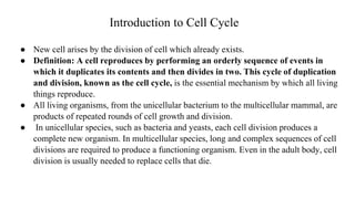
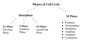
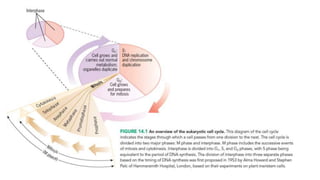
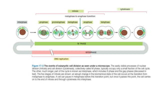
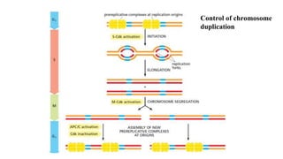
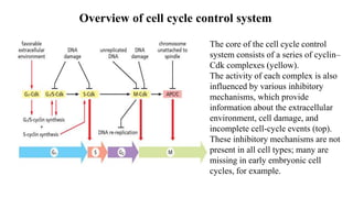
SlideShare a Scribd company logo
Module I
Cell biology II
S.Y.Biotech
Introduction to Cell Cycle
● New cell arises by the division of cell which already exists.
● Definition: A cell reproduces by performing an orderly sequence of events in
which it duplicates its contents and then divides in two. This cycle of duplication
and division, known as the cell cycle, is the essential mechanism by which all living
things reproduce.
● All living organisms, from the unicellular bacterium to the multicellular mammal, are
products of repeated rounds of cell growth and division.
● In unicellular species, such as bacteria and yeasts, each cell division produces a
complete new organism. In multicellular species, long and complex sequences of cell
divisions are required to produce a functioning organism. Even in the adult body, cell
division is usually needed to replace cells that die.
Phases of Cell Cycle
Interphase M Phase
G1 Phase
First Gap
Phase
S Phase
Synthesis
Phase
G2 Phase
second Gap
Phase
● Prophase
● Prometaphase
● Metaphase
● Anaphase
● Telophase
● Cytokinesis
Eukaryotic cell cycle
● The eukaryotic cell cycle is traditionally divided into four sequential phases: G1, S,
G2, and M. G1, S, and G2 together are called interphase.
● Interphase require 23 hours while M phase require 1 hour.
● Gap Phases:
➔ To allow time for growth, most cell cycles have gap phases—a G1 phase between
M phase and S phase and a G2 phase between S phase and mitosis. They also
provide time for the cell to monitor the internal and external environment.
➔ They ensure that conditions are suitable and preparations are complete before
the cell commits itself to the major upheavals of S phase and mitosis. The G1
phase is especially important in this respect. Its length can vary greatly depending on
external conditions and extracellular signals from other cells.
➔ G0 Phase:
❖ If extracellular conditions are unfavorable, for
example, cells delay progress through G1 and
may even enter a specialized resting state known
as G0 (G zero), in which they can remain for
days, weeks, or even years before resuming
proliferation. Indeed, many cells remain
permanently in G0 until they or the organism dies.
❖ If extracellular conditions are favorable and
signals to grow and divide are present, cells in
early G1 or G0 progress through a commitment
point near the end of G1 known as Start (in
yeasts) or the restriction point (in mammalian
cells).
● S phase:
➔ DNA replication occurs during a period of the cell cycle termed S phase (Synthesis Phase)
.
➔ The linear chromosomes of eukaryotic cells are vast and dynamic assemblies of DNA and
protein, and their duplication is a complex process that takes up a major fraction of the cell
cycle.
➔ Duration of S phase is about 8 hours.
➔ S phase is also the period when the cell synthesizes the additional histones that will be
needed as the cell doubles the number of nucleosomes in its chromosomes ensuring that
the daughter cells inherit all features of chromosome structure.
DNA synthesis:
1. Helix Unwinding
2. Extension of Primer
3. Complementary Strand
Synthesis
4. Formation of new copy of
DNA
● M Phase
1. Prophase:
Prometaphase:
Metaphase
Anaphase
Telophase
Cytokinesis
● After mitosis the cell is divided into two
daughter cells by a separate process
called cytokinesis.
● The first hint of cytokinesis in most
animal cells appears during anaphase as
an indentation of the cell surface in a
narrow band around the cell. As time
progresses, the indentation deepens to
form a furrow that moves inward toward
the center of the cell.
● The plane of the furrow lies in the same
plane previously occupied by the
chromosomes of the metaphase plate, so
that the two sets of chromosomes are
ultimately partitioned into different cells.
Cell Cycle Control System
● The control system is rigidly programmed to provide a fixed amount of time for the
completion of each cell-cycle event.
● The cell-cycle control system is based on a connected series of biochemical switches, each
of which initiates a specific cell-cycle event.
● Features of cell cycle control system:
1. The biochemical switches are generally binary (on/off) and launch events in a complete,
irreversible fashion. It would clearly be disastrous, for example, if events like chromosome
condensation or nuclear-envelope breakdown were only partially initiated or started but not
completed
2. Robust and reliable system,because backup mechanisms and other features allow the
system to operate effectively under a variety of conditions and even if some components
fail.
3. Highly adaptable and can be modified to suit specific cell types or to respond to specific
intracellular or extracellular signals.
Checkpoints
1. G1-S Checkpoint: It is a Start (or the
restriction) point in late G1, where the
cell commits to cell-cycle entry and
chromosome duplication.
2. G2-M Checkpoint: where the
control system triggers the early
mitotic events that lead to
chromosome alignment on the mitotic
spindle in metaphase.
3. Metaphase to anaphase transition:
where the control system stimulates
sister-chromatid separation, leading to
the completion of mitosis and
cytokinesis.
Key Components of Control System
Cyclins Cdk (Cyclin
dependent kinases)
● Central components of the cell-cycle control system are members of a family of
protein kinases known as cyclin-dependent kinases (Cdks).
● The activities of these kinases rise and fall as the cell progresses through the cycle,
leading to cyclical changes in the phosphorylation of intracellular proteins that
initiate or regulate the major events of the cell cycle.
● Cyclical changes in Cdk activity are controlled by a complex array of enzymes and
other proteins. The most important of these Cdk regulators are proteins known
as cyclins.
● Cdks, as their name implies, are dependent on cyclins for their activity: unless they
are bound tightly to a cyclin, they have no protein kinase activity
Fig: Two key components of the cell-cycle
control system:
When cyclin forms a complex with Cdk,
the protein kinase is activated to trigger
specific cell-cycle events. Without cyclin,
Cdk is inactive.
Cyclins were originally named because they undergo a cycle of synthesis and
degradation in each cell cycle. The levels of the Cdk proteins, by contrast, are
constant. Cyclical changes in cyclin protein levels result in the cyclic assembly and
activation of cyclin–Cdk complexes at specific stages of the cell cycle.
Classes of Cyclins
G1S Cyclins S Cyclins M Cyclins
Cyclin E: Activate
Cdks in late G1 and
thereby help trigger
progression through
Start, resulting in a
commitment to cell-
cycle entry.
Their levels fall in S
phase.
Cyclin A: Bind Cdks
soon after progression
through Start and help
stimulate chromosome
duplication. S-cyclin
levels remain elevated
until mitosis, and these
cyclins also contribute to
the control of some early
mitotic events.
Cyclin B:
Activate Cdks that
stimulate entry
into mitosis at the
G2/M transition.
M-cyclin levels
fall in mid-
mitosis.
G1 Cyclins
Cyclin D:
Helps govern
the activities of
the G1/S-
cyclins, which
control
progression
through Start in
late G1.
Cyclin–Cdk complexes of the cell-cycle control system.
The concentrations of the three major cyclin types oscillate during the cell cycle, while the
concentrations of Cdks (not shown) do not change and exceed cyclin amounts.
In late G1, rising G1/S-cyclin levels lead to the formation of G1/S-Cdk complexes that trigger
progression through the Start transition. S-Cdk complexes form at the start of S phase and
trigger DNA replication, as well as some early mitotic events. M-Cdk complexes form during G2
but are held in an inactive state; they are activated at the end of G2 and trigger entry into mitosis at
the G2/M transition.
Classes of CDKs
G1 Cdk G1-S Cdk S Cdk M Cdk
● Cdk 4 and
Cdk 6
● Interact
with G1
cyclins
● Cdk 2
● Interact
with G1-S
cyclins
● Cdk 2 and
Cdk 1
● Interact
with S
cyclins
● Cdk 1
● Interact
with M
cyclins
Activation Mechanism of Cdk by cyclins
The enzyme is shown in three
states. (A) In the inactive state,
without cyclin bound, the active site
is blocked by a region of the protein
called the T-loop (red). (B) The
binding of cyclin causes the T-loop
to move out of the active site,
resulting in partial activation of the
Cdk2. (C) Phosphorylation of
Cdk2 (by CAK) at a threonine
residue in the T-loop further
activates the enzyme by changing
the shape of the T-loop, improving
the ability of the enzyme to bind
its protein substrates.
Suppression of Cdk activity
1. By Inhibitory phosphorylation:
The active cyclin–Cdk complex
is turned off when the kinase
Wee1 phosphorylates two
closely spaced sites above the
active site. Removal of these
phosphates by the phosphatase
Cdc25 activates the cyclin-Cdk
complex. For simplicity, only one
inhibitory phosphate is shown.
2.By Cdk Inhibitor Proteins:
Binding of Cdk inhibitor proteins
(CKIs) inactivates cyclin–Cdk
complexes. The three-dimensional
structure of a cyclin–Cdk–CKI
complex reveals that CKI binding
stimulates a large rearrangement in
the structure of the Cdk active site,
rendering it inactive.Cells use CKIs
primarily to help govern the activities
of G1/S- and S-Cdks early in the cell
cycle.
Regulation of Metaphase to anaphase transition
● Activation of specific cyclin–Cdk complexes drives progression through the Start and
G2/M transitions, progression through the metaphase-to-anaphase transition is
triggered not by protein phosphorylation but by protein destruction, leading to the
final stages of cell division.
● The key regulator of the metaphase-to-anaphase transition is the anaphase
promoting complex, or cyclosome (APC/C), It is a large mutisubunit complex and a
member of the ubiquitin ligase family of enzymes.They polyubiquitylate specific
target proteins, resulting in their destruction in proteasomes.
Targets Of APC/C
Securin S and M cyclins
It protects the protein linkages
that hold sister-chromatid pairs
together in early mitosis.
Destruction of securin in
metaphase activates a protease
that separates the sisters and
unleashes anaphase,
Destroying these cyclins inactivates most
Cdks in the cell. As a result, the many
proteins phosphorylated by Cdks from S
phase to early mitosis are
dephosphorylated by various phosphatases
in the anaphase cell.
This dephosphorylation of Cdk targets
is required for the completion of M
phase, including the final steps in
mitosis and then cytokinesis.
APC/C is activated by
Cdc20 subunit. Then this
APC/C acts on S and M
cyclin for their
degradation.
SCF (Skp, Cullin, F-box containing complex)
● SCF is a multisubunit protein complex. It is a ubiquitin ligase protein.
● It has many functions in the cell, but its major role in the cell cycle is to ubiquitylate
certain CKI proteins in late G1, thereby helping to control the activation of S-
Cdks and DNA replication.
● SCF is also responsible for the destruction of G1/S-cyclins in early S phase.
● SCF activity depends on substrate-binding subunits called F-box proteins. Unlike
APC/C activity, however, SCF activity is constant during the cell cycle.
Ubiquitylation by SCF is controlled instead by changes in the phosphorylation state
of its target proteins, as F-box subunits recognize only specifically phosphorylated
proteins.
The activity of the ubiquitin ligase
SCF depends on substrate-binding
subunits called F-box proteins, of
which there are many different
types. The phosphorylation of a
target protein, such as the CKI
shown, allows the target to be
recognized by a specific F-box
subunit.
Cell Cycle Control at S Phase
The central event of chromosome duplication—DNA replication—poses two problems for
the cell.
1. Replication must occur with extreme accuracy to minimize the risk of mutations
in the next cell generation.
2. Every nucleotide in the genome must be copied once, and only once, to prevent
the damaging effects of gene amplification.
Control of chromosome
duplication
Control of chromosome duplication
● Preparations for DNA replication begin in late mitosis and G1, when the DNA
helicases are loaded by multiple proteins at the replication origin, forming the
prereplicative complex (preRC).
● In late mitosis and early G1, the proteins Cdc6 and Cdt1 collaborate with the
ORC(Origin Recognition Complex) to load the inactive DNA helicases around
the DNA next to the origin.
● S-Cdk activation leads to activation of the DNA helicases, which unwind the DNA at
origins to initiate DNA replication. Two replication forks move out from each origin
until the entire chromosome is duplicated. Duplicated chromosomes are then
segregated in M phase.
● S-Cdk activation in S phase also prevents assembly of new preRCs at any origin until
the following G1—thereby ensuring that each origin is activated only once in each
cell cycle.
Control of initiation
of replication
Control of the initiation of DNA replication:
● The replication origin is bound by the ORC(Origin recognition complex) throughout the
cell cycle.
● In early G1, Cdc6 associates with the ORC, and these proteins bind the DNA helicase,
which contains six closely related subunits called Mcm proteins. The helicase also
associates with a protein called Cdt1.
● Using energy provided by ATP hydrolysis, the ORC and Cdc6 proteins load two copies
of the DNA helicase, in an inactive form, around the DNA next to the origin, thereby
forming the prereplicative complex (preRC).
● At the onset of S phase, S-Cdk stimulates the assembly of several initiator proteins on
each DNA helicase, while another protein kinase, DDK, phosphorylates subunits of the
DNA helicase. As a result, the DNA helicases are activated and unwind the DNA.
● DNA polymerase and other replication proteins are recruited to the origin, and DNA
replication begins. The ORC is displaced by the replication machinery and then rebinds.
● S-Cdk and other mechanisms also inactivate the preRC components ORC, Cdc6, and
Cdt1, thereby preventing formation of new preRCs at the origins until the end of mitosis.
Ensuring that DNA is replicated once per cell cycle:
1. At the same time as S-Cdk initiates DNA replication, several mechanisms prevent
assembly of new preRCs. S-Cdk phosphorylates and thereby inhibits the ORC
and Cdc6 proteins.
2. Inactivation of the APC/C in late G1 also helps turn off preRC assembly. In late
mitosis and early G1, the APC/C triggers the destruction of a Cdt1 inhibitor
called geminin, thereby allowing Cdt1 to be active.
3. When the APC/C is turned off in late G1, geminin accumulates and inhibits the Cdt1
that is not associated with DNA.
4. Also, the association of Cdt1 with a protein at active replication forks stimulates Cdt1
destruction.
5. In these various ways, preRC formation is prevented from S phase to mitosis, thereby
ensuring that each origin is fired only once per cell cycle.
Cohesin holds sister chromatids together
Cohesin is a protein complex with
four subunits. (A) Two subunits,
Smc1 and Smc3, are coiled-coil
proteins with an ATPase domain
at one end; (B) two additional
subunits, Scc1 and Scc3, connect
the ATPase head domains,
forming a ring structure that may
encircle the sister chromatids as
shown in (C). The ATPase
domains are required for cohesin
loading on the DNA.
Overview of cell cycle control system
The core of the cell cycle control
system consists of a series of cyclin–
Cdk complexes (yellow).
The activity of each complex is also
influenced by various inhibitory
mechanisms, which provide
information about the extracellular
environment, cell damage, and
incomplete cell-cycle events (top).
These inhibitory mechanisms are not
present in all cell types; many are
missing in early embryonic cell
cycles, for example.
The Cell Cycle.pptx

More Related Content

Similar to The Cell Cycle.pptx

Cell cycle regulation ppt
Cell cycle regulation  pptCell cycle regulation  ppt
Cell cycle regulation ppt
manojsiddartha bolthajira
 
Cell Cycle- an essential part of cell growth
Cell Cycle- an essential part of cell growthCell Cycle- an essential part of cell growth
Cell Cycle- an essential part of cell growth
haritha shankar
 
Unit 4-cell cycle and cell division
Unit 4-cell cycle and cell divisionUnit 4-cell cycle and cell division
Unit 4-cell cycle and cell division
Komal Kp
 
Eukayotic_cell_cycle Prashant.ppt
Eukayotic_cell_cycle Prashant.pptEukayotic_cell_cycle Prashant.ppt
Eukayotic_cell_cycle Prashant.ppt
drpvczback
 
Introduction to the cell cycle
Introduction to the cell cycleIntroduction to the cell cycle
Introduction to the cell cycleElsa von Licy
 
Cell cycle presentation
Cell cycle presentationCell cycle presentation
Cell cycle presentationIbnu Alias
 
Cell cycle and it's checkpoints
Cell cycle and it's checkpointsCell cycle and it's checkpoints
Cell cycle and it's checkpoints
Ritisha Gupta
 
Cell cycle and therapeutic implications
Cell cycle and therapeutic implications Cell cycle and therapeutic implications
Cell cycle and therapeutic implications
Priyanka Malekar
 
Proteins Of Cell Cycle.pptx
Proteins Of Cell Cycle.pptxProteins Of Cell Cycle.pptx
Proteins Of Cell Cycle.pptx
SanaSidhikKV1
 
CELL CYCLE & ITS REGULATION.pptx
CELL CYCLE & ITS REGULATION.pptxCELL CYCLE & ITS REGULATION.pptx
CELL CYCLE & ITS REGULATION.pptx
AlthamishAzzu
 
cell cycle.pdf
cell cycle.pdfcell cycle.pdf
cell cycle.pdf
ashharnomani
 
Cell cycle checkpoints in yeast
Cell cycle checkpoints in yeast Cell cycle checkpoints in yeast
Cell cycle checkpoints in yeast
mehraseema
 
Cell cycle checkpoints in yeast
Cell cycle checkpoints in yeast Cell cycle checkpoints in yeast
Cell cycle checkpoints in yeast
mehraseema
 
Cell division cycle in yeast
Cell division cycle in yeast Cell division cycle in yeast
Cell division cycle in yeast
mehraseema
 
Cell Cycle and Division Human Anatomy and Physiology
Cell Cycle and Division Human Anatomy and PhysiologyCell Cycle and Division Human Anatomy and Physiology
Cell Cycle and Division Human Anatomy and Physiology
MahendraYadav550460
 
Cell cycle and growth regulation
Cell cycle and growth regulation Cell cycle and growth regulation
Cell cycle and growth regulation
Dr. Archana Balakrishnan
 
cell cycle 1st sem.pptx
cell cycle 1st sem.pptxcell cycle 1st sem.pptx
cell cycle 1st sem.pptx
dikshasingh649964
 
Molecular event during Cell cycle By KK Sahu Sir
Molecular event during Cell cycle By KK Sahu SirMolecular event during Cell cycle By KK Sahu Sir
Molecular event during Cell cycle By KK Sahu Sir
KAUSHAL SAHU
 
3. Cell Cycle.pptx
3. Cell Cycle.pptx3. Cell Cycle.pptx
3. Cell Cycle.pptx
SukhdeepSingh962560
 
The Cell cycle
The Cell cycleThe Cell cycle
The Cell cycle
Maha Hammady
 

Similar to The Cell Cycle.pptx (20)

Cell cycle regulation ppt
Cell cycle regulation  pptCell cycle regulation  ppt
Cell cycle regulation ppt
 
Cell Cycle- an essential part of cell growth
Cell Cycle- an essential part of cell growthCell Cycle- an essential part of cell growth
Cell Cycle- an essential part of cell growth
 
Unit 4-cell cycle and cell division
Unit 4-cell cycle and cell divisionUnit 4-cell cycle and cell division
Unit 4-cell cycle and cell division
 
Eukayotic_cell_cycle Prashant.ppt
Eukayotic_cell_cycle Prashant.pptEukayotic_cell_cycle Prashant.ppt
Eukayotic_cell_cycle Prashant.ppt
 
Introduction to the cell cycle
Introduction to the cell cycleIntroduction to the cell cycle
Introduction to the cell cycle
 
Cell cycle presentation
Cell cycle presentationCell cycle presentation
Cell cycle presentation
 
Cell cycle and it's checkpoints
Cell cycle and it's checkpointsCell cycle and it's checkpoints
Cell cycle and it's checkpoints
 
Cell cycle and therapeutic implications
Cell cycle and therapeutic implications Cell cycle and therapeutic implications
Cell cycle and therapeutic implications
 
Proteins Of Cell Cycle.pptx
Proteins Of Cell Cycle.pptxProteins Of Cell Cycle.pptx
Proteins Of Cell Cycle.pptx
 
CELL CYCLE & ITS REGULATION.pptx
CELL CYCLE & ITS REGULATION.pptxCELL CYCLE & ITS REGULATION.pptx
CELL CYCLE & ITS REGULATION.pptx
 
cell cycle.pdf
cell cycle.pdfcell cycle.pdf
cell cycle.pdf
 
Cell cycle checkpoints in yeast
Cell cycle checkpoints in yeast Cell cycle checkpoints in yeast
Cell cycle checkpoints in yeast
 
Cell cycle checkpoints in yeast
Cell cycle checkpoints in yeast Cell cycle checkpoints in yeast
Cell cycle checkpoints in yeast
 
Cell division cycle in yeast
Cell division cycle in yeast Cell division cycle in yeast
Cell division cycle in yeast
 
Cell Cycle and Division Human Anatomy and Physiology
Cell Cycle and Division Human Anatomy and PhysiologyCell Cycle and Division Human Anatomy and Physiology
Cell Cycle and Division Human Anatomy and Physiology
 
Cell cycle and growth regulation
Cell cycle and growth regulation Cell cycle and growth regulation
Cell cycle and growth regulation
 
cell cycle 1st sem.pptx
cell cycle 1st sem.pptxcell cycle 1st sem.pptx
cell cycle 1st sem.pptx
 
Molecular event during Cell cycle By KK Sahu Sir
Molecular event during Cell cycle By KK Sahu SirMolecular event during Cell cycle By KK Sahu Sir
Molecular event during Cell cycle By KK Sahu Sir
 
3. Cell Cycle.pptx
3. Cell Cycle.pptx3. Cell Cycle.pptx
3. Cell Cycle.pptx
 
The Cell cycle
The Cell cycleThe Cell cycle
The Cell cycle
 

More from Alisha Shaikh

cell cloning- Therapeutic and reproductive cloning
cell cloning- Therapeutic and reproductive cloningcell cloning- Therapeutic and reproductive cloning
cell cloning- Therapeutic and reproductive cloning
Alisha Shaikh
 
Glycogenesis and glycogenolysis pathways
Glycogenesis and glycogenolysis pathwaysGlycogenesis and glycogenolysis pathways
Glycogenesis and glycogenolysis pathways
Alisha Shaikh
 
_ETC and Oxidative phosphorylation.pptx
_ETC and  Oxidative phosphorylation.pptx_ETC and  Oxidative phosphorylation.pptx
_ETC and Oxidative phosphorylation.pptx
Alisha Shaikh
 
_Cell Signalling .pptx
_Cell Signalling .pptx_Cell Signalling .pptx
_Cell Signalling .pptx
Alisha Shaikh
 
_Meiosis.pptx
_Meiosis.pptx_Meiosis.pptx
_Meiosis.pptx
Alisha Shaikh
 
_Mitosis.pptx
_Mitosis.pptx_Mitosis.pptx
_Mitosis.pptx
Alisha Shaikh
 
Different types of vectors.pptx
Different types of vectors.pptxDifferent types of vectors.pptx
Different types of vectors.pptx
Alisha Shaikh
 
_Site directed Mutagenesis.pptx
_Site directed Mutagenesis.pptx_Site directed Mutagenesis.pptx
_Site directed Mutagenesis.pptx
Alisha Shaikh
 
_Sequencing Methods .pptx
_Sequencing Methods .pptx_Sequencing Methods .pptx
_Sequencing Methods .pptx
Alisha Shaikh
 
_Biogas .pptx
_Biogas .pptx_Biogas .pptx
_Biogas .pptx
Alisha Shaikh
 
_PCR and its different types.pptx
_PCR and its different types.pptx_PCR and its different types.pptx
_PCR and its different types.pptx
Alisha Shaikh
 
Spectroscopy.pptx
Spectroscopy.pptxSpectroscopy.pptx
Spectroscopy.pptx
Alisha Shaikh
 
Chromatographic Techniques.pptx
Chromatographic Techniques.pptxChromatographic Techniques.pptx
Chromatographic Techniques.pptx
Alisha Shaikh
 
_centrifugation .pptx
_centrifugation .pptx_centrifugation .pptx
_centrifugation .pptx
Alisha Shaikh
 
Marine Biotechnology.pptx
Marine Biotechnology.pptxMarine Biotechnology.pptx
Marine Biotechnology.pptx
Alisha Shaikh
 
Molecular Diagnostics.pptx
Molecular Diagnostics.pptxMolecular Diagnostics.pptx
Molecular Diagnostics.pptx
Alisha Shaikh
 

More from Alisha Shaikh (16)

cell cloning- Therapeutic and reproductive cloning
cell cloning- Therapeutic and reproductive cloningcell cloning- Therapeutic and reproductive cloning
cell cloning- Therapeutic and reproductive cloning
 
Glycogenesis and glycogenolysis pathways
Glycogenesis and glycogenolysis pathwaysGlycogenesis and glycogenolysis pathways
Glycogenesis and glycogenolysis pathways
 
_ETC and Oxidative phosphorylation.pptx
_ETC and  Oxidative phosphorylation.pptx_ETC and  Oxidative phosphorylation.pptx
_ETC and Oxidative phosphorylation.pptx
 
_Cell Signalling .pptx
_Cell Signalling .pptx_Cell Signalling .pptx
_Cell Signalling .pptx
 
_Meiosis.pptx
_Meiosis.pptx_Meiosis.pptx
_Meiosis.pptx
 
_Mitosis.pptx
_Mitosis.pptx_Mitosis.pptx
_Mitosis.pptx
 
Different types of vectors.pptx
Different types of vectors.pptxDifferent types of vectors.pptx
Different types of vectors.pptx
 
_Site directed Mutagenesis.pptx
_Site directed Mutagenesis.pptx_Site directed Mutagenesis.pptx
_Site directed Mutagenesis.pptx
 
_Sequencing Methods .pptx
_Sequencing Methods .pptx_Sequencing Methods .pptx
_Sequencing Methods .pptx
 
_Biogas .pptx
_Biogas .pptx_Biogas .pptx
_Biogas .pptx
 
_PCR and its different types.pptx
_PCR and its different types.pptx_PCR and its different types.pptx
_PCR and its different types.pptx
 
Spectroscopy.pptx
Spectroscopy.pptxSpectroscopy.pptx
Spectroscopy.pptx
 
Chromatographic Techniques.pptx
Chromatographic Techniques.pptxChromatographic Techniques.pptx
Chromatographic Techniques.pptx
 
_centrifugation .pptx
_centrifugation .pptx_centrifugation .pptx
_centrifugation .pptx
 
Marine Biotechnology.pptx
Marine Biotechnology.pptxMarine Biotechnology.pptx
Marine Biotechnology.pptx
 
Molecular Diagnostics.pptx
Molecular Diagnostics.pptxMolecular Diagnostics.pptx
Molecular Diagnostics.pptx
 

Recently uploaded

Lateral Ventricles.pdf very easy good diagrams comprehensive
Lateral Ventricles.pdf very easy good diagrams comprehensiveLateral Ventricles.pdf very easy good diagrams comprehensive
Lateral Ventricles.pdf very easy good diagrams comprehensive
silvermistyshot
 
BLOOD AND BLOOD COMPONENT- introduction to blood physiology
BLOOD AND BLOOD COMPONENT- introduction to blood physiologyBLOOD AND BLOOD COMPONENT- introduction to blood physiology
BLOOD AND BLOOD COMPONENT- introduction to blood physiology
NoelManyise1
 
Chapter 12 - climate change and the energy crisis
Chapter 12 - climate change and the energy crisisChapter 12 - climate change and the energy crisis
Chapter 12 - climate change and the energy crisis
tonzsalvador2222
 
general properties of oerganologametal.ppt
general properties of oerganologametal.pptgeneral properties of oerganologametal.ppt
general properties of oerganologametal.ppt
IqrimaNabilatulhusni
 
Deep Behavioral Phenotyping in Systems Neuroscience for Functional Atlasing a...
Deep Behavioral Phenotyping in Systems Neuroscience for Functional Atlasing a...Deep Behavioral Phenotyping in Systems Neuroscience for Functional Atlasing a...
Deep Behavioral Phenotyping in Systems Neuroscience for Functional Atlasing a...
Ana Luísa Pinho
 
erythropoiesis-I_mechanism& clinical significance.pptx
erythropoiesis-I_mechanism& clinical significance.pptxerythropoiesis-I_mechanism& clinical significance.pptx
erythropoiesis-I_mechanism& clinical significance.pptx
muralinath2
 
bordetella pertussis.................................ppt
bordetella pertussis.................................pptbordetella pertussis.................................ppt
bordetella pertussis.................................ppt
kejapriya1
 
Nutraceutical market, scope and growth: Herbal drug technology
Nutraceutical market, scope and growth: Herbal drug technologyNutraceutical market, scope and growth: Herbal drug technology
Nutraceutical market, scope and growth: Herbal drug technology
Lokesh Patil
 
Richard's aventures in two entangled wonderlands
Richard's aventures in two entangled wonderlandsRichard's aventures in two entangled wonderlands
Richard's aventures in two entangled wonderlands
Richard Gill
 
S.1 chemistry scheme term 2 for ordinary level
S.1 chemistry scheme term 2 for ordinary levelS.1 chemistry scheme term 2 for ordinary level
S.1 chemistry scheme term 2 for ordinary level
ronaldlakony0
 
Comparing Evolved Extractive Text Summary Scores of Bidirectional Encoder Rep...
Comparing Evolved Extractive Text Summary Scores of Bidirectional Encoder Rep...Comparing Evolved Extractive Text Summary Scores of Bidirectional Encoder Rep...
Comparing Evolved Extractive Text Summary Scores of Bidirectional Encoder Rep...
University of Maribor
 
Salas, V. (2024) "John of St. Thomas (Poinsot) on the Science of Sacred Theol...
Salas, V. (2024) "John of St. Thomas (Poinsot) on the Science of Sacred Theol...Salas, V. (2024) "John of St. Thomas (Poinsot) on the Science of Sacred Theol...
Salas, V. (2024) "John of St. Thomas (Poinsot) on the Science of Sacred Theol...
Studia Poinsotiana
 
PRESENTATION ABOUT PRINCIPLE OF COSMATIC EVALUATION
PRESENTATION ABOUT PRINCIPLE OF COSMATIC EVALUATIONPRESENTATION ABOUT PRINCIPLE OF COSMATIC EVALUATION
PRESENTATION ABOUT PRINCIPLE OF COSMATIC EVALUATION
ChetanK57
 
THE IMPORTANCE OF MARTIAN ATMOSPHERE SAMPLE RETURN.
THE IMPORTANCE OF MARTIAN ATMOSPHERE SAMPLE RETURN.THE IMPORTANCE OF MARTIAN ATMOSPHERE SAMPLE RETURN.
THE IMPORTANCE OF MARTIAN ATMOSPHERE SAMPLE RETURN.
Sérgio Sacani
 
role of pramana in research.pptx in science
role of pramana in research.pptx in sciencerole of pramana in research.pptx in science
role of pramana in research.pptx in science
sonaliswain16
 
platelets_clotting_biogenesis.clot retractionpptx
platelets_clotting_biogenesis.clot retractionpptxplatelets_clotting_biogenesis.clot retractionpptx
platelets_clotting_biogenesis.clot retractionpptx
muralinath2
 
Mammalian Pineal Body Structure and Also Functions
Mammalian Pineal Body Structure and Also FunctionsMammalian Pineal Body Structure and Also Functions
Mammalian Pineal Body Structure and Also Functions
YOGESH DOGRA
 
如何办理(uvic毕业证书)维多利亚大学毕业证本科学位证书原版一模一样
如何办理(uvic毕业证书)维多利亚大学毕业证本科学位证书原版一模一样如何办理(uvic毕业证书)维多利亚大学毕业证本科学位证书原版一模一样
如何办理(uvic毕业证书)维多利亚大学毕业证本科学位证书原版一模一样
yqqaatn0
 
extra-chromosomal-inheritance[1].pptx.pdfpdf
extra-chromosomal-inheritance[1].pptx.pdfpdfextra-chromosomal-inheritance[1].pptx.pdfpdf
extra-chromosomal-inheritance[1].pptx.pdfpdf
DiyaBiswas10
 
Comparative structure of adrenal gland in vertebrates
Comparative structure of adrenal gland in vertebratesComparative structure of adrenal gland in vertebrates
Comparative structure of adrenal gland in vertebrates
sachin783648
 

Recently uploaded (20)

Lateral Ventricles.pdf very easy good diagrams comprehensive
Lateral Ventricles.pdf very easy good diagrams comprehensiveLateral Ventricles.pdf very easy good diagrams comprehensive
Lateral Ventricles.pdf very easy good diagrams comprehensive
 
BLOOD AND BLOOD COMPONENT- introduction to blood physiology
BLOOD AND BLOOD COMPONENT- introduction to blood physiologyBLOOD AND BLOOD COMPONENT- introduction to blood physiology
BLOOD AND BLOOD COMPONENT- introduction to blood physiology
 
Chapter 12 - climate change and the energy crisis
Chapter 12 - climate change and the energy crisisChapter 12 - climate change and the energy crisis
Chapter 12 - climate change and the energy crisis
 
general properties of oerganologametal.ppt
general properties of oerganologametal.pptgeneral properties of oerganologametal.ppt
general properties of oerganologametal.ppt
 
Deep Behavioral Phenotyping in Systems Neuroscience for Functional Atlasing a...
Deep Behavioral Phenotyping in Systems Neuroscience for Functional Atlasing a...Deep Behavioral Phenotyping in Systems Neuroscience for Functional Atlasing a...
Deep Behavioral Phenotyping in Systems Neuroscience for Functional Atlasing a...
 
erythropoiesis-I_mechanism& clinical significance.pptx
erythropoiesis-I_mechanism& clinical significance.pptxerythropoiesis-I_mechanism& clinical significance.pptx
erythropoiesis-I_mechanism& clinical significance.pptx
 
bordetella pertussis.................................ppt
bordetella pertussis.................................pptbordetella pertussis.................................ppt
bordetella pertussis.................................ppt
 
Nutraceutical market, scope and growth: Herbal drug technology
Nutraceutical market, scope and growth: Herbal drug technologyNutraceutical market, scope and growth: Herbal drug technology
Nutraceutical market, scope and growth: Herbal drug technology
 
Richard's aventures in two entangled wonderlands
Richard's aventures in two entangled wonderlandsRichard's aventures in two entangled wonderlands
Richard's aventures in two entangled wonderlands
 
S.1 chemistry scheme term 2 for ordinary level
S.1 chemistry scheme term 2 for ordinary levelS.1 chemistry scheme term 2 for ordinary level
S.1 chemistry scheme term 2 for ordinary level
 
Comparing Evolved Extractive Text Summary Scores of Bidirectional Encoder Rep...
Comparing Evolved Extractive Text Summary Scores of Bidirectional Encoder Rep...Comparing Evolved Extractive Text Summary Scores of Bidirectional Encoder Rep...
Comparing Evolved Extractive Text Summary Scores of Bidirectional Encoder Rep...
 
Salas, V. (2024) "John of St. Thomas (Poinsot) on the Science of Sacred Theol...
Salas, V. (2024) "John of St. Thomas (Poinsot) on the Science of Sacred Theol...Salas, V. (2024) "John of St. Thomas (Poinsot) on the Science of Sacred Theol...
Salas, V. (2024) "John of St. Thomas (Poinsot) on the Science of Sacred Theol...
 
PRESENTATION ABOUT PRINCIPLE OF COSMATIC EVALUATION
PRESENTATION ABOUT PRINCIPLE OF COSMATIC EVALUATIONPRESENTATION ABOUT PRINCIPLE OF COSMATIC EVALUATION
PRESENTATION ABOUT PRINCIPLE OF COSMATIC EVALUATION
 
THE IMPORTANCE OF MARTIAN ATMOSPHERE SAMPLE RETURN.
THE IMPORTANCE OF MARTIAN ATMOSPHERE SAMPLE RETURN.THE IMPORTANCE OF MARTIAN ATMOSPHERE SAMPLE RETURN.
THE IMPORTANCE OF MARTIAN ATMOSPHERE SAMPLE RETURN.
 
role of pramana in research.pptx in science
role of pramana in research.pptx in sciencerole of pramana in research.pptx in science
role of pramana in research.pptx in science
 
platelets_clotting_biogenesis.clot retractionpptx
platelets_clotting_biogenesis.clot retractionpptxplatelets_clotting_biogenesis.clot retractionpptx
platelets_clotting_biogenesis.clot retractionpptx
 
Mammalian Pineal Body Structure and Also Functions
Mammalian Pineal Body Structure and Also FunctionsMammalian Pineal Body Structure and Also Functions
Mammalian Pineal Body Structure and Also Functions
 
如何办理(uvic毕业证书)维多利亚大学毕业证本科学位证书原版一模一样
如何办理(uvic毕业证书)维多利亚大学毕业证本科学位证书原版一模一样如何办理(uvic毕业证书)维多利亚大学毕业证本科学位证书原版一模一样
如何办理(uvic毕业证书)维多利亚大学毕业证本科学位证书原版一模一样
 
extra-chromosomal-inheritance[1].pptx.pdfpdf
extra-chromosomal-inheritance[1].pptx.pdfpdfextra-chromosomal-inheritance[1].pptx.pdfpdf
extra-chromosomal-inheritance[1].pptx.pdfpdf
 
Comparative structure of adrenal gland in vertebrates
Comparative structure of adrenal gland in vertebratesComparative structure of adrenal gland in vertebrates
Comparative structure of adrenal gland in vertebrates
 

The Cell Cycle.pptx

  • 1. Module I Cell biology II S.Y.Biotech
  • 2. Introduction to Cell Cycle ● New cell arises by the division of cell which already exists. ● Definition: A cell reproduces by performing an orderly sequence of events in which it duplicates its contents and then divides in two. This cycle of duplication and division, known as the cell cycle, is the essential mechanism by which all living things reproduce. ● All living organisms, from the unicellular bacterium to the multicellular mammal, are products of repeated rounds of cell growth and division. ● In unicellular species, such as bacteria and yeasts, each cell division produces a complete new organism. In multicellular species, long and complex sequences of cell divisions are required to produce a functioning organism. Even in the adult body, cell division is usually needed to replace cells that die.
  • 3. Phases of Cell Cycle Interphase M Phase G1 Phase First Gap Phase S Phase Synthesis Phase G2 Phase second Gap Phase ● Prophase ● Prometaphase ● Metaphase ● Anaphase ● Telophase ● Cytokinesis
  • 4.
  • 5. Eukaryotic cell cycle ● The eukaryotic cell cycle is traditionally divided into four sequential phases: G1, S, G2, and M. G1, S, and G2 together are called interphase. ● Interphase require 23 hours while M phase require 1 hour. ● Gap Phases: ➔ To allow time for growth, most cell cycles have gap phases—a G1 phase between M phase and S phase and a G2 phase between S phase and mitosis. They also provide time for the cell to monitor the internal and external environment. ➔ They ensure that conditions are suitable and preparations are complete before the cell commits itself to the major upheavals of S phase and mitosis. The G1 phase is especially important in this respect. Its length can vary greatly depending on external conditions and extracellular signals from other cells.
  • 6. ➔ G0 Phase: ❖ If extracellular conditions are unfavorable, for example, cells delay progress through G1 and may even enter a specialized resting state known as G0 (G zero), in which they can remain for days, weeks, or even years before resuming proliferation. Indeed, many cells remain permanently in G0 until they or the organism dies. ❖ If extracellular conditions are favorable and signals to grow and divide are present, cells in early G1 or G0 progress through a commitment point near the end of G1 known as Start (in yeasts) or the restriction point (in mammalian cells).
  • 7. ● S phase: ➔ DNA replication occurs during a period of the cell cycle termed S phase (Synthesis Phase) . ➔ The linear chromosomes of eukaryotic cells are vast and dynamic assemblies of DNA and protein, and their duplication is a complex process that takes up a major fraction of the cell cycle. ➔ Duration of S phase is about 8 hours. ➔ S phase is also the period when the cell synthesizes the additional histones that will be needed as the cell doubles the number of nucleosomes in its chromosomes ensuring that the daughter cells inherit all features of chromosome structure.
  • 8. DNA synthesis: 1. Helix Unwinding 2. Extension of Primer 3. Complementary Strand Synthesis 4. Formation of new copy of DNA
  • 9. ● M Phase 1. Prophase:
  • 14. Cytokinesis ● After mitosis the cell is divided into two daughter cells by a separate process called cytokinesis. ● The first hint of cytokinesis in most animal cells appears during anaphase as an indentation of the cell surface in a narrow band around the cell. As time progresses, the indentation deepens to form a furrow that moves inward toward the center of the cell. ● The plane of the furrow lies in the same plane previously occupied by the chromosomes of the metaphase plate, so that the two sets of chromosomes are ultimately partitioned into different cells.
  • 15.
  • 16. Cell Cycle Control System ● The control system is rigidly programmed to provide a fixed amount of time for the completion of each cell-cycle event. ● The cell-cycle control system is based on a connected series of biochemical switches, each of which initiates a specific cell-cycle event. ● Features of cell cycle control system: 1. The biochemical switches are generally binary (on/off) and launch events in a complete, irreversible fashion. It would clearly be disastrous, for example, if events like chromosome condensation or nuclear-envelope breakdown were only partially initiated or started but not completed 2. Robust and reliable system,because backup mechanisms and other features allow the system to operate effectively under a variety of conditions and even if some components fail. 3. Highly adaptable and can be modified to suit specific cell types or to respond to specific intracellular or extracellular signals.
  • 17. Checkpoints 1. G1-S Checkpoint: It is a Start (or the restriction) point in late G1, where the cell commits to cell-cycle entry and chromosome duplication. 2. G2-M Checkpoint: where the control system triggers the early mitotic events that lead to chromosome alignment on the mitotic spindle in metaphase. 3. Metaphase to anaphase transition: where the control system stimulates sister-chromatid separation, leading to the completion of mitosis and cytokinesis.
  • 18. Key Components of Control System Cyclins Cdk (Cyclin dependent kinases) ● Central components of the cell-cycle control system are members of a family of protein kinases known as cyclin-dependent kinases (Cdks). ● The activities of these kinases rise and fall as the cell progresses through the cycle, leading to cyclical changes in the phosphorylation of intracellular proteins that initiate or regulate the major events of the cell cycle. ● Cyclical changes in Cdk activity are controlled by a complex array of enzymes and other proteins. The most important of these Cdk regulators are proteins known as cyclins. ● Cdks, as their name implies, are dependent on cyclins for their activity: unless they are bound tightly to a cyclin, they have no protein kinase activity
  • 19. Fig: Two key components of the cell-cycle control system: When cyclin forms a complex with Cdk, the protein kinase is activated to trigger specific cell-cycle events. Without cyclin, Cdk is inactive. Cyclins were originally named because they undergo a cycle of synthesis and degradation in each cell cycle. The levels of the Cdk proteins, by contrast, are constant. Cyclical changes in cyclin protein levels result in the cyclic assembly and activation of cyclin–Cdk complexes at specific stages of the cell cycle.
  • 20. Classes of Cyclins G1S Cyclins S Cyclins M Cyclins Cyclin E: Activate Cdks in late G1 and thereby help trigger progression through Start, resulting in a commitment to cell- cycle entry. Their levels fall in S phase. Cyclin A: Bind Cdks soon after progression through Start and help stimulate chromosome duplication. S-cyclin levels remain elevated until mitosis, and these cyclins also contribute to the control of some early mitotic events. Cyclin B: Activate Cdks that stimulate entry into mitosis at the G2/M transition. M-cyclin levels fall in mid- mitosis. G1 Cyclins Cyclin D: Helps govern the activities of the G1/S- cyclins, which control progression through Start in late G1.
  • 21. Cyclin–Cdk complexes of the cell-cycle control system. The concentrations of the three major cyclin types oscillate during the cell cycle, while the concentrations of Cdks (not shown) do not change and exceed cyclin amounts. In late G1, rising G1/S-cyclin levels lead to the formation of G1/S-Cdk complexes that trigger progression through the Start transition. S-Cdk complexes form at the start of S phase and trigger DNA replication, as well as some early mitotic events. M-Cdk complexes form during G2 but are held in an inactive state; they are activated at the end of G2 and trigger entry into mitosis at the G2/M transition.
  • 22. Classes of CDKs G1 Cdk G1-S Cdk S Cdk M Cdk ● Cdk 4 and Cdk 6 ● Interact with G1 cyclins ● Cdk 2 ● Interact with G1-S cyclins ● Cdk 2 and Cdk 1 ● Interact with S cyclins ● Cdk 1 ● Interact with M cyclins
  • 23.
  • 24. Activation Mechanism of Cdk by cyclins The enzyme is shown in three states. (A) In the inactive state, without cyclin bound, the active site is blocked by a region of the protein called the T-loop (red). (B) The binding of cyclin causes the T-loop to move out of the active site, resulting in partial activation of the Cdk2. (C) Phosphorylation of Cdk2 (by CAK) at a threonine residue in the T-loop further activates the enzyme by changing the shape of the T-loop, improving the ability of the enzyme to bind its protein substrates.
  • 25. Suppression of Cdk activity 1. By Inhibitory phosphorylation: The active cyclin–Cdk complex is turned off when the kinase Wee1 phosphorylates two closely spaced sites above the active site. Removal of these phosphates by the phosphatase Cdc25 activates the cyclin-Cdk complex. For simplicity, only one inhibitory phosphate is shown.
  • 26. 2.By Cdk Inhibitor Proteins: Binding of Cdk inhibitor proteins (CKIs) inactivates cyclin–Cdk complexes. The three-dimensional structure of a cyclin–Cdk–CKI complex reveals that CKI binding stimulates a large rearrangement in the structure of the Cdk active site, rendering it inactive.Cells use CKIs primarily to help govern the activities of G1/S- and S-Cdks early in the cell cycle.
  • 27. Regulation of Metaphase to anaphase transition ● Activation of specific cyclin–Cdk complexes drives progression through the Start and G2/M transitions, progression through the metaphase-to-anaphase transition is triggered not by protein phosphorylation but by protein destruction, leading to the final stages of cell division. ● The key regulator of the metaphase-to-anaphase transition is the anaphase promoting complex, or cyclosome (APC/C), It is a large mutisubunit complex and a member of the ubiquitin ligase family of enzymes.They polyubiquitylate specific target proteins, resulting in their destruction in proteasomes.
  • 28. Targets Of APC/C Securin S and M cyclins It protects the protein linkages that hold sister-chromatid pairs together in early mitosis. Destruction of securin in metaphase activates a protease that separates the sisters and unleashes anaphase, Destroying these cyclins inactivates most Cdks in the cell. As a result, the many proteins phosphorylated by Cdks from S phase to early mitosis are dephosphorylated by various phosphatases in the anaphase cell. This dephosphorylation of Cdk targets is required for the completion of M phase, including the final steps in mitosis and then cytokinesis.
  • 29. APC/C is activated by Cdc20 subunit. Then this APC/C acts on S and M cyclin for their degradation.
  • 30. SCF (Skp, Cullin, F-box containing complex) ● SCF is a multisubunit protein complex. It is a ubiquitin ligase protein. ● It has many functions in the cell, but its major role in the cell cycle is to ubiquitylate certain CKI proteins in late G1, thereby helping to control the activation of S- Cdks and DNA replication. ● SCF is also responsible for the destruction of G1/S-cyclins in early S phase. ● SCF activity depends on substrate-binding subunits called F-box proteins. Unlike APC/C activity, however, SCF activity is constant during the cell cycle. Ubiquitylation by SCF is controlled instead by changes in the phosphorylation state of its target proteins, as F-box subunits recognize only specifically phosphorylated proteins.
  • 31. The activity of the ubiquitin ligase SCF depends on substrate-binding subunits called F-box proteins, of which there are many different types. The phosphorylation of a target protein, such as the CKI shown, allows the target to be recognized by a specific F-box subunit.
  • 32. Cell Cycle Control at S Phase The central event of chromosome duplication—DNA replication—poses two problems for the cell. 1. Replication must occur with extreme accuracy to minimize the risk of mutations in the next cell generation. 2. Every nucleotide in the genome must be copied once, and only once, to prevent the damaging effects of gene amplification.
  • 34. Control of chromosome duplication ● Preparations for DNA replication begin in late mitosis and G1, when the DNA helicases are loaded by multiple proteins at the replication origin, forming the prereplicative complex (preRC). ● In late mitosis and early G1, the proteins Cdc6 and Cdt1 collaborate with the ORC(Origin Recognition Complex) to load the inactive DNA helicases around the DNA next to the origin. ● S-Cdk activation leads to activation of the DNA helicases, which unwind the DNA at origins to initiate DNA replication. Two replication forks move out from each origin until the entire chromosome is duplicated. Duplicated chromosomes are then segregated in M phase. ● S-Cdk activation in S phase also prevents assembly of new preRCs at any origin until the following G1—thereby ensuring that each origin is activated only once in each cell cycle.
  • 36. Control of the initiation of DNA replication: ● The replication origin is bound by the ORC(Origin recognition complex) throughout the cell cycle. ● In early G1, Cdc6 associates with the ORC, and these proteins bind the DNA helicase, which contains six closely related subunits called Mcm proteins. The helicase also associates with a protein called Cdt1. ● Using energy provided by ATP hydrolysis, the ORC and Cdc6 proteins load two copies of the DNA helicase, in an inactive form, around the DNA next to the origin, thereby forming the prereplicative complex (preRC). ● At the onset of S phase, S-Cdk stimulates the assembly of several initiator proteins on each DNA helicase, while another protein kinase, DDK, phosphorylates subunits of the DNA helicase. As a result, the DNA helicases are activated and unwind the DNA. ● DNA polymerase and other replication proteins are recruited to the origin, and DNA replication begins. The ORC is displaced by the replication machinery and then rebinds. ● S-Cdk and other mechanisms also inactivate the preRC components ORC, Cdc6, and Cdt1, thereby preventing formation of new preRCs at the origins until the end of mitosis.
  • 37. Ensuring that DNA is replicated once per cell cycle: 1. At the same time as S-Cdk initiates DNA replication, several mechanisms prevent assembly of new preRCs. S-Cdk phosphorylates and thereby inhibits the ORC and Cdc6 proteins. 2. Inactivation of the APC/C in late G1 also helps turn off preRC assembly. In late mitosis and early G1, the APC/C triggers the destruction of a Cdt1 inhibitor called geminin, thereby allowing Cdt1 to be active. 3. When the APC/C is turned off in late G1, geminin accumulates and inhibits the Cdt1 that is not associated with DNA. 4. Also, the association of Cdt1 with a protein at active replication forks stimulates Cdt1 destruction. 5. In these various ways, preRC formation is prevented from S phase to mitosis, thereby ensuring that each origin is fired only once per cell cycle.
  • 38. Cohesin holds sister chromatids together Cohesin is a protein complex with four subunits. (A) Two subunits, Smc1 and Smc3, are coiled-coil proteins with an ATPase domain at one end; (B) two additional subunits, Scc1 and Scc3, connect the ATPase head domains, forming a ring structure that may encircle the sister chromatids as shown in (C). The ATPase domains are required for cohesin loading on the DNA.
  • 39. Overview of cell cycle control system The core of the cell cycle control system consists of a series of cyclin– Cdk complexes (yellow). The activity of each complex is also influenced by various inhibitory mechanisms, which provide information about the extracellular environment, cell damage, and incomplete cell-cycle events (top). These inhibitory mechanisms are not present in all cell types; many are missing in early embryonic cell cycles, for example.