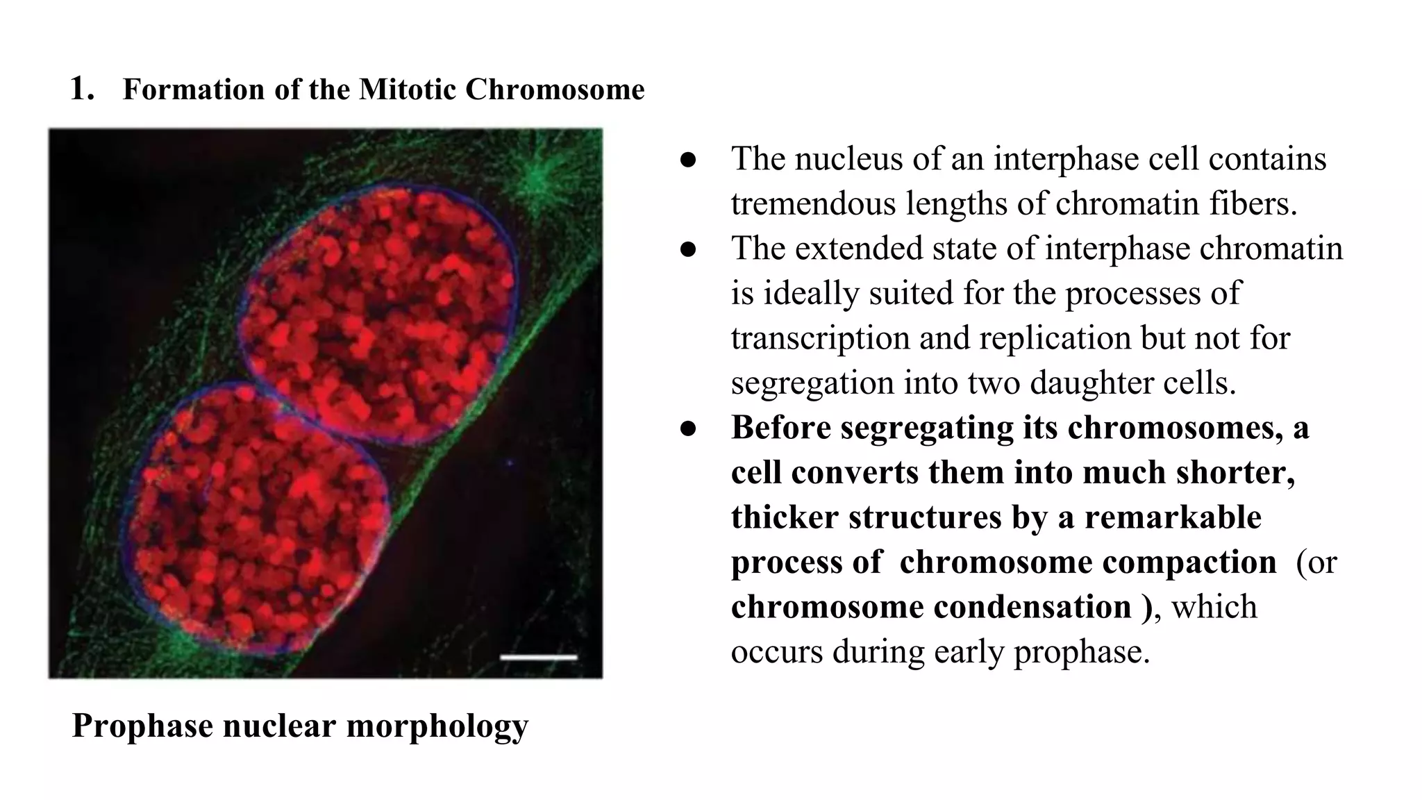
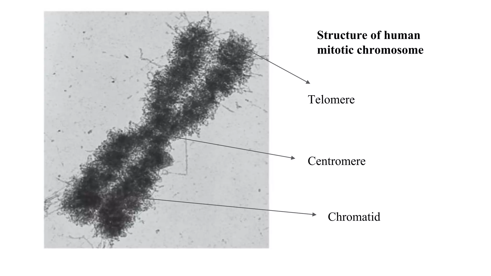
The cell division in Plants and animals
1. Mitosis
Cell division is a process in which one cell divides in two daughter cells.
Types of cell division
Cell Division
Mitosis Meiosis
Mitosis is the process of cell
division where a cell divides
in two daughter cells which
have identical number of
chromosomes compared to
the parent cell.
2n
Meiosis is the process of cell
division where a cell divides
in two daughter cells which
have half number of
chromosomes than parent
cell.
2n
2n
2n
n
n
Mitosis
● Mitosis is a process of nuclear division in which the replicated DNA molecules of
each chromosome are faithfully segregated into two nuclei.
● Mitosis is usually accompanied by cytokinesis , a process by which a dividing cell
splits in two, partitioning the cytoplasm into two cellular packages.
● The two daughter cells resulting from mitosis and cytokinesis possess a genetic
content identical to each other and to the mother cell from which they arose.
● Mitosis, therefore, maintains the chromosome number and generates new cells
for the growth and maintenance of an organism.
● Mitosis is a stage of the cell cycle when the cell devotes virtually all of its energy
to a single activity—chromosome segregation. As a result, most metabolic
activities of the cell, including transcription and translation, are reduced during
mitosis, and the cell becomes relatively unresponsive to external stimuli.
Overview of mitosis
Prophase
During the first stage of mitosis, prophase , the duplicated chromosomes are
prepared for segregation and the mitotic machinery is assembled.
Major events in Prophase:
1. Formation of the Mitotic Chromosome
2. Function of Centromeres and Kinetochores
3. Formation of the Mitotic Spindle
4. The Dissolution of the Nuclear Envelope and Partitioning of Cytoplasmic Organelles
1. Formation of the Mitotic Chromosome
● The nucleus of an interphase cell contains
tremendous lengths of chromatin fibers.
● The extended state of interphase chromatin
is ideally suited for the processes of
transcription and replication but not for
segregation into two daughter cells.
● Before segregating its chromosomes, a
cell converts them into much shorter,
thicker structures by a remarkable
process of chromosome compaction (or
chromosome condensation ), which
occurs during early prophase.
Prophase nuclear morphology
● Chromosome compaction/condensation is done by multiprotein complexes called
condensins.
● In the presence of a topoisomerase and ATP, condensin is able to bind to DNA and
curl the DNA into positively supercoiled loops.
● Chromosome compaction at prophase requires topoisomerase II, which along with
condensin is present as part of the mitotic chromosome scaffold.
● Condensin is activated at the onset of mitosis by phosphorylation of several of its
subunits by the cyclin–Cdk responsible for driving cells from G2 into mitosis. Thus
condensin is one of the targets through which Cdks are able to trigger cell cycle activities.
● As the result of compaction, the chromosomes of a mitotic cell appear as distinct,
rod‐like structures.
● Close examination of mitotic chromosomes reveals each of them to be composed of two
mirror image, “sister” chromatids.
Just after replication, the DNA helices of a pair of
sister chromatids would be held in association by
cohesin molecules that encircled the sister DNA
helices, as shown at the left of the drawing.
As the cell entered mitosis, the compaction
process would begin, aided by condensin
molecules, as shown in the right part of the drawing.
In this model, condensin brings about chromosome
compaction by forming a ring around supercoiled
loops of DNA within chromatin. Cohesin molecules
would continue to hold the DNA of sister chromatids
together.
It is proposed (but not shown in this drawing) that
cooperative interactions between condensin
molecules would then organize the supercoiled
loops into larger coils, which are then folded into
a mitotic chromosome fiber.
Structure of human
mitotic chromosome
Centromere
Chromatid
Telomere
Release of cohesin
● In vertebrates, cohesin is released from the
chromosomes in two distinct stages. Most of the
cohesin is removed from the arms of the
chromosomes as they become compacted during
prophase.
● Dissociation is induced by phosphorylation of
cohesin associated proteins by two important
mitotic enzymes called Polo‐like kinase and
Aurora B kinase. The associated cohesins are
subsequently removed from chromosome arms
through the action of the protein WAPL.
● In the wake of this event, the chromatids of each
mitotic chromosome are held relatively loosely
along their extended arms, but much more tightly
at their centromeres
several metaphase chromosomes showing the paired
identical chromatids associated loosely along their
length and joined tightly at the centromere.
2. Centromeres and kinetochores
● The most notable landmark on a mitotic chromosome is an indentation or primary
constriction, which marks the position of the centromere.
● The centromere is the residence of highly repeated DNA sequences that serve as the binding
sites for specific proteins.
● Kinetochores are proteinaceous, button‐like structures of a mitotic chromosome at the
outer surface of the centromere of each chromatid.
● Most of the 100‐plus proteins that make up the kinetochore assemble at the centromere at early
prophase.
● Kinetochore proteins are thought to be recruited to the centromere because of the presence
there of the novel nucleosomes containing the histone variant CENP‐A .
● The kinetochore functions as the site of (1) attachment of the chromosome to the dynamic
microtubules of the mitotic spindle, (2) the residence of several motor proteins involved
in chromosome motility , and (3) a key component in the signaling pathway of an
important mitotic checkpoint.
Kinetochore and microtubule attachment
Fig:
( a ) Electron micrograph of a section through one of the kinetochores of a mammalian metaphase chromosome, showing
its three‐layered (trilaminar) structure. Microtubules of the mitotic spindle can be seen to terminate at the
kinetochore.
( b ) Schematic representation of the kinetochore, which contains an electron‐dense inner and outer plate separated
by a lightly staining interzone. Proposed functions of the inner and outer plates are indicated in part a . The inner plate
contains a variety of proteins attached to the centromeric heterochromatin of the chromosome. Associated with the outer
plate is the fibrous corona, which binds motor proteins involved in chromosome movement.
( c ) A schematic model showing a proposed disposition of several of the proteins found at the outer surface of the
kinetochore. Among the motor proteins associated with the kinetochore, cytoplasmic dynein moves toward the minus
end of a microtubule, whereas CENP‐E moves toward the plus end. These motors may also play a role in tethering the
microtubule to the kinetochore. The protein labeled “depolymerase” is a member of the kinesin superfamily that
functions in depolymerization of microtubules rather than motility. In this drawing, the depolymerases are in an inactive
state (the microtubule is not depolymerizing). Ndc80 is a protein complex consisting of four different protein subunits
that form a 57‐nm‐long, rod‐shaped molecule extending outward from the body of the kinetochore. Globular domains at
either end of the complex mediate attachment to the microtubule and kinetochore. These Ndc80 fibrils have been
implicated as couplers of the kinetochore to the plus end of a dynamic microtubules
3. Formation of mitotic spindle
● Microtubule assembly in animal cells is initiated by a special
microtubule‐organizing structure, the centrosome.
● As a cell progresses past G2 and into mitosis, the microtubules of the cytoskeleton
undergo sweeping disassembly in preparation for their reassembly as components of a
complex micro‐sized machine called the mitotic spindle .
● The rapid disassembly of the interphase cytoskeleton is thought to be accomplished by
the inactivation of proteins that stabilize microtubules (e.g., microtubule‐associated
proteins, or MAPs) and the activation of proteins that destabilize these polymers.
● To understand the formation of the mitotic spindle, we need to first understand the
centrosome cycle as it progresses in concert with the cell cycle
Centrosome cycle
During G1 , the centrosome contains a single
pair of centrioles that are no longer as tightly
associated as they were during mitosis.
During S phase, daughter procentrioles
form adjacent to maternal centrioles so
that two pairs of centrioles become visible
within the centrosome. The daughter
procentrioles continue to elongate during G2
phase, and at the beginning of mitosis, the
centrosome splits, with each pair of centrioles
becoming part of its own centrosome. As they
separate, the centrosomes organize the
microtubule fibers that make up the mitotic
spindle
Process of microtubule formation
● The first stage in the formation of the mitotic spindle in a typical animal cell is the
appearance of microtubules in a “sunburst” arrangement, or aster, around each
centrosome during early prophase.
● Microtubules grow by addition of subunits to their plus ends, while their minus ends
remain associated with the pericentriolar material (PCM) of the centrosome.
Phosphorylation of proteins of the PCM by Polo‐like kinase is thought to play a key
role in stimulating nucleation of spindle microtubules during prophase.
● The process of aster formation is followed by separation of the centrosomes from one
another and their subsequent movement around the nucleus toward opposite ends of the cell.
Centrosome separation is driven by motor proteins associated with the adjacent microtubules.
● As the centrosomes separate, the microtubules stretching between them increase in
number and elongate. Eventually, the two centrosomes reach points opposite one
another, thus establishing the two poles of a bipolar mitotic spindle . Following mitosis,
one centrosome will be distributed to each daughter cell.
Astral Microtubules
4. The Dissolution of the Nuclear Envelope and Partitioning of Cytoplasmic Organelles
● Dissolution of nuclear envelope: In most eukaryotic cells, the mitotic spindle is assembled
in the cytoplasm and the chromosomes are compacted in the nucleoplasm. Interaction
between the spindle and chromosomes is made possible by the breakdown of the nuclear
envelope at the end of prophase.
● The three major components of the nuclear envelope—the nuclear pore complexes,
nuclear lamina, and nuclear membranes—are disassembled in separate processes. All
of these processes are thought to be initiated by phosphorylation of key substrates by mitotic
kinases, particularly cyclin B–Cdk1.
● The nuclear pore complexes are disassembled as the interactions between nucleoporin
subcomplexes are disrupted, and the subcomplexes dissociate into the surrounding
medium.
● The nuclear lamina is disassembled by depolymerization of the lamin filaments.
● The integrity of the nuclear membranes is first disrupted mechanically as holes are torn into
the nuclear envelope by cytoplasmic dynein molecules associated with the outer nuclear
membrane.
Partitioning of Cytoplasmic Organelles
Some of the membranous organelles of the cytoplasm remain relatively intact through
mitosis; these include mitochondria, lysosomes, and peroxisomes, as well as the
chloroplasts of a plant cell but Golgi complex and endoplasmic reticulum are
partitioned during mitosis
Prometaphase
● The dissolution of the nuclear envelope
marks the start of the second phase of
mitosis, prometaphase , during which
mitotic spindle assembly is completed
and the chromosomes are moved into
position at the center of the cell.
● At the beginning of prometaphase,
compacted chromosomes are scattered
throughout the space that was the
nuclear region
Mitotic spindle assembly in prometaphase
As the microtubules of the spindle penetrate into the central region of the cell, the free
(plus) ends of the microtubules are seen to grow and shrink in a dynamic fashion.
Those microtubules that contact a kinetochore are “captured” and stabilized.
Fig: steps in
chromosome‐
microtubule
interactions during
prometaphase.
Step I
A kinetochore has made contact with the sidewall of a microtubule and is capable of utilizing
kinetochore‐bound motors to slide in either direction along the microtubule.
Step II
A chromosome has become attached to the plus end of a microtubule from one spindle pole
(an end‐on attachment forming a mono‐oriented chromosome).
Step III
The chromosome has become attached in an end‐on orientation to microtubules from both
poles (forming a bi‐oriented chromosome).
Step IV
The bi‐oriented chromosome has been moved to the center of the cell and will become part of
the metaphase plate. Chromosomes at this stage are under tension (as indicated by the space
between the chromatids) due to the opposing pulling forces exerted by the microtubules from
opposite poles.
The chromosome in step 3a has both of its kinetochores attached to microtubules from the
same spindle pole.
● The chromosomes of a prometaphase cell are moved by a process called congression
toward the center of the mitotic spindle, midway between the poles. The forces required
for chromosome movements during prometaphase are generated by motor proteins
associated with both the kinetochores and arms of the chromosomes.
● Microtubule dynamics also play a key role in facilitating chromosome movements
during prometaphase. As the chromosomes congress toward the center of the mitotic
spindle, the longer microtubules attached to one kinetochore are shortened, while
the shorter microtubules attached to the sister kinetochore are elongated. These
changes in microtubule length are thought to be governed by differences in pulling
force (tension) on the two sister kinetochores.
● Shortening and elongation of microtubules occur primarily by loss or gain of subunits
at the plus end of the microtubule.
● Each chromosome moves into
position along a plane at the center
of the spindle, so that microtubules
from each pole are equivalent in length.
Metaphase
● Once all of the chromosomes have become aligned at the spindle equator—with
one chromatid of each chromosome connected by its kinetochore to microtubules
from one pole and its sister chromatid connected by its kinetochore to microtubules
from the opposite pole—the cell has reached the stage of metaphase.
● The plane of alignment of the chromosomes at metaphase is referred to as the
metaphase plate .
● The mitotic spindle of the metaphase cell contains a highly organized array of
microtubules that is ideally suited for the task of separating the duplicated
chromatids positioned at the center of the cell.
Each spindle pole contains a pair of centrioles surrounded by amorphous pericentriolar material
at which the microtubules are nucleated. Three types of spindle microtubules—astral, chromosomal,
and polar spindle microtubules. All of the spindle microtubules, which can number in the thousands,
have their minus ends pointed toward the centrosome. Although not shown here, spindles may also
contain shorter microtubules that do not make contact with either a kinetochore or a spindle pole.
● Astral microtubules that radiate outward from the centrosome into the region outside
the body of the spindle. They help position the spindle apparatus in the cell and may
help determine the plane of cytokinesis.
● Chromosomal (or kinetochore ) microtubules that extend between the centrosome
and the kinetochores of the chromosomes. In mammalian cells, each kinetochore is
attached to a bundle of 20–30 microtubules, which forms a spindle fiber (or k‐fiber
). During metaphase, the chromosomal microtubules exert a pulling force on the
kinetochores. As a result, the chromosomes are maintained in the equatorial plane by a
“tug‐of‐war” between balanced pulling forces exerted by chromosomal spindle fibers
from opposite poles.
● Polar (or interpolar ) microtubules that extend from the centrosome past the
chromosomes. Polar microtubules from one centrosome overlap with their counterparts
from the opposite centrosome. The polar microtubules form a structural basket that
maintains the mechanical integrity of the spindle.
Types of Microtubules
Tubulin flux through the microtubules of the
mitotic spindle at metaphase.
• Even though the microtubules appear
stationary at this stage, injection of
fluorescently labeled tubulin subunits
indicates that the components of the spindle
are in a dynamic state of flux.
• Subunits are incorporated preferentially at
the kinetochores of the chromosomal
microtubules and the equatorial ends of the
polar microtubules, and they are lost
preferentially from the minus ends of the
microtubules in the region of the poles.
• Tubulin subunits move through the
microtubules of a metaphase spindle at a
rate of about 1 μm/min.
Anaphase
● Anaphase begins when the sister chromatids
of each chromosome split apart and start
their movement toward opposite poles.
● The movement of chromosomes toward
opposite poles is very slow relative to other
types of cellular movements, proceeding at
approximately 1 μm per minute.
● The slow rate of chromosome movement
ensures that the chromosomes segregate
accurately and without entanglement.
The Role of Proteolysis in Progression through Mitosis
Anaphase promoting complex , or APC ,
plays a key role in regulating events that
occur during mitosis. The APC contains
about a dozen core subunits, in addition to an
“adaptor protein” that plays a key role in
determining which proteins serve as the APC
substrate. Two alternate versions of this
adaptor protein—Cdc20 and Cdh1—
determine substrate selection during
mitosis. APC complexes containing one or
the other of these adaptors are known as
APC Cdc20 or APC Cdh1.
Anaphase A and Anaphase B
The movement of the chromosomes toward the poles is referred to as anaphase A to
distinguish it from a separate but simultaneous movement, called anaphase B , in which
the two spindle poles move farther apart.
The elongation of the mitotic spindle during anaphase B is accompanied by the net
addition of tubulin subunits to the plus ends of the polar microtubules.
Thus, subunits can be preferentially added to polar microtubules and removed from
chromosomal microtubules at the same time in different regions of the same mitotic
spindle
Microtubule dynamics
during anaphase
Tubulin subunits are lost from both
ends of the chromosomal microtubules,
resulting in shortening of chromosomal
fibers and movement of the chromosomes
toward the poles during anaphase A.
Meanwhile, tubulin subunits are added
to the plus ends of polar microtubules,
which also slide across one another,
leading to separation of the poles during
anaphase B.
• The primary difference in microtubule dynamics between metaphase and anaphase is
that subunits are added to the plus ends of microtubules during metaphase, keeping
the length of the chromosomal fibers constant, whereas subunits are lost from the plus
ends during anaphase, resulting in shortening of the chromosomal fibers.
The Spindle assembly checkpoint/Metaphase to anaphase transition checkpoint
● Cells possess checkpoint mechanisms that monitor the status of events during the
cell cycle. One of these checkpoints operates at the transition between metaphase
and anaphase. The spindle assembly checkpoint (SAC) , as it is called, is best
revealed when a chromosome fails to become aligned properly at the metaphase plate.
● When this happens, the checkpoint mechanism delays the onset of anaphase until the
misplaced chromosome has assumed its proper position along the spindle equator.
● Unattached kinetochores assemble a complex of proteins that mediates the
spindle assembly checkpoint. The presence of these proteins at an unattached
kinetochore sends a “wait” signal to the cell cycle machinery that prevents the cell
from continuing on into anaphase. Once the wayward chromosome becomes attached
to spindle fibers from both spindle poles and becomes properly aligned at the
metaphase plate, the signaling complex leaves the kinetochore, which turns off the
“wait” signal and allows the cell to progress into anaphase.
The spindle assembly checkpoint.
Fluorescence micrograph of a mammalian
cell in late prometaphase labeled with
antibodies against the spindle checkpoint
protein Mad2 (pink) and tubulin of the
microtubules (green). The chromosomes
appear blue. Only one of the chromosomes
of this cell is seen to contain Mad2, and
this chromosome has not yet become
aligned at the metaphase plate. The
presence of Mad2 on the kinetochore of
this chromosome is sufficient to prevent
the initiation of anaphase.
Telophase
● As the chromosomes near their respective
poles, they tend to collect in a mass, which
marks the beginning of the final stage of
mitosis, or telophase
● During telophase, daughter cells return to
the interphase condition: The mitotic
spindle disassembles, the nuclear
envelope reforms, and the chromosomes
become more and more dispersed.
Cytokinesis
● Mitosis accomplishes the segregation of duplicated
chromosomes into daughter nuclei, but the cell is
divided into two daughter cells by a separate process
called cytokinesis.
● The first hint of cytokinesis in most animal cells
appears during anaphase as an indentation of the cell
surface in a narrow band around the cell.
● As time progresses, the indentation deepens to form
a furrow that moves inward toward the center of
the cell.
● The plane of the furrow lies in the same plane
previously occupied by the chromosomes of the
metaphase plate, so that the two sets of chromosomes
are ultimately partitioned into different cells.
Cytokinesis in Animals
Contractile ring theory-Animals
● The force required to cleave a cell is generated
in a thin band of contractile cytoplasm
located in the cortex , just beneath the
plasma membrane of the furrow.
● Microscopic examination of the cortex beneath
the furrow of a cleaving cell reveals the
presence of large numbers of actin filaments
● These unbranched actin filaments are assembled
in the cell cortex by the action of a formin
protein, which may also anchor the filaments to
the overlying plasma membrane.
Cytokinesis in Plants
Formation of cell plate-Plants
● Plant cells, which are enclosed by a relatively
inextensible cell wall, undergo cytokinesis by
a very different mechanism.
● plant cells must construct an extracellular wall
inside a living cell. Wall formation starts in
the center of the cell and grows outward to
meet the existing lateral walls.
● The formation of a new cell wall begins with
the construction of a simpler precursor,which
is called the cell plate.
● The plane in which the cell plate forms is
perpendicular to the axis of the mitotic
spindle.

_Mitosis.pptx

  • 1.
    The cell divisionin Plants and animals 1. Mitosis
  • 2.
    Cell division isa process in which one cell divides in two daughter cells. Types of cell division Cell Division Mitosis Meiosis Mitosis is the process of cell division where a cell divides in two daughter cells which have identical number of chromosomes compared to the parent cell. 2n Meiosis is the process of cell division where a cell divides in two daughter cells which have half number of chromosomes than parent cell. 2n 2n 2n n n
  • 3.
    Mitosis ● Mitosis isa process of nuclear division in which the replicated DNA molecules of each chromosome are faithfully segregated into two nuclei. ● Mitosis is usually accompanied by cytokinesis , a process by which a dividing cell splits in two, partitioning the cytoplasm into two cellular packages. ● The two daughter cells resulting from mitosis and cytokinesis possess a genetic content identical to each other and to the mother cell from which they arose. ● Mitosis, therefore, maintains the chromosome number and generates new cells for the growth and maintenance of an organism. ● Mitosis is a stage of the cell cycle when the cell devotes virtually all of its energy to a single activity—chromosome segregation. As a result, most metabolic activities of the cell, including transcription and translation, are reduced during mitosis, and the cell becomes relatively unresponsive to external stimuli.
  • 4.
  • 5.
    Prophase During the firststage of mitosis, prophase , the duplicated chromosomes are prepared for segregation and the mitotic machinery is assembled. Major events in Prophase: 1. Formation of the Mitotic Chromosome 2. Function of Centromeres and Kinetochores 3. Formation of the Mitotic Spindle 4. The Dissolution of the Nuclear Envelope and Partitioning of Cytoplasmic Organelles
  • 6.
    1. Formation ofthe Mitotic Chromosome ● The nucleus of an interphase cell contains tremendous lengths of chromatin fibers. ● The extended state of interphase chromatin is ideally suited for the processes of transcription and replication but not for segregation into two daughter cells. ● Before segregating its chromosomes, a cell converts them into much shorter, thicker structures by a remarkable process of chromosome compaction (or chromosome condensation ), which occurs during early prophase. Prophase nuclear morphology
  • 7.
    ● Chromosome compaction/condensationis done by multiprotein complexes called condensins. ● In the presence of a topoisomerase and ATP, condensin is able to bind to DNA and curl the DNA into positively supercoiled loops. ● Chromosome compaction at prophase requires topoisomerase II, which along with condensin is present as part of the mitotic chromosome scaffold. ● Condensin is activated at the onset of mitosis by phosphorylation of several of its subunits by the cyclin–Cdk responsible for driving cells from G2 into mitosis. Thus condensin is one of the targets through which Cdks are able to trigger cell cycle activities. ● As the result of compaction, the chromosomes of a mitotic cell appear as distinct, rod‐like structures. ● Close examination of mitotic chromosomes reveals each of them to be composed of two mirror image, “sister” chromatids.
  • 8.
    Just after replication,the DNA helices of a pair of sister chromatids would be held in association by cohesin molecules that encircled the sister DNA helices, as shown at the left of the drawing. As the cell entered mitosis, the compaction process would begin, aided by condensin molecules, as shown in the right part of the drawing. In this model, condensin brings about chromosome compaction by forming a ring around supercoiled loops of DNA within chromatin. Cohesin molecules would continue to hold the DNA of sister chromatids together. It is proposed (but not shown in this drawing) that cooperative interactions between condensin molecules would then organize the supercoiled loops into larger coils, which are then folded into a mitotic chromosome fiber.
  • 9.
    Structure of human mitoticchromosome Centromere Chromatid Telomere
  • 10.
    Release of cohesin ●In vertebrates, cohesin is released from the chromosomes in two distinct stages. Most of the cohesin is removed from the arms of the chromosomes as they become compacted during prophase. ● Dissociation is induced by phosphorylation of cohesin associated proteins by two important mitotic enzymes called Polo‐like kinase and Aurora B kinase. The associated cohesins are subsequently removed from chromosome arms through the action of the protein WAPL. ● In the wake of this event, the chromatids of each mitotic chromosome are held relatively loosely along their extended arms, but much more tightly at their centromeres several metaphase chromosomes showing the paired identical chromatids associated loosely along their length and joined tightly at the centromere.
  • 11.
    2. Centromeres andkinetochores ● The most notable landmark on a mitotic chromosome is an indentation or primary constriction, which marks the position of the centromere. ● The centromere is the residence of highly repeated DNA sequences that serve as the binding sites for specific proteins. ● Kinetochores are proteinaceous, button‐like structures of a mitotic chromosome at the outer surface of the centromere of each chromatid. ● Most of the 100‐plus proteins that make up the kinetochore assemble at the centromere at early prophase. ● Kinetochore proteins are thought to be recruited to the centromere because of the presence there of the novel nucleosomes containing the histone variant CENP‐A . ● The kinetochore functions as the site of (1) attachment of the chromosome to the dynamic microtubules of the mitotic spindle, (2) the residence of several motor proteins involved in chromosome motility , and (3) a key component in the signaling pathway of an important mitotic checkpoint.
  • 12.
  • 13.
    Fig: ( a )Electron micrograph of a section through one of the kinetochores of a mammalian metaphase chromosome, showing its three‐layered (trilaminar) structure. Microtubules of the mitotic spindle can be seen to terminate at the kinetochore. ( b ) Schematic representation of the kinetochore, which contains an electron‐dense inner and outer plate separated by a lightly staining interzone. Proposed functions of the inner and outer plates are indicated in part a . The inner plate contains a variety of proteins attached to the centromeric heterochromatin of the chromosome. Associated with the outer plate is the fibrous corona, which binds motor proteins involved in chromosome movement. ( c ) A schematic model showing a proposed disposition of several of the proteins found at the outer surface of the kinetochore. Among the motor proteins associated with the kinetochore, cytoplasmic dynein moves toward the minus end of a microtubule, whereas CENP‐E moves toward the plus end. These motors may also play a role in tethering the microtubule to the kinetochore. The protein labeled “depolymerase” is a member of the kinesin superfamily that functions in depolymerization of microtubules rather than motility. In this drawing, the depolymerases are in an inactive state (the microtubule is not depolymerizing). Ndc80 is a protein complex consisting of four different protein subunits that form a 57‐nm‐long, rod‐shaped molecule extending outward from the body of the kinetochore. Globular domains at either end of the complex mediate attachment to the microtubule and kinetochore. These Ndc80 fibrils have been implicated as couplers of the kinetochore to the plus end of a dynamic microtubules
  • 14.
    3. Formation ofmitotic spindle ● Microtubule assembly in animal cells is initiated by a special microtubule‐organizing structure, the centrosome. ● As a cell progresses past G2 and into mitosis, the microtubules of the cytoskeleton undergo sweeping disassembly in preparation for their reassembly as components of a complex micro‐sized machine called the mitotic spindle . ● The rapid disassembly of the interphase cytoskeleton is thought to be accomplished by the inactivation of proteins that stabilize microtubules (e.g., microtubule‐associated proteins, or MAPs) and the activation of proteins that destabilize these polymers. ● To understand the formation of the mitotic spindle, we need to first understand the centrosome cycle as it progresses in concert with the cell cycle
  • 15.
    Centrosome cycle During G1, the centrosome contains a single pair of centrioles that are no longer as tightly associated as they were during mitosis. During S phase, daughter procentrioles form adjacent to maternal centrioles so that two pairs of centrioles become visible within the centrosome. The daughter procentrioles continue to elongate during G2 phase, and at the beginning of mitosis, the centrosome splits, with each pair of centrioles becoming part of its own centrosome. As they separate, the centrosomes organize the microtubule fibers that make up the mitotic spindle
  • 16.
    Process of microtubuleformation ● The first stage in the formation of the mitotic spindle in a typical animal cell is the appearance of microtubules in a “sunburst” arrangement, or aster, around each centrosome during early prophase. ● Microtubules grow by addition of subunits to their plus ends, while their minus ends remain associated with the pericentriolar material (PCM) of the centrosome. Phosphorylation of proteins of the PCM by Polo‐like kinase is thought to play a key role in stimulating nucleation of spindle microtubules during prophase. ● The process of aster formation is followed by separation of the centrosomes from one another and their subsequent movement around the nucleus toward opposite ends of the cell. Centrosome separation is driven by motor proteins associated with the adjacent microtubules. ● As the centrosomes separate, the microtubules stretching between them increase in number and elongate. Eventually, the two centrosomes reach points opposite one another, thus establishing the two poles of a bipolar mitotic spindle . Following mitosis, one centrosome will be distributed to each daughter cell.
  • 17.
  • 18.
    4. The Dissolutionof the Nuclear Envelope and Partitioning of Cytoplasmic Organelles ● Dissolution of nuclear envelope: In most eukaryotic cells, the mitotic spindle is assembled in the cytoplasm and the chromosomes are compacted in the nucleoplasm. Interaction between the spindle and chromosomes is made possible by the breakdown of the nuclear envelope at the end of prophase. ● The three major components of the nuclear envelope—the nuclear pore complexes, nuclear lamina, and nuclear membranes—are disassembled in separate processes. All of these processes are thought to be initiated by phosphorylation of key substrates by mitotic kinases, particularly cyclin B–Cdk1. ● The nuclear pore complexes are disassembled as the interactions between nucleoporin subcomplexes are disrupted, and the subcomplexes dissociate into the surrounding medium. ● The nuclear lamina is disassembled by depolymerization of the lamin filaments. ● The integrity of the nuclear membranes is first disrupted mechanically as holes are torn into the nuclear envelope by cytoplasmic dynein molecules associated with the outer nuclear membrane.
  • 19.
    Partitioning of CytoplasmicOrganelles Some of the membranous organelles of the cytoplasm remain relatively intact through mitosis; these include mitochondria, lysosomes, and peroxisomes, as well as the chloroplasts of a plant cell but Golgi complex and endoplasmic reticulum are partitioned during mitosis
  • 20.
    Prometaphase ● The dissolutionof the nuclear envelope marks the start of the second phase of mitosis, prometaphase , during which mitotic spindle assembly is completed and the chromosomes are moved into position at the center of the cell. ● At the beginning of prometaphase, compacted chromosomes are scattered throughout the space that was the nuclear region
  • 21.
    Mitotic spindle assemblyin prometaphase As the microtubules of the spindle penetrate into the central region of the cell, the free (plus) ends of the microtubules are seen to grow and shrink in a dynamic fashion. Those microtubules that contact a kinetochore are “captured” and stabilized. Fig: steps in chromosome‐ microtubule interactions during prometaphase.
  • 22.
    Step I A kinetochorehas made contact with the sidewall of a microtubule and is capable of utilizing kinetochore‐bound motors to slide in either direction along the microtubule. Step II A chromosome has become attached to the plus end of a microtubule from one spindle pole (an end‐on attachment forming a mono‐oriented chromosome). Step III The chromosome has become attached in an end‐on orientation to microtubules from both poles (forming a bi‐oriented chromosome). Step IV The bi‐oriented chromosome has been moved to the center of the cell and will become part of the metaphase plate. Chromosomes at this stage are under tension (as indicated by the space between the chromatids) due to the opposing pulling forces exerted by the microtubules from opposite poles. The chromosome in step 3a has both of its kinetochores attached to microtubules from the same spindle pole.
  • 23.
    ● The chromosomesof a prometaphase cell are moved by a process called congression toward the center of the mitotic spindle, midway between the poles. The forces required for chromosome movements during prometaphase are generated by motor proteins associated with both the kinetochores and arms of the chromosomes. ● Microtubule dynamics also play a key role in facilitating chromosome movements during prometaphase. As the chromosomes congress toward the center of the mitotic spindle, the longer microtubules attached to one kinetochore are shortened, while the shorter microtubules attached to the sister kinetochore are elongated. These changes in microtubule length are thought to be governed by differences in pulling force (tension) on the two sister kinetochores. ● Shortening and elongation of microtubules occur primarily by loss or gain of subunits at the plus end of the microtubule.
  • 24.
    ● Each chromosomemoves into position along a plane at the center of the spindle, so that microtubules from each pole are equivalent in length.
  • 25.
    Metaphase ● Once allof the chromosomes have become aligned at the spindle equator—with one chromatid of each chromosome connected by its kinetochore to microtubules from one pole and its sister chromatid connected by its kinetochore to microtubules from the opposite pole—the cell has reached the stage of metaphase. ● The plane of alignment of the chromosomes at metaphase is referred to as the metaphase plate . ● The mitotic spindle of the metaphase cell contains a highly organized array of microtubules that is ideally suited for the task of separating the duplicated chromatids positioned at the center of the cell.
  • 26.
    Each spindle polecontains a pair of centrioles surrounded by amorphous pericentriolar material at which the microtubules are nucleated. Three types of spindle microtubules—astral, chromosomal, and polar spindle microtubules. All of the spindle microtubules, which can number in the thousands, have their minus ends pointed toward the centrosome. Although not shown here, spindles may also contain shorter microtubules that do not make contact with either a kinetochore or a spindle pole.
  • 27.
    ● Astral microtubulesthat radiate outward from the centrosome into the region outside the body of the spindle. They help position the spindle apparatus in the cell and may help determine the plane of cytokinesis. ● Chromosomal (or kinetochore ) microtubules that extend between the centrosome and the kinetochores of the chromosomes. In mammalian cells, each kinetochore is attached to a bundle of 20–30 microtubules, which forms a spindle fiber (or k‐fiber ). During metaphase, the chromosomal microtubules exert a pulling force on the kinetochores. As a result, the chromosomes are maintained in the equatorial plane by a “tug‐of‐war” between balanced pulling forces exerted by chromosomal spindle fibers from opposite poles. ● Polar (or interpolar ) microtubules that extend from the centrosome past the chromosomes. Polar microtubules from one centrosome overlap with their counterparts from the opposite centrosome. The polar microtubules form a structural basket that maintains the mechanical integrity of the spindle. Types of Microtubules
  • 28.
    Tubulin flux throughthe microtubules of the mitotic spindle at metaphase. • Even though the microtubules appear stationary at this stage, injection of fluorescently labeled tubulin subunits indicates that the components of the spindle are in a dynamic state of flux. • Subunits are incorporated preferentially at the kinetochores of the chromosomal microtubules and the equatorial ends of the polar microtubules, and they are lost preferentially from the minus ends of the microtubules in the region of the poles. • Tubulin subunits move through the microtubules of a metaphase spindle at a rate of about 1 μm/min.
  • 29.
    Anaphase ● Anaphase beginswhen the sister chromatids of each chromosome split apart and start their movement toward opposite poles. ● The movement of chromosomes toward opposite poles is very slow relative to other types of cellular movements, proceeding at approximately 1 μm per minute. ● The slow rate of chromosome movement ensures that the chromosomes segregate accurately and without entanglement.
  • 30.
    The Role ofProteolysis in Progression through Mitosis Anaphase promoting complex , or APC , plays a key role in regulating events that occur during mitosis. The APC contains about a dozen core subunits, in addition to an “adaptor protein” that plays a key role in determining which proteins serve as the APC substrate. Two alternate versions of this adaptor protein—Cdc20 and Cdh1— determine substrate selection during mitosis. APC complexes containing one or the other of these adaptors are known as APC Cdc20 or APC Cdh1.
  • 31.
    Anaphase A andAnaphase B The movement of the chromosomes toward the poles is referred to as anaphase A to distinguish it from a separate but simultaneous movement, called anaphase B , in which the two spindle poles move farther apart. The elongation of the mitotic spindle during anaphase B is accompanied by the net addition of tubulin subunits to the plus ends of the polar microtubules. Thus, subunits can be preferentially added to polar microtubules and removed from chromosomal microtubules at the same time in different regions of the same mitotic spindle
  • 32.
    Microtubule dynamics during anaphase Tubulinsubunits are lost from both ends of the chromosomal microtubules, resulting in shortening of chromosomal fibers and movement of the chromosomes toward the poles during anaphase A. Meanwhile, tubulin subunits are added to the plus ends of polar microtubules, which also slide across one another, leading to separation of the poles during anaphase B.
  • 33.
    • The primarydifference in microtubule dynamics between metaphase and anaphase is that subunits are added to the plus ends of microtubules during metaphase, keeping the length of the chromosomal fibers constant, whereas subunits are lost from the plus ends during anaphase, resulting in shortening of the chromosomal fibers.
  • 34.
    The Spindle assemblycheckpoint/Metaphase to anaphase transition checkpoint ● Cells possess checkpoint mechanisms that monitor the status of events during the cell cycle. One of these checkpoints operates at the transition between metaphase and anaphase. The spindle assembly checkpoint (SAC) , as it is called, is best revealed when a chromosome fails to become aligned properly at the metaphase plate. ● When this happens, the checkpoint mechanism delays the onset of anaphase until the misplaced chromosome has assumed its proper position along the spindle equator. ● Unattached kinetochores assemble a complex of proteins that mediates the spindle assembly checkpoint. The presence of these proteins at an unattached kinetochore sends a “wait” signal to the cell cycle machinery that prevents the cell from continuing on into anaphase. Once the wayward chromosome becomes attached to spindle fibers from both spindle poles and becomes properly aligned at the metaphase plate, the signaling complex leaves the kinetochore, which turns off the “wait” signal and allows the cell to progress into anaphase.
  • 35.
    The spindle assemblycheckpoint. Fluorescence micrograph of a mammalian cell in late prometaphase labeled with antibodies against the spindle checkpoint protein Mad2 (pink) and tubulin of the microtubules (green). The chromosomes appear blue. Only one of the chromosomes of this cell is seen to contain Mad2, and this chromosome has not yet become aligned at the metaphase plate. The presence of Mad2 on the kinetochore of this chromosome is sufficient to prevent the initiation of anaphase.
  • 36.
    Telophase ● As thechromosomes near their respective poles, they tend to collect in a mass, which marks the beginning of the final stage of mitosis, or telophase ● During telophase, daughter cells return to the interphase condition: The mitotic spindle disassembles, the nuclear envelope reforms, and the chromosomes become more and more dispersed.
  • 37.
    Cytokinesis ● Mitosis accomplishesthe segregation of duplicated chromosomes into daughter nuclei, but the cell is divided into two daughter cells by a separate process called cytokinesis. ● The first hint of cytokinesis in most animal cells appears during anaphase as an indentation of the cell surface in a narrow band around the cell. ● As time progresses, the indentation deepens to form a furrow that moves inward toward the center of the cell. ● The plane of the furrow lies in the same plane previously occupied by the chromosomes of the metaphase plate, so that the two sets of chromosomes are ultimately partitioned into different cells.
  • 38.
    Cytokinesis in Animals Contractilering theory-Animals ● The force required to cleave a cell is generated in a thin band of contractile cytoplasm located in the cortex , just beneath the plasma membrane of the furrow. ● Microscopic examination of the cortex beneath the furrow of a cleaving cell reveals the presence of large numbers of actin filaments ● These unbranched actin filaments are assembled in the cell cortex by the action of a formin protein, which may also anchor the filaments to the overlying plasma membrane.
  • 39.
    Cytokinesis in Plants Formationof cell plate-Plants ● Plant cells, which are enclosed by a relatively inextensible cell wall, undergo cytokinesis by a very different mechanism. ● plant cells must construct an extracellular wall inside a living cell. Wall formation starts in the center of the cell and grows outward to meet the existing lateral walls. ● The formation of a new cell wall begins with the construction of a simpler precursor,which is called the cell plate. ● The plane in which the cell plate forms is perpendicular to the axis of the mitotic spindle.