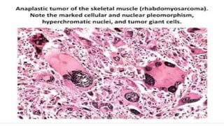
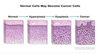
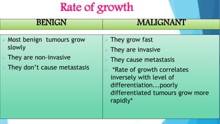
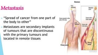
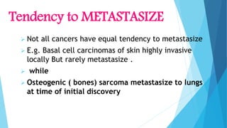
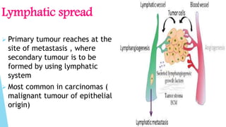
DIFFERENCE BETWEEN BENIGN AND
MALIGNANT NEOPLASM
NEOPLASM:
 literally means new
growth.
 An abnormal tissue mass
that grows by cellular
proliferation more rapidly
than normal tissue mass ,
and continuous to grow after
the stimuli that initiated the
new growth cease
DEFINITION
NAMING
 BENINGN
Use cell type and suffix “ oma”
E.g. fibroma
 MALIGNANT
Use cell type and suffix
“sarcoma or carcinoma”
Ways to differentiate B/W BENIGN and
MALIGNANT NEOPLASM
 There are four different features by
which benign and malignant
neoplasms can be distinguished
 Differentiation and Anaplasia
 Rate of growth
 Local invasion
 Metastasis
Differentiation and ANAPLASIA
 Benign
Composed of well differentiated cell
e.g: lipoma, chondroma
Mitoses are rare and of normal
configuration.
MALIGNANT
 Range from well
differentiated to
completely
undifferentiated
 Adenocarcinomas is an
example of well
differentiated malignant
neoplasm.
ANAPLASIA
Malignant neoplasms that are
composed of undifferentiated
cells are said to be
anaplastic.
FEATURES OF ANAPLASTIC CELLS
 Show pleomorphism ( variation in size
and shape)
 Nuclei are hyper chromatic ( dark
staining)
 Increase nuclear to cytoplasmic ratio
1:1 ( 1:4 or 1:6 normal)
 Giant cells ( considerably larger than
normal cells)
 Mitoses are numerous
DYSPLASIA
 Disorderly but non-
neoplastic
proliferation is
called dysplasia...
 Also called pre
malignant
Features of DYSPLASTIC cell
 They show pleomorphism
 Nuclei are hyperchromatic
 Mitoses abundant than normal figures
 But not synonymous with cancer
because mild to moderate dysplasia
could be regressed if inciting causes
are removed.
EXCEPTIONS
 Some malignant tumours grow
slowly
 E.g.: Acute childhood
“leukaemia” initiate during
foetal development yet
manifest full blown cancer years
later
LOCAL INVASION
Benign
 Remain localized
don’t have
capacity to
infiltrate, invade
or metastasize
 They have capsule
(fibrous) that
separates them
from host tissues
Rate of growth
BENIGN
 Most benign tumours grow
slowly
 They are non-invasive
 They don’t cause metastasis
MALIGNANT
 They grow fast
 They are invasive
 They cause metastasis
 *Rate of growth correlates
inversely with level of
differentiation...poorly
differentiated tumours grow more
rapidly*
NOTE
 Lack of capsule does not always mean ,
tumour is malignant. Some benign
tumours lack capsule
 E.g. Benign vascular neoplasm of dermis
Malignant Neoplasm
 Malignant neoplasms invade surrounding
tissues
 They don’t have well defined capsule
CUT SECTION OF INVASIVE DUCTAL
CARCINOMA OF BREAST
Metastasis
 “Spread of cancer from one part of
the body to other”
 Metastases are secondary implants
of tumours that are discontinuous
with the primary tumours and
located in remote tissues
Tendency to METASTASIZE
 Not all cancers have equal tendency to metastasize
 E.g. Basal cell carcinomas of skin highly invasive
locally But rarely metastasize .
 while
 Osteogenic ( bones) sarcoma metastasize to lungs
at time of initial discovery
Ways of dissemination of
malignant tumours
 There are 3 pathways of dissemination
 1: seeding within the body
 2: Lymphatic spread
 3: Haematogenous spread
Seeding within the body
 Spread by seeding occurs when
neoplasm invade a natural body
cavity e.g.: Cancers of ovary
Lymphatic spread
 Primary tumour reaches at the
site of metastasis , where
secondary tumour is to be
formed by using lymphatic
system
 Most common in carcinomas (
malignant tumour of epithelial
origin)
SKIP METASTASES
 In some cases the cancer cells
seem to traverse the
lymphatic channels from
primary nodes to be trapped
in subsequent lymph nodes
producing so called skip
metastases.
Haematogenous spread
 Primary tumour reaches at the site of metastasis
where secondary tumour is to be formed by using
Blood circulatory system
 This spread is favoured by sarcomas ( malignant
cancer of parenchymal origin)
 Liver and lungs are most frequently involved in
secondary site of haematogenous dissemination .
A LIVER STUDDED
WITH METASTATIC
CANCER
Summary
 Benign and malignant tumours can be distinguished from one
another based on the degree of differentiation , rate of growth,
local invasiveness, and distant spread
 Benign tumours resemble the tissue of origin and are well
differentiated; malignant tumours are poorly or completely
undifferentiated(anaplastic).
 Benign tumours are slow growing ; Malignant tumours generally
grow faster
 Benign tumours have capsule; Malignant tumours don't have
capsule .
 Benign tumours remain localized to the site of origin ; Malignant
tumours are locally invasive and metastasize to distant sites.
DIFFERENCE BETWEEN BENIGN AND MALIGNANT NEOPLASM (CANCER)

DIFFERENCE BETWEEN BENIGN AND MALIGNANT NEOPLASM (CANCER)

  • 1.
    DIFFERENCE BETWEEN BENIGNAND MALIGNANT NEOPLASM
  • 2.
    NEOPLASM:  literally meansnew growth.  An abnormal tissue mass that grows by cellular proliferation more rapidly than normal tissue mass , and continuous to grow after the stimuli that initiated the new growth cease DEFINITION
  • 3.
    NAMING  BENINGN Use celltype and suffix “ oma” E.g. fibroma  MALIGNANT Use cell type and suffix “sarcoma or carcinoma”
  • 4.
    Ways to differentiateB/W BENIGN and MALIGNANT NEOPLASM  There are four different features by which benign and malignant neoplasms can be distinguished  Differentiation and Anaplasia  Rate of growth  Local invasion  Metastasis
  • 5.
    Differentiation and ANAPLASIA Benign Composed of well differentiated cell e.g: lipoma, chondroma Mitoses are rare and of normal configuration.
  • 6.
    MALIGNANT  Range fromwell differentiated to completely undifferentiated  Adenocarcinomas is an example of well differentiated malignant neoplasm.
  • 7.
    ANAPLASIA Malignant neoplasms thatare composed of undifferentiated cells are said to be anaplastic.
  • 8.
    FEATURES OF ANAPLASTICCELLS  Show pleomorphism ( variation in size and shape)  Nuclei are hyper chromatic ( dark staining)  Increase nuclear to cytoplasmic ratio 1:1 ( 1:4 or 1:6 normal)  Giant cells ( considerably larger than normal cells)  Mitoses are numerous
  • 10.
    DYSPLASIA  Disorderly butnon- neoplastic proliferation is called dysplasia...  Also called pre malignant
  • 12.
    Features of DYSPLASTICcell  They show pleomorphism  Nuclei are hyperchromatic  Mitoses abundant than normal figures  But not synonymous with cancer because mild to moderate dysplasia could be regressed if inciting causes are removed.
  • 13.
    EXCEPTIONS  Some malignanttumours grow slowly  E.g.: Acute childhood “leukaemia” initiate during foetal development yet manifest full blown cancer years later
  • 14.
    LOCAL INVASION Benign  Remainlocalized don’t have capacity to infiltrate, invade or metastasize  They have capsule (fibrous) that separates them from host tissues
  • 15.
    Rate of growth BENIGN Most benign tumours grow slowly  They are non-invasive  They don’t cause metastasis MALIGNANT  They grow fast  They are invasive  They cause metastasis  *Rate of growth correlates inversely with level of differentiation...poorly differentiated tumours grow more rapidly*
  • 16.
    NOTE  Lack ofcapsule does not always mean , tumour is malignant. Some benign tumours lack capsule  E.g. Benign vascular neoplasm of dermis
  • 17.
    Malignant Neoplasm  Malignantneoplasms invade surrounding tissues  They don’t have well defined capsule
  • 18.
    CUT SECTION OFINVASIVE DUCTAL CARCINOMA OF BREAST
  • 19.
    Metastasis  “Spread ofcancer from one part of the body to other”  Metastases are secondary implants of tumours that are discontinuous with the primary tumours and located in remote tissues
  • 20.
    Tendency to METASTASIZE Not all cancers have equal tendency to metastasize  E.g. Basal cell carcinomas of skin highly invasive locally But rarely metastasize .  while  Osteogenic ( bones) sarcoma metastasize to lungs at time of initial discovery
  • 21.
    Ways of disseminationof malignant tumours  There are 3 pathways of dissemination  1: seeding within the body  2: Lymphatic spread  3: Haematogenous spread
  • 22.
    Seeding within thebody  Spread by seeding occurs when neoplasm invade a natural body cavity e.g.: Cancers of ovary
  • 23.
    Lymphatic spread  Primarytumour reaches at the site of metastasis , where secondary tumour is to be formed by using lymphatic system  Most common in carcinomas ( malignant tumour of epithelial origin)
  • 24.
    SKIP METASTASES  Insome cases the cancer cells seem to traverse the lymphatic channels from primary nodes to be trapped in subsequent lymph nodes producing so called skip metastases.
  • 25.
    Haematogenous spread  Primarytumour reaches at the site of metastasis where secondary tumour is to be formed by using Blood circulatory system  This spread is favoured by sarcomas ( malignant cancer of parenchymal origin)  Liver and lungs are most frequently involved in secondary site of haematogenous dissemination .
  • 26.
    A LIVER STUDDED WITHMETASTATIC CANCER
  • 27.
    Summary  Benign andmalignant tumours can be distinguished from one another based on the degree of differentiation , rate of growth, local invasiveness, and distant spread  Benign tumours resemble the tissue of origin and are well differentiated; malignant tumours are poorly or completely undifferentiated(anaplastic).  Benign tumours are slow growing ; Malignant tumours generally grow faster  Benign tumours have capsule; Malignant tumours don't have capsule .  Benign tumours remain localized to the site of origin ; Malignant tumours are locally invasive and metastasize to distant sites.