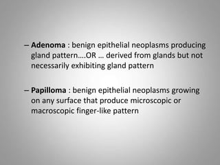
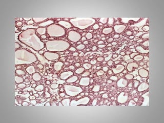
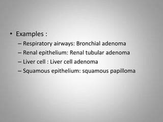
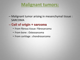
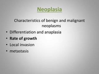
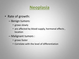
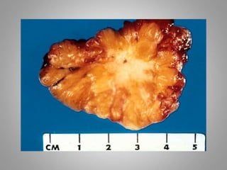
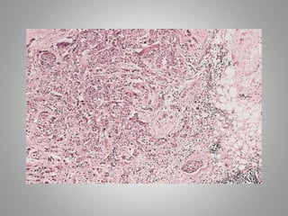
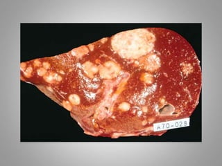
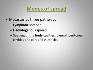
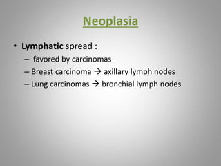
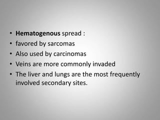
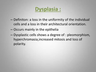
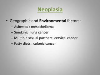
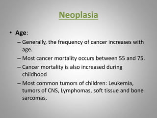
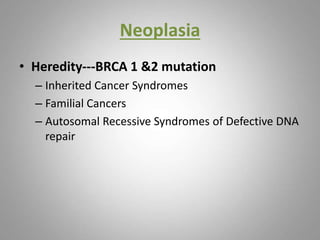
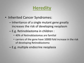
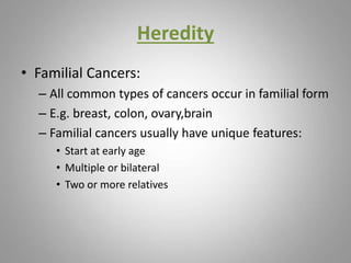
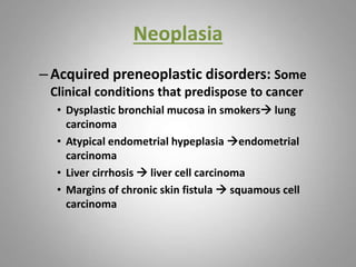
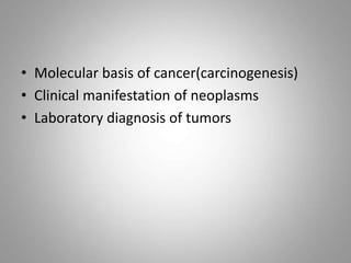
This document provides an overview of neoplasia (new abnormal growths or tumors). It defines neoplasia and discusses the classification of tumors as benign or malignant based on characteristics like differentiation, growth rate, invasion and metastasis potential. It also covers tumor nomenclature, risk factors for cancer like age, environment, heredity and pre-existing conditions. The document summarizes key concepts around epidemiology, molecular basis of cancer development, and laboratory diagnosis of tumors.
1. The document discusses neoplasia (abnormal growth of cells) and cancer. It defines key terms like neoplasm, tumor, benign and malignant tumors.
2. It describes how tumors are classified based on cell of origin, biological behavior, appearance and other features. Examples of different tumor types are provided.
3. The key differences between benign and malignant tumors are growth rate, invasion of surrounding tissue, metastasis, and differentiation of cells. Malignant tumors tend to grow and spread more rapidly.
This document provides an overview of neoplasia (new growths) and tumor nomenclature. It defines key terms like neoplasia, tumor, cancer, and oncology. It discusses the biology of tumor growth and characteristics of benign versus malignant neoplasms. Specifically, it covers topics like differentiation, anaplasia, rate of growth, invasion, and metastasis. The document also provides details on tumor naming conventions based on cell/tissue of origin and examples of exceptions to typical naming rules.
Neoplasms can be benign or malignant, with the key distinguishing characteristics including differentiation, growth rate, local invasion, and metastasis. Well-differentiated neoplasms with slow growth, encapsulation and no invasion or metastasis are typically benign, while poorly differentiated neoplasms with rapid uncontrolled growth, invasiveness and ability to metastasize are malignant. Nomenclature of neoplasms is based on the tissue of origin, structure, embryonic layer, and presence of multiple cell types.
This document provides an overview of neoplasia (abnormal growths) and cancer. It defines key terms like tumour, neoplasm, and neoplasia. It classifies tumours and outlines the differences between benign and malignant tumours. Malignant tumours are poorly differentiated, invade locally, metastasize, and show features of anaplasia. The document also discusses cancer risk factors, molecular basis of cancer involving genetic mutations, carcinogenic agents like chemicals and radiation, tumour growth and angiogenesis, staging and grading of tumours, and routes of tumour spread.
1) Neoplasia refers to abnormal and uncontrolled cell growth, which can be benign (non-cancerous) or malignant (cancerous). The study of tumors is called oncology.
2) Tumors have two components - tumor parenchyma consisting of neoplastic cells, and reactive stroma made of connective tissue. The characteristics of the parenchymal cells determine if a tumor is benign or malignant.
3) Benign tumors are self-limited, do not invade or metastasize. Malignant tumors can invade surrounding tissues and spread to other parts of the body.
This document discusses neoplasms (abnormal masses of cells). It defines benign and malignant neoplasms, with benign being slow-growing and non-metastasizing, while malignant are fast-growing and can metastasize. Malignant neoplasms vary in differentiation from well to poorly differentiated. The prognosis depends on factors like growth rate, size, site, cell type, differentiation, metastasis, and response to therapy.
Type of Tumors بلال العميسي( مختبرات طبية).pptxssuser668f10
This document discusses the characteristics and classification of neoplasms. It defines neoplasia as new abnormal growth and distinguishes between benign and malignant tumors. The key differences are that benign tumors grow slowly, remain localized, and do not invade other tissues or spread to other parts of the body, while malignant tumors grow rapidly, invade surrounding tissues, and metastasize to distant sites. The document further classifies tumors based on tissue of origin and other histological features, and describes the cellular and molecular changes involved in the development and progression of cancer.
The document provides information on cancer and tumor biology. It defines cancer as uncontrolled cell proliferation that can be benign or malignant. Malignant tumors are able to invade surrounding tissues and metastasize to distant sites. Some key differences between benign and malignant tumors include variations in cell shape and size, ploidy, mitotic rate, loss of specialization, and organization. The causes of cancer include chemical, physical, and infectious factors that introduce mutations in DNA. The development of cancer involves multiple genetic alterations that progress cancer from normal to invasive stages.
This document provides an overview of neoplasia (new growths) and cancer. It defines neoplasia as abnormal cell growth triggered by mutations affecting a single cell and its progeny. Neoplasms have two components - neoplastic cells that form the tumor and reactive stroma including connective tissue. Tumors are classified as benign or malignant based on their biological behavior and morphology. Malignant tumors invade, destroy structures, and metastasize, while benign tumors remain localized. Grading of cancers is based on differentiation, and staging incorporates tumor size, lymph node involvement, and metastasis. Laboratory diagnosis of cancer involves histologic, cytologic, molecular, and tumor marker methods.
Neoplasms are characterized as either benign or malignant tumors. Benign tumors remain localized and do not invade surrounding tissue, while malignant tumors grow uncontrollably, invade surrounding areas, and metastasize to distant sites. Cancer development occurs through genetic mutations that alter cell growth and behavior over time. Malignant tumors are further classified by their site of origin, structure, and degree of differentiation through grading and staging systems.
This document provides an overview of neoplasia and tumor characteristics and classification. It defines neoplasia as new abnormal cell growth that can be benign or malignant. Benign tumors remain localized, while malignant tumors invade surrounding tissues and metastasize to distant sites. Tumors are classified based on tissue of origin, degree of differentiation, growth patterns, and whether they are benign or malignant. Malignant tumors are generally less differentiated, grow and spread more rapidly, and invade locally and metastasize, making them more difficult to treat. The document also discusses cellular adaptations, causes of cancer, tumor properties, grading, staging, and nomenclature used in tumor classification.
This document provides an overview of neoplasia (new growth or tumor). It defines a neoplasm as an abnormal mass of tissue with growth that exceeds and is uncoordinated with normal tissues. Neoplasms can be benign (non-cancerous) or malignant (cancerous). Malignant tumors are characterized by a lack of differentiation, local invasion, and metastasis (spread) to distant sites. The grading and staging of cancers provides information on the aggressiveness and extent of disease, which helps guide treatment decisions.
Neoplasia refers to new or abnormal growth of tissue. Benign tumors remain localized and do not invade other tissues or spread to other parts of the body, while malignant tumors are invasive and metastatic. Malignant tumors are collectively referred to as cancer. The classification and diagnosis of tumors involves examining their histological features such as differentiation, growth rate, size, invasion and metastasis. Laboratory diagnosis of cancer involves histological examination of biopsies, cytological examination of cell samples, molecular analysis and identification of tumor markers. Staging of cancer determines the extent of disease spread and guides treatment decisions.
1. The document discusses neoplasia, defining it as new growth resulting from genetic alterations in cells that cause uncontrolled proliferation.
2. It classifies tumors as benign, malignant, or premalignant and discusses their naming conventions based on tissue of origin and characteristics.
3. The key differences between benign and malignant tumors are that benign tumors are slow-growing, localized, well-differentiated and do not metastasize, while malignant tumors are poorly differentiated, invade surrounding tissues, grow quickly and can metastasize to distant sites.
Neoplasia refers to abnormal cell growth. Tumors can be benign or malignant, classified based on histologic features and biological behavior. All tumors contain proliferating neoplastic cells and supportive stroma. Tumors are named based on cell of origin, with suffixes indicating benign (-oma) or malignant (-sarcoma, -carcinoma) nature. Malignant tumors are characterized by rapid growth, local invasion, and potential for metastasis, while benign tumors are slow-growing and localized. Cancer development involves accumulation of genetic alterations that disrupt normal cell growth regulation.
This document discusses neoplasia and provides definitions and classifications of different types of tumors. Some key points include:
- Neoplasms can be classified as benign or malignant based on their ability to invade surrounding tissues and metastasize. Malignant tumors are less differentiated.
- Epithelial tumors are further classified based on cell of origin and growth pattern (e.g. adenoma, papilloma, polyp). Mesenchymal tumors are classified as sarcomas.
- Environmental and genetic factors can affect cancer risk and distribution. Cancer incidence generally increases with age. Certain inherited syndromes confer higher cancer susceptibility.
- Precancerous conditions like dysplasia are characterized by cellular
This document discusses neoplasia and the differences between benign and malignant tumors. Benign tumors are slow-growing, localized masses composed of well-differentiated cells that lack the ability to metastasize. Malignant tumors vary in differentiation and growth rate, have the ability to invade surrounding tissues and metastasize to distant sites, and can ultimately lead to patient death if not treated. The prognosis of a patient with a tumor depends on factors like the tumor's growth rate, size, site, cell type, presence of metastasis, and the patient's overall health.
This document provides information on neoplasia (new growth) and tumor nomenclature. It defines neoplasia as abnormal and uncontrolled cell growth that exceeds normal tissues. Tumors are named based on their cell or tissue of origin, with benign tumors ending in "-oma" and malignant tumors called carcinomas for epithelial cells and sarcomas for mesenchymal cells. Common sites for teratomas are the gonads and along midline fusion lines. Hamartomas contain normal tissues for the organ, while choristomas contain ectopic tissues. Environmental exposures like coal tar were found to induce skin cancer in rabbits.
Neoplasia & carcinogenesis.pptx dr.jawahar singh.pptx 1jawahar singh
This document summarizes a presentation on neoplasia and carcinogenesis. It begins with definitions of neoplasia and discusses the nomenclature and classification of neoplasms as benign or malignant. The key differences between benign and malignant tumors are described. The document then covers the molecular basis of cancer, outlining a multistep process of carcinogenesis involving oncogenes, tumor suppressor genes, DNA repair genes, and genes regulating apoptosis. It describes the hallmarks of cancer and discusses epidemiology, etiology, tumor immunity, and clinical aspects of neoplasia.
Unit 18 (A); Paediatric Oncologyppt.pptxRashidUllah7
This document discusses cancer terminology, types, causes, symptoms, and medical management. It focuses on defining cancer and describing the etiology, pathophysiology, signs and symptoms. It discusses diagnostic tests and the medical and nursing approaches to treating patients with cancer, with specifics on breast cancer and ovarian cancer. Key points covered include the classification and naming of cancers based on origin, the differences between benign and malignant tumors, genetics and environmental factors that can cause cancer, and common nursing diagnoses and interventions for cancer patients.
Principles of Oncology discusses the study, diagnosis, and treatment of tumors (neoplasms). It defines key terms like benign and malignant, carcinomas and sarcomas, and describes methods of examining and categorizing tumors microscopically and visually. Imaging, biopsies, and tumor markers are used to diagnose cancers before discussing common treatment techniques like surgery, chemotherapy, radiation therapy, and immunotherapy.
This document defines neoplasia and discusses the classification and characteristics of benign and malignant tumors. Some key points:
- Neoplasia refers to abnormal and uncontrolled cell growth. Benign tumors are non-cancerous, while malignant tumors are cancers that can invade nearby tissue and metastasize.
- Tumors are named based on the cell/tissue type and growth pattern. Malignant tumors ending in "-carcinoma" involve epithelial cells, while "-sarcomas" involve connective tissue.
- The characteristics that distinguish benign and malignant tumors are differentiation, growth rate, invasion, and metastasis. Malignant tumors tend to be less differentiated, grow faster, invade surrounding tissue, and metastas
Neoplasia refers to abnormal cell growth or tumor formation. There are two basic components of tumors - the parenchyma made up of transformed cells that determine behavior, and the stroma that provides support. Tumors can be benign or malignant. Benign tumors are non-invasive and self-limited, while malignant tumors are invasive and metastatic. The rate of growth, presence of cancer stem cells, clinical features, gross appearance, microscopic features, local invasion, and metastasis help characterize tumors as benign or malignant.
This document discusses the classification of tumors based on their cell of origin, behavior, appearance, and degree of differentiation. There are two main classifications - behavioral and histogenetic. The behavioral classification categorizes tumors as benign or malignant based on their biological behavior such as growth rate, invasion, and metastasis. The histogenetic classification categorizes tumors based on their presumed cell and tissue of origin, such as epithelium or connective tissue. Within these classifications, tumors can be further characterized by attributes like histology, site of origin, and prognosis to aid in diagnosis, treatment, and analysis of outcomes.
This document provides information on tumors of the salivary glands. It discusses the anatomy and histology of salivary glands, classification of salivary gland tumors, and specifics on certain tumor types including pleomorphic adenoma and Warthin's tumor. Pleomorphic adenoma is the most common benign salivary gland tumor, characterized by epithelial and mesenchymal differentiation. Warthin's tumor commonly occurs bilaterally in the parotid glands of older smoking males. The document covers epidemiology, etiology, histogenesis, clinical features, investigation, pathology and treatment of various salivary gland tumors.
Neoplasia refers to abnormal growth of cells. Benign tumors remain localized, do not invade other tissues or metastasize. They are well-differentiated and grow slowly. Malignant tumors are poorly differentiated, invade surrounding tissues and spread to distant sites. They grow rapidly and can be fatal if not treated. The key differences between benign and malignant tumors are their ability to invade other tissues and metastasize to other parts of the body.
This document discusses neoplasia and tumor biology. It begins with definitions of neoplasia and benign versus malignant tumors. It describes the molecular basis of carcinogenesis including genetic changes that lead to autonomous growth. The progression of malignancy is explained in terms of differentiation, dysplasia, anaplasia and metastatic ability. Risk factors for cancer development including heredity, environment, viral exposure and DNA repair defects are outlined. The key genetic changes involved in malignant transformation are deregulated growth signals, evasion of growth inhibition, apoptosis resistance, replication immortality, angiogenesis and invasion.
Detailed Description about soft tissue sarcoma.
Deals with topics including etiology, histopathology,clinical presentation ,staging and prognostic factors and management methods including surgery and adjuvent therapy .
Soft tissue sarcomas are a heterogeneous group of malignant tumours derived from primitive mesenchymal cells.
They are aggressive tumours which are locally invasive and recurrent.
They are named based on the cell of origin .
They require multimodal treatment including surgery and certain adjuvent therapies
Neonatal tetanus is caused by Clostridium tetani bacteria entering the body through an unsterilized umbilical cord. It causes painful muscle spasms and can be fatal, especially in developing countries where proper umbilical cord care may not be practiced. Prevention focuses on immunizing pregnant women and promoting clean delivery techniques like sterilizing cutting and tying instruments and keeping the umbilical cord clean and dry. Treatment involves controlling spasms, administering antitoxins, antibiotics, and supportive care like oxygen and IV fluids. With immunization programs, many countries have eliminated neonatal tetanus as a major public health problem.
This document defines acute rheumatic fever as an indirect complication of group A streptococcal pharyngitis that primarily affects the heart, central nervous system, joints, and skin. It discusses the major manifestations of acute rheumatic fever such as carditis, migratory polyarthritis, Sydenham's chorea, erythema marginatum, and subcutaneous nodules. The modified Jones criteria for diagnosing acute rheumatic fever requires two major manifestations or one major and two minor manifestations along with evidence of a prior streptococcal infection. Treatment involves antibiotics, salicylates or steroids depending on symptoms, and long-term antibiotic prophylaxis to prevent recurrences.
TORCH infections refer to a group of congenital infections transmitted from mother to fetus, including toxoplasmosis, rubella, cytomegalovirus, herpes simplex virus, and syphilis. These infections can cause fetal death, neonatal loss, or long-term injuries. Symptoms may not appear at birth but develop later. While prevention focuses on prenatal care and immunization, treatment depends on the specific infection but aims to prevent complications in the fetus or newborn.
The document provides information about the nervous system and various types of headaches. It discusses the organization of the nervous system including the central nervous system (CNS), peripheral nervous system, and divisions of the nervous system. It then summarizes assessment of the neurological system including history, physical exam, and diagnostic tests. Specific types of headaches discussed include migraine headaches, which often have an aura phase and are exacerbated by light and sound, and cluster headaches, which occur in distinct cluster periods with pain-free intervals in between. Treatment options for both migraine and cluster headaches are also outlined.
- The document discusses pediatrics HIV, including definitions of HIV and AIDS, epidemiology, etiology, transmission, risk factors, pathogenesis, natural history and clinical manifestations, diagnosis, staging, management including ART, OI prophylaxis, infant feeding recommendations, and contraindications for various treatments. Some key points include that vertical transmission is the most common route of transmission in pediatrics, ART should be started as early as possible in all HIV-infected children, and cotrimoxazole preventive therapy is recommended for HIV-exposed infants from 4-6 weeks of age.
This document discusses meningitis and encephalitis. It provides information on the etiology, pathogenesis, clinical features, diagnosis and treatment of these conditions. For meningitis, common causes include bacteria like S. pneumoniae and viruses. Risk factors include lack of immunity and crowding. Signs include fever, headache and neck stiffness. CSF analysis shows neutrophil predominance and elevated proteins. Treatment involves antibiotics, anticonvulsants, corticosteroids and supportive care. Encephalitis is caused by viruses, bacteria, fungi or parasites invading the brain tissue. It presents with altered mental status and seizures. Diagnosis involves CSF analysis, imaging and EEG. Treatment focuses on supportive care and antivirals or antibiotics depending
This document provides an overview of the female reproductive system, including common pathologies that can affect each organ. It discusses disorders of the lower genital tract and cervix, uterus, fallopian tubes, ovaries, and pregnancy-related conditions. Some key topics covered include infections, endometrial diseases, cervical intraepithelial neoplasia, ovarian cysts and tumors, and trophoblastic diseases. The document aims to describe the clinical presentations, risk factors, pathological findings, and management considerations for various female reproductive system disorders.
The male reproductive system is susceptible to several congenital malformations, infections, tumors, and other issues. The penis can be affected by hypospadias, epispadias, phimosis, balanitis and STDs, as well as benign tumors like condyloma and malignant tumors such as penile cancer. The testes and epididymis can have problems like cryptorchidism, atrophy, epididymo-orchitis, and testicular torsion. The scrotum can swell due to testicular tumors, infections, torsion, hernias, varicocele, hydrocele and other causes. The prostate is prone to prostatitis, benign prostatic hyperplasia
Tissue renewal and repair is essential for survival. There are three main processes: regeneration, repair through scar formation, and fibrosis. Regeneration replaces lost structures, while repair and scarring are needed when tissue damage is too severe for regeneration alone. The ability of tissues to repair depends on their proliferative capacity, with some tissues like skin able to continuously regenerate and others like heart unable to regenerate. Wound healing involves inflammation, new tissue growth, remodeling, and can result in regeneration, scarring, or complications like dehiscence, ulcers, hypertrophic scarring, or contractures if the repair process is abnormal.
Pathology is the study of disease through scientific methods. This document discusses several core concepts in pathology including cellular adaptations, injury, and death. It describes different types of cellular changes like hyperplasia, hypertrophy, atrophy, and metaplasia that can occur as adaptive responses to stressors. The document also examines the morphology and mechanisms of the two main types of cell death: necrosis and apoptosis. Necrosis is unprogrammed cell death due to external factors, while apoptosis is a tightly regulated genetic program of cell suicide.
Acute and chronic inflammation can be summarized as follows:
1. Acute inflammation is an initial rapid response to infection or tissue damage characterized by vascular changes like vasodilation and increased permeability, cellular events like leukocyte extravasation, and aims to eliminate the cause and promote healing.
2. Chronic inflammation is prolonged inflammation that can last weeks or months, features mononuclear cell infiltration, ongoing tissue destruction and attempts at repair simultaneously, and can lead to fibrosis or scarring.
3. Causes of chronic inflammation include persistent infections, hypersensitivity reactions, exposure to toxic agents, and autoimmunity. Granulomatous inflammation is a distinctive chronic pattern featuring macrophage aggregation. Systemic effects include fever
Introducing STATA is a document that summarizes the key features and capabilities of the statistical software package STATA. It describes STATA as a powerful and flexible package for statistical analysis, data management, and publication-quality graphing. The document outlines STATA's main interface elements including its command window, results window, variable window, and other windows. It also explains how to import and export data, use do-files to record analyses, and manage variables through labeling, generating new variables, and replacing values.
Epi Info is a free public domain software program developed by the CDC for data entry, analysis, and reporting. It allows users to develop electronic data entry forms, enter and analyze data, and produce statistics, graphs, tables, and maps. The main applications in Epi Info include designing data entry forms, entering data using these forms, and analyzing the entered data using a wide range of statistical tests. Sample size calculations can also be performed using Epi Info to determine the necessary sample sizes for different study designs like population surveys, case control studies, and cross-sectional or cohort studies.
This document provides an overview of SPSS and how to prepare and modify data files in SPSS. It discusses the SPSS windows interface, defining variables, entering and saving data, and modifying existing data files through actions like sorting, splitting, selecting cases, and merging files. The document is intended as an instruction manual or guide for using SPSS for data management and analysis.
A workshop hosted by the South African Journal of Science aimed at postgraduate students and early career researchers with little or no experience in writing and publishing journal articles.
How to Manage Your Lost Opportunities in Odoo 17 CRMCeline George
Odoo 17 CRM allows us to track why we lose sales opportunities with "Lost Reasons." This helps analyze our sales process and identify areas for improvement. Here's how to configure lost reasons in Odoo 17 CRM
Executive Directors Chat Leveraging AI for Diversity, Equity, and InclusionTechSoup
Let’s explore the intersection of technology and equity in the final session of our DEI series. Discover how AI tools, like ChatGPT, can be used to support and enhance your nonprofit's DEI initiatives. Participants will gain insights into practical AI applications and get tips for leveraging technology to advance their DEI goals.
Walmart Business+ and Spark Good for Nonprofits.pdfTechSoup
"Learn about all the ways Walmart supports nonprofit organizations.
You will hear from Liz Willett, the Head of Nonprofits, and hear about what Walmart is doing to help nonprofits, including Walmart Business and Spark Good. Walmart Business+ is a new offer for nonprofits that offers discounts and also streamlines nonprofits order and expense tracking, saving time and money.
The webinar may also give some examples on how nonprofits can best leverage Walmart Business+.
The event will cover the following::
Walmart Business + (https://business.walmart.com/plus) is a new shopping experience for nonprofits, schools, and local business customers that connects an exclusive online shopping experience to stores. Benefits include free delivery and shipping, a 'Spend Analytics” feature, special discounts, deals and tax-exempt shopping.
Special TechSoup offer for a free 180 days membership, and up to $150 in discounts on eligible orders.
Spark Good (walmart.com/sparkgood) is a charitable platform that enables nonprofits to receive donations directly from customers and associates.
Answers about how you can do more with Walmart!"
How to Fix the Import Error in the Odoo 17Celine George
An import error occurs when a program fails to import a module or library, disrupting its execution. In languages like Python, this issue arises when the specified module cannot be found or accessed, hindering the program's functionality. Resolving import errors is crucial for maintaining smooth software operation and uninterrupted development processes.
Strategies for Effective Upskilling is a presentation by Chinwendu Peace in a Your Skill Boost Masterclass organisation by the Excellence Foundation for South Sudan on 08th and 09th June 2024 from 1 PM to 3 PM on each day.
Main Java[All of the Base Concepts}.docxadhitya5119
This is part 1 of my Java Learning Journey. This Contains Custom methods, classes, constructors, packages, multithreading , try- catch block, finally block and more.
How to Add Chatter in the odoo 17 ERP ModuleCeline George
In Odoo, the chatter is like a chat tool that helps you work together on records. You can leave notes and track things, making it easier to talk with your team and partners. Inside chatter, all communication history, activity, and changes will be displayed.
3. Neoplasia
• Cancer is one of the leading causes of death
worldwide.
• Emotional and physical suffering by the
patient.
• Different mortality rate …..
– Some are curable
– Others are fatal
4. Neoplasia
• Neoplasia = new growth
• Neoplasm = tumor
• Tumor = swelling
• The study of tumors = Oncology
– Oncos = tumor + ology = study of
5. Neoplasia
• Definition:
– is an abnormal mass of tissue,
– the growth of which is uncoordinated with that of normal
tissues,
– and that persists in the same excessive manner after the
cessation of the stimulus which evoked the change
– With the loss of responsiveness to normal growth
controls
8. Neoplasia
• Benign tumors :
– Will remain localized
– Cannot spread to distant sites
– Generally can be locally excised
– Patient generally survives
9. Neoplasia
• Malignant neoplasms:
– Can invade and destroy adjacent structure
– Can spread to distant sites
– Cause death (if not treated )
10. Neoplasia
• All tumors benign and malignant have two
basic components:
–Parechyma: made up of neoplastic cells
–Stroma: made up of non-neoplastic, host-
derived connective tissue and blood vessels
11. Neoplasia
• The parenchyma:
– Determines the biological behavior of the tumor
– From which the tumor derives its name
12. Neoplasia
• The stroma:
– Carries the blood supply
– Provides support for the growth of the
parenchyma
13. • In some tumors, the stromal support is scant
and so the neoplasm is soft and fleshy.
• In other cases the parenchymal cells
stimulate the formation of an abundant
collagenous stroma, referred to as
desmoplasia.
17. • Benign tumors arising from mesenchymal
tissue,
• cell of origin + oma
– Benign tumor arising in fibrous tissue:
Fibro + oma = Fibroma
- Benign tumor arising in fatty tissue:
Lipo + oma = lipoma
18. • Benign tumor arising in cartilage
chondro + oma = chondroma
• Benign tumor arising in smooth muscle
Leiomyo + oma = leiomyoma
• Benign tumor arising in skeletal muscle
Rhabdomyo + oma = rhabdomyoma
19. • epithelial benign tumors are classified on the
basis of :
– The cell of origin
– Microscopic pattern
– Macroscopic pattern
20. – Adenoma : benign epithelial neoplasms producing
gland pattern….OR … derived from glands but not
necessarily exhibiting gland pattern
– Papilloma : benign epithelial neoplasms growing
on any surface that produce microscopic or
macroscopic finger-like pattern
21.
22.
23. • Polyp : a mass that projects above a mucosal
surface to form a macroscopically visible
structure.
e.g. - colonic polyp
- nasal polyp
27. Malignant tumors:
– Malignant tumor arising in mesenchymal tissue :
SARCOMA
– Cell of origin + sarcoma
• From fibrous tissue: Fibrosarcoma
• From bone : Osteosarcoma
• From cartilage : chondrosarcoma
28. Malignant neoplasm of epithelial cells origin derived
from any of the three germ layer , are called
Carcinomas.
Cell of origin + Carcinoma
Squamous cells + carcinoma = squamous cell
carcinoma
Adenocarcinoma: One with a glandular growth
pattern microscopically.
34. (3) Special nomenclature
① Blastoma: tumors rigging in immature tissue
or nervous tissue, most of them are malignant
e.g.medulloblastoma,retinoblastoma,
nephroblastoma,Neuroblastoma
② Some tumors attaching the suffix-oma.
But malignant
e. g. seminoma, lymphoma, melanoma,
dysgerminoma, glioma, hepatoma
35. ③ Some malignant tumors, but called disease.
e. g. leukemias, paget’s disease
④ Some malignant tumors nominated by
scientists’ name
e. g. Hodgkin’s disease, Ewing’s tumor
⑤ Mixed tumors: tumors which derived from one
germ layer may undergo divergent
differentiation creating mixed tumors
e. g. mixed tumor of salivary gland
37. Neoplasia
1. Differentiation and anaplasia:
• Differentiation means : the extent to which
the parenchymal cells of the tumor resemble
their normal counterparts morphologically
and functionally
38. Neoplasia
• well differentiated = closely resemble their
normal counterparts
• Moderately differentiated
• Poorly differentiated
• Undifferentiated ( Anaplasia )
39. Neoplasia
• Benign tumors = well differentiated
• Malignant tumors =
well differentiated -----> anaplastic
52. Neoplasia
• Metastasis :
– Definition : the development of secondary
implants discontinuous with the primary tumor,
possibly in remote tissues.
53.
54. Neoplasia
• Metastasis :
– Cancers have different ability to metastasize
– Approximately 30% patients present with clinically
evident metastases.
– Generally, the more anaplastic and the larger the
primary tumor, the more likely is metastasis
55. Benign Malignant:
• Slow growing,
• capsulated,
• Non-invasive
• do not metastasize,
• well differentiated,
• suffix “oma” eg.
Fibroma.
• Fast growing,
• non capsulated,
• Invasive & Infiltrate
• Metastasize.
• poorly differentiated,
• Suffix “Carcinoma” or
“Sarcoma”
56. Modes of spread
• Metastasis : three pathways
– Lymphatic spread :
– Hematogenous spread :
– Seeding of the body cavities: pleural, peritoneal
cavities and cerebral ventricles
58. • Hematogenous spread :
• favored by sarcomas
• Also used by carcinomas
• Veins are more commonly invaded
• The liver and lungs are the most frequently
involved secondary sites.
59. Dysplasia :
– Definiton: a loss in the uniformity of the individual
cells and a loss in their architectural orientation.
– Occurs mainly in the epithelia
– Dysplastic cells shows a degree of : pleomorphism,
hyperchromasia,increased mitosis and loss of
polarity.
60. • Dysplasia does not mean cancer
• Dyplasia does not necessarily progress to
cancer
• Dysplasia may be reversible
• If dysplastic changes involve the entire
thickness of the epithelium it is called :
CARCINOMA IN-SITU
61.
62. • Carcinoma in-situ
– Definition: an intraepithelial malignancy in which
malignant cells involve the entire thickness of the
epithelium without penetration of the basement
membrane.
– Applicable only to epithelial neoplasms.
63.
64.
65. Teratoma:
– Teratoma contains recognizable mature or
immature cells or tissues representative of more
than one germ-cell layer and some times all
three.
– Teratomas originate from totipotential cells such
as those normally present in the ovary and testis.
66. • Such cells have the capacity to differentiate into any
of the cell types found in the adult body.
• So they may give rise to neoplasms that mimic
bone, epithelium, muscle, fat, nerve and other
tissues.
• Most common sites are: ovary, testis
67. Epidemiology
– Will help to discover aetiology
– Planning of preventive measures
– To know what is common and what is rare.
– Development of screening methods for early
diagnosis
68.
69. Neoplasia
• Factors affecting incidence of cancer
– Geographic and Environmental
– Age
– Heredity
– Acquired preneoplastic disorders
70. Neoplasia
• Geographic and Environmental factors:
– Asbestos : mesothelioma
– Smoking : lung cancer
– Multiple sexual partners: cervical cancer
– Fatty diets : colonic cancer
71. Neoplasia
• Age:
– Generally, the frequency of cancer increases with
age.
– Most cancer mortality occurs between 55 and 75.
– Cancer mortality is also increased during
childhood
– Most common tumors of children: Leukemia,
tumors of CNS, Lymphomas, soft tissue and bone
sarcomas.
72. Neoplasia
• Heredity---BRCA 1 &2 mutation
– Inherited Cancer Syndromes
– Familial Cancers
– Autosomal Recessive Syndromes of Defective DNA
repair
73. Heredity
• Inherited Cancer Syndromes:
– Inheritance of a single mutant gene greatly
increases the risk of developing neoplasm
– E.g. Retinoblastoma in children :
• 40% of Retinoblastomas are familial
• carriers of the gene have 10000 fold increase in the risk
of developing Retinoblastoma
– E.g. multiple endocrine neoplasia
74. Heredity
• Familial Cancers:
– All common types of cancers occur in familial form
– E.g. breast, colon, ovary,brain
– Familial cancers usually have unique features:
• Start at early age
• Multiple or bilateral
• Two or more relatives
75. Heredity
– Autosomal Recessive Syndromes of Defective DNA
repair :
• Small group of autosomal recessive disorders
• Characterized by DNA instability
76. Neoplasia
–Acquired preneoplastic disorders: Some
Clinical conditions that predispose to cancer
• Dysplastic bronchial mucosa in smokers lung
carcinoma
• Atypical endometrial hypeplasia endometrial
carcinoma
• Liver cirrhosis liver cell carcinoma
• Margins of chronic skin fistula squamous cell
carcinoma
77. • Molecular basis of cancer(carcinogenesis)
• Clinical manifestation of neoplasms
• Laboratory diagnosis of tumors