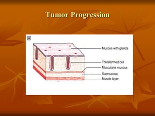
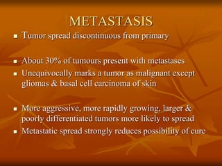
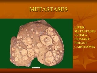
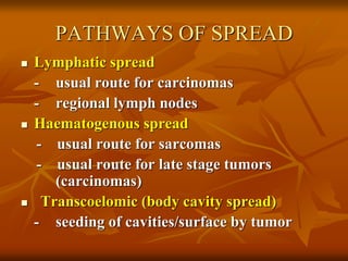
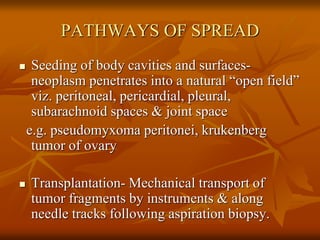
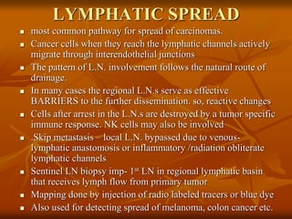
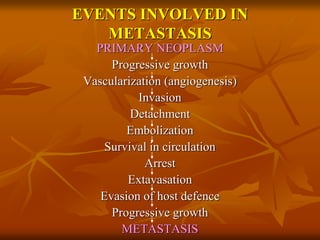
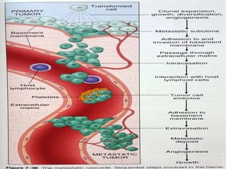
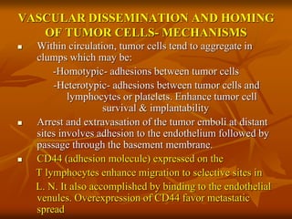
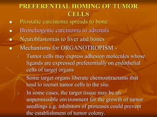
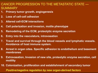
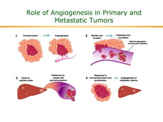
NEOPLASIA:
METASTASIS
DR. ROOPAM JAIN
Additional Dean
Profssor & Head
Dept. of Pathology & Blood Bank
Tumor Progression
METASTASIS
 Tumor spread discontinuous from primary
 About 30% of tumours present with metastases
 Unequivocally marks a tumor as malignant except
gliomas & basal cell carcinoma of skin
 More aggressive, more rapidly growing, larger &
poorly differentiated tumors more likely to spread
 Metastatic spread strongly reduces possibility of cure
METASTASES
LIVER
METASTASES
FROM A
PRIMARY
BREAST
CARCINOMA
PATHWAYS OF SPREAD
 Lymphatic spread
- usual route for carcinomas
- regional lymph nodes
 Haematogenous spread
- usual route for sarcomas
- usual route for late stage tumors
(carcinomas)
 Transcoelomic (body cavity spread)
- seeding of cavities/surface by tumor
PATHWAYS OF SPREAD
 Seeding of body cavities and surfaces-
neoplasm penetrates into a natural “open field”
viz. peritoneal, pericardial, pleural,
subarachnoid spaces & joint space
e.g. pseudomyxoma peritonei, krukenberg
tumor of ovary
 Transplantation- Mechanical transport of
tumor fragments by instruments & along
needle tracks following aspiration biopsy.
LYMPHATIC SPREAD
 most common pathway for spread of carcinomas.
 Cancer cells when they reach the lymphatic channels actively
migrate through interendothelial junctions
 The pattern of L.N. involvement follows the natural route of
drainage.
 In many cases the regional L.N.s serve as effective
BARRIERS to the further dissemination. so, reactive changes
 Cells after arrest in the L.N.s are destroyed by a tumor specific
immune response. NK cells may also be involved
 .Skip metastasis – local L.N. bypassed due to venous-
lymphatic anastomosis or inflammatory /radiation obliterate
lymphatic channels
 Sentinel LN biopsy imp- 1st LN in regional lymphatic basin
that receives lymph flow from primary tumor
 Mapping done by injection of radio labeled tracers or blue dye
 Also used for detecting spread of melanoma, colon cancer etc.
HEMATOGENOUS SPREAD
 Typical of sarcomas
 Arteries less readily penetrated than veins due to thicker wall
 Arterial spread occur when lung is involved via pulmonary
capillary bed or A-V shunt
 Venous invasion the tumor cells follow the venous drainage
of the primary site.
 Thus, in such spreads liver (portal area) and lungs (caval area)
would be most frequently secondarily involved.
 Cancers in close proximity to the vertebral column spread
through paravertebral veins. i. e. thyroid & lung
 Certain cancers may involve veins themselves. E.g. Renal cell
ca often invades the renal vein due to which it grows in a
snakelike fashion sometimes reaching upto the right side of the
heart.
EVENTS INVOLVED IN
METASTASIS
PRIMARY NEOPLASM
Progressive growth
Vascularization (angiogenesis)
Invasion
Detachment
Embolization
Survival in circulation
Arrest
Extavasation
Evasion of host defence
Progressive growth
METASTASIS
EVENTS INVOLVED IN
METASTASIS...
INVASION OF THE ECM
(1) Detachment of tumor cells from each other- Normal
cells are glued to each other by transmembrane
glycoproteins e.g. Cadherins.
E- cadherins linked to cytoskeleton by catenins
( in Adenocarcinoma of the colon there is down regulation of the E-
cadherin expression)
(2) Attachment to matrix components- Normal
epithelial cells express high affinity receptors-
integrin family for the basement membrane
LAMININ that are polarized to their basal surface.
Also cancer cells have receptors for fibronectin &
collagen.
INVASION OF THE ECM(contd.)
(3) Degradation of ECM- It is achieved by proteolytic enzymes
produced by tumor cells (or even host cells like stromal
fibroblasts & infiltrating macrophages)
3 classes of proteases- Serine, Cysteine, Metalloproteinase.
E.g.–type IV collagenase which is a metalloproteinase
–Cathepsin D which is a cysteine protease
–Urokinase type plasminogen activator which is
a serine protease
(4) Migration of tumor cells- i.e. locomotion. It is mediated by
tumor cell derived cytokines.
-cleavage products of some ECM components have Growth
promoting , angiogenic & chemotactic activities for tumor
cells
-Collagen IV degradation also produce endostatin & tumstatin -
antiangiogenic
EVENTS INVOLVED IN
METASTASIS...
Vascular dissemination & Homing of tumor cells
 After invasion of the vascular or lymphatic channels
the cells may either grow at the site of penetration or
because of their loss of cohesiveness, be swept away
as individual cells or small embolic clumps.
 Here they are vulnerable to the immune (CMI and
humoral) as well as non-immune (NK cells, activated
macrophages) responses.
VASCULAR DISSEMINATION AND HOMING
OF TUMOR CELLS- MECHANISMS
 Within circulation, tumor cells tend to aggregate in
clumps which may be:
-Homotypic- adhesions between tumor cells
-Heterotypic- adhesions between tumor cells and
lymphocytes or platelets. Enhance tumor cell
survival & implantability
 Arrest and extravasation of the tumor emboli at distant
sites involves adhesion to the endothelium followed by
passage through the basement membrane.
 CD44 (adhesion molecule) expressed on the
T lymphocytes enhance migration to selective sites in
L. N. It also accomplished by binding to the endothelial
venules. Overexpression of CD44 favor metastatic
spread
EVENTS…
 The cells that survive then arrest in capillary
beds of distant organs either by adherence
to the endothelial cells or attachment to the
vascular basement membrane.
 Thereafter the metastases must avoid the
host immune response, develop its own
vascularization in the same way and
produce a secondary implant.
PREFERENTIAL HOMING OF TUMOR
CELLS
 Prostatic carcinoma spreads to bone
 Bronchogenic carcinoma to adrenals
 Neuroblastomas to liver and bones
 Mechanisms for ORGANOTROPISM -
1. Tumor cells may express adhesion molecules whose
ligands are expressed preferentially on endothelial
cells of target organs
2. Some target organs liberate chemoattractants that
tend to recruit tumor cells to the site.
3. In some cases, the target tissue may be an
unpermissible environment for the growth of tumor
seedlings e.g. inhibitors of proteases could prevent
the establishment of tumor colony.
CANCER PROGRESSION TO THE METASTATIC STATE ---
SUMMARY
1. Primary tumor growth, angiogenesis
2. Loss of cell-cell adhesion
3. Altered cell-ECM interactions
4. Cell polarization and invasion, motile phenotype
5. Remodeling of the ECM, proteolytic enzyme secretion
6. Entry into the vasculature, intravasation
7. Travel and survival through the blood vessels and lymphatic vessels.
Avoidance of host immune system.
8. Arrest in organ sites. Specific adhesion to endothelium and basement
membranes
9. Extravasation, Invasion of new site, proteolytic enzyme secretion, cell
motility
10. Colonization, proliferation and establishment of secondary tumor
Positive/negative regulation by new organ-derived factors
Role of Angiogenesis in Primary and
Metastatic Tumors

Neoplasia: Metastasis

  • 1.
    NEOPLASIA: METASTASIS DR. ROOPAM JAIN AdditionalDean Profssor & Head Dept. of Pathology & Blood Bank
  • 2.
  • 3.
    METASTASIS  Tumor spreaddiscontinuous from primary  About 30% of tumours present with metastases  Unequivocally marks a tumor as malignant except gliomas & basal cell carcinoma of skin  More aggressive, more rapidly growing, larger & poorly differentiated tumors more likely to spread  Metastatic spread strongly reduces possibility of cure
  • 4.
  • 5.
    PATHWAYS OF SPREAD Lymphatic spread - usual route for carcinomas - regional lymph nodes  Haematogenous spread - usual route for sarcomas - usual route for late stage tumors (carcinomas)  Transcoelomic (body cavity spread) - seeding of cavities/surface by tumor
  • 6.
    PATHWAYS OF SPREAD Seeding of body cavities and surfaces- neoplasm penetrates into a natural “open field” viz. peritoneal, pericardial, pleural, subarachnoid spaces & joint space e.g. pseudomyxoma peritonei, krukenberg tumor of ovary  Transplantation- Mechanical transport of tumor fragments by instruments & along needle tracks following aspiration biopsy.
  • 7.
    LYMPHATIC SPREAD  mostcommon pathway for spread of carcinomas.  Cancer cells when they reach the lymphatic channels actively migrate through interendothelial junctions  The pattern of L.N. involvement follows the natural route of drainage.  In many cases the regional L.N.s serve as effective BARRIERS to the further dissemination. so, reactive changes  Cells after arrest in the L.N.s are destroyed by a tumor specific immune response. NK cells may also be involved  .Skip metastasis – local L.N. bypassed due to venous- lymphatic anastomosis or inflammatory /radiation obliterate lymphatic channels  Sentinel LN biopsy imp- 1st LN in regional lymphatic basin that receives lymph flow from primary tumor  Mapping done by injection of radio labeled tracers or blue dye  Also used for detecting spread of melanoma, colon cancer etc.
  • 8.
    HEMATOGENOUS SPREAD  Typicalof sarcomas  Arteries less readily penetrated than veins due to thicker wall  Arterial spread occur when lung is involved via pulmonary capillary bed or A-V shunt  Venous invasion the tumor cells follow the venous drainage of the primary site.  Thus, in such spreads liver (portal area) and lungs (caval area) would be most frequently secondarily involved.  Cancers in close proximity to the vertebral column spread through paravertebral veins. i. e. thyroid & lung  Certain cancers may involve veins themselves. E.g. Renal cell ca often invades the renal vein due to which it grows in a snakelike fashion sometimes reaching upto the right side of the heart.
  • 9.
    EVENTS INVOLVED IN METASTASIS PRIMARYNEOPLASM Progressive growth Vascularization (angiogenesis) Invasion Detachment Embolization Survival in circulation Arrest Extavasation Evasion of host defence Progressive growth METASTASIS
  • 11.
    EVENTS INVOLVED IN METASTASIS... INVASIONOF THE ECM (1) Detachment of tumor cells from each other- Normal cells are glued to each other by transmembrane glycoproteins e.g. Cadherins. E- cadherins linked to cytoskeleton by catenins ( in Adenocarcinoma of the colon there is down regulation of the E- cadherin expression) (2) Attachment to matrix components- Normal epithelial cells express high affinity receptors- integrin family for the basement membrane LAMININ that are polarized to their basal surface. Also cancer cells have receptors for fibronectin & collagen.
  • 12.
    INVASION OF THEECM(contd.) (3) Degradation of ECM- It is achieved by proteolytic enzymes produced by tumor cells (or even host cells like stromal fibroblasts & infiltrating macrophages) 3 classes of proteases- Serine, Cysteine, Metalloproteinase. E.g.–type IV collagenase which is a metalloproteinase –Cathepsin D which is a cysteine protease –Urokinase type plasminogen activator which is a serine protease (4) Migration of tumor cells- i.e. locomotion. It is mediated by tumor cell derived cytokines. -cleavage products of some ECM components have Growth promoting , angiogenic & chemotactic activities for tumor cells -Collagen IV degradation also produce endostatin & tumstatin - antiangiogenic
  • 13.
    EVENTS INVOLVED IN METASTASIS... Vasculardissemination & Homing of tumor cells  After invasion of the vascular or lymphatic channels the cells may either grow at the site of penetration or because of their loss of cohesiveness, be swept away as individual cells or small embolic clumps.  Here they are vulnerable to the immune (CMI and humoral) as well as non-immune (NK cells, activated macrophages) responses.
  • 15.
    VASCULAR DISSEMINATION ANDHOMING OF TUMOR CELLS- MECHANISMS  Within circulation, tumor cells tend to aggregate in clumps which may be: -Homotypic- adhesions between tumor cells -Heterotypic- adhesions between tumor cells and lymphocytes or platelets. Enhance tumor cell survival & implantability  Arrest and extravasation of the tumor emboli at distant sites involves adhesion to the endothelium followed by passage through the basement membrane.  CD44 (adhesion molecule) expressed on the T lymphocytes enhance migration to selective sites in L. N. It also accomplished by binding to the endothelial venules. Overexpression of CD44 favor metastatic spread
  • 16.
    EVENTS…  The cellsthat survive then arrest in capillary beds of distant organs either by adherence to the endothelial cells or attachment to the vascular basement membrane.  Thereafter the metastases must avoid the host immune response, develop its own vascularization in the same way and produce a secondary implant.
  • 17.
    PREFERENTIAL HOMING OFTUMOR CELLS  Prostatic carcinoma spreads to bone  Bronchogenic carcinoma to adrenals  Neuroblastomas to liver and bones  Mechanisms for ORGANOTROPISM - 1. Tumor cells may express adhesion molecules whose ligands are expressed preferentially on endothelial cells of target organs 2. Some target organs liberate chemoattractants that tend to recruit tumor cells to the site. 3. In some cases, the target tissue may be an unpermissible environment for the growth of tumor seedlings e.g. inhibitors of proteases could prevent the establishment of tumor colony.
  • 18.
    CANCER PROGRESSION TOTHE METASTATIC STATE --- SUMMARY 1. Primary tumor growth, angiogenesis 2. Loss of cell-cell adhesion 3. Altered cell-ECM interactions 4. Cell polarization and invasion, motile phenotype 5. Remodeling of the ECM, proteolytic enzyme secretion 6. Entry into the vasculature, intravasation 7. Travel and survival through the blood vessels and lymphatic vessels. Avoidance of host immune system. 8. Arrest in organ sites. Specific adhesion to endothelium and basement membranes 9. Extravasation, Invasion of new site, proteolytic enzyme secretion, cell motility 10. Colonization, proliferation and establishment of secondary tumor Positive/negative regulation by new organ-derived factors
  • 19.
    Role of Angiogenesisin Primary and Metastatic Tumors