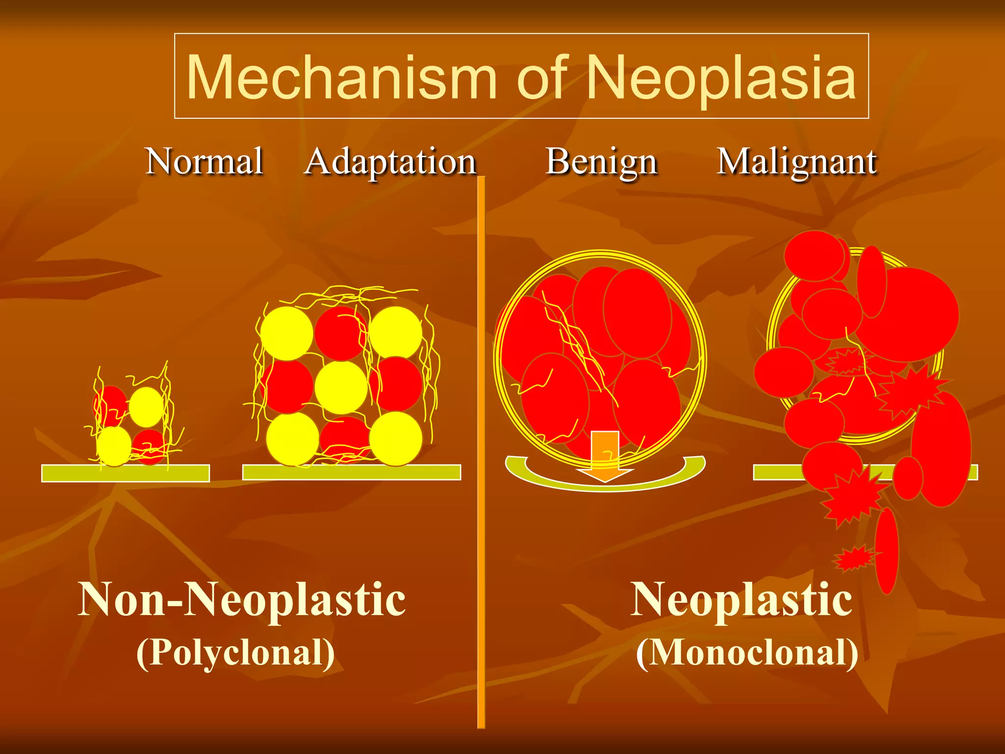
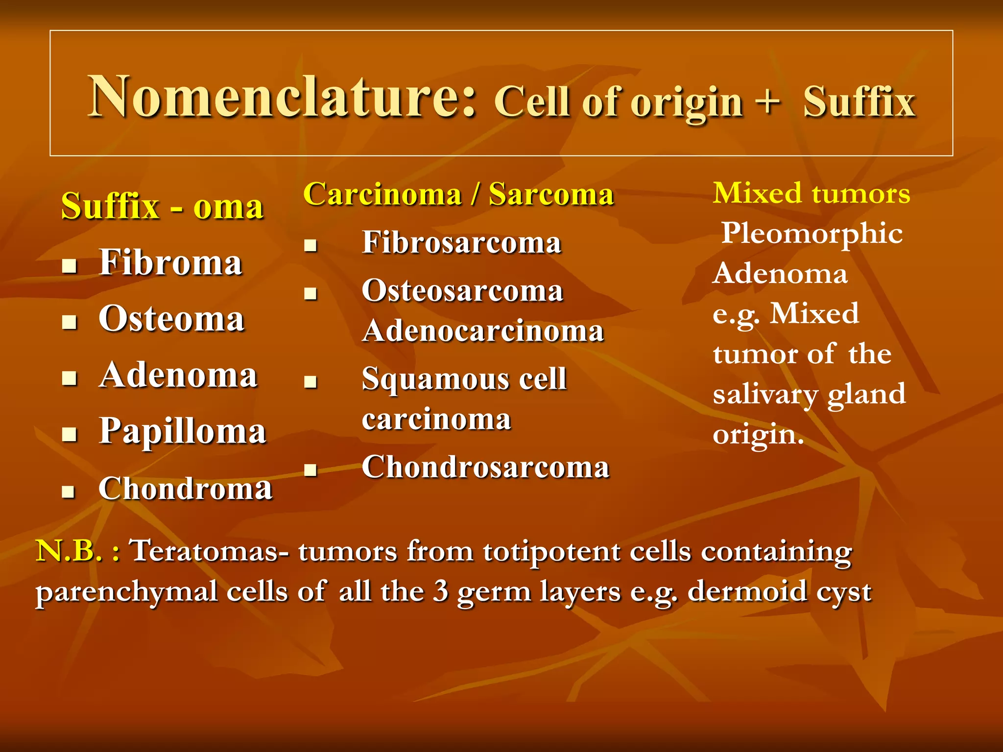
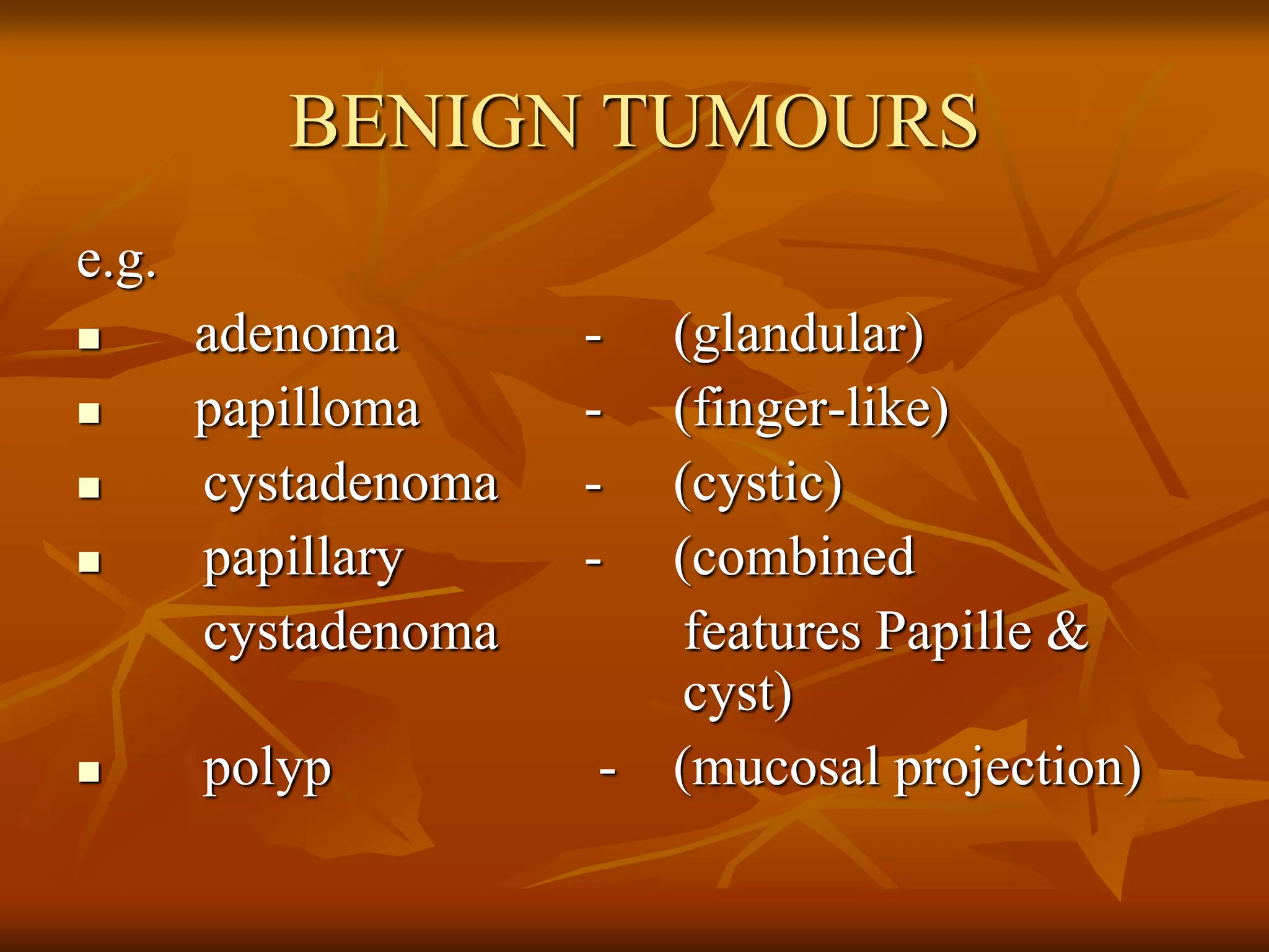
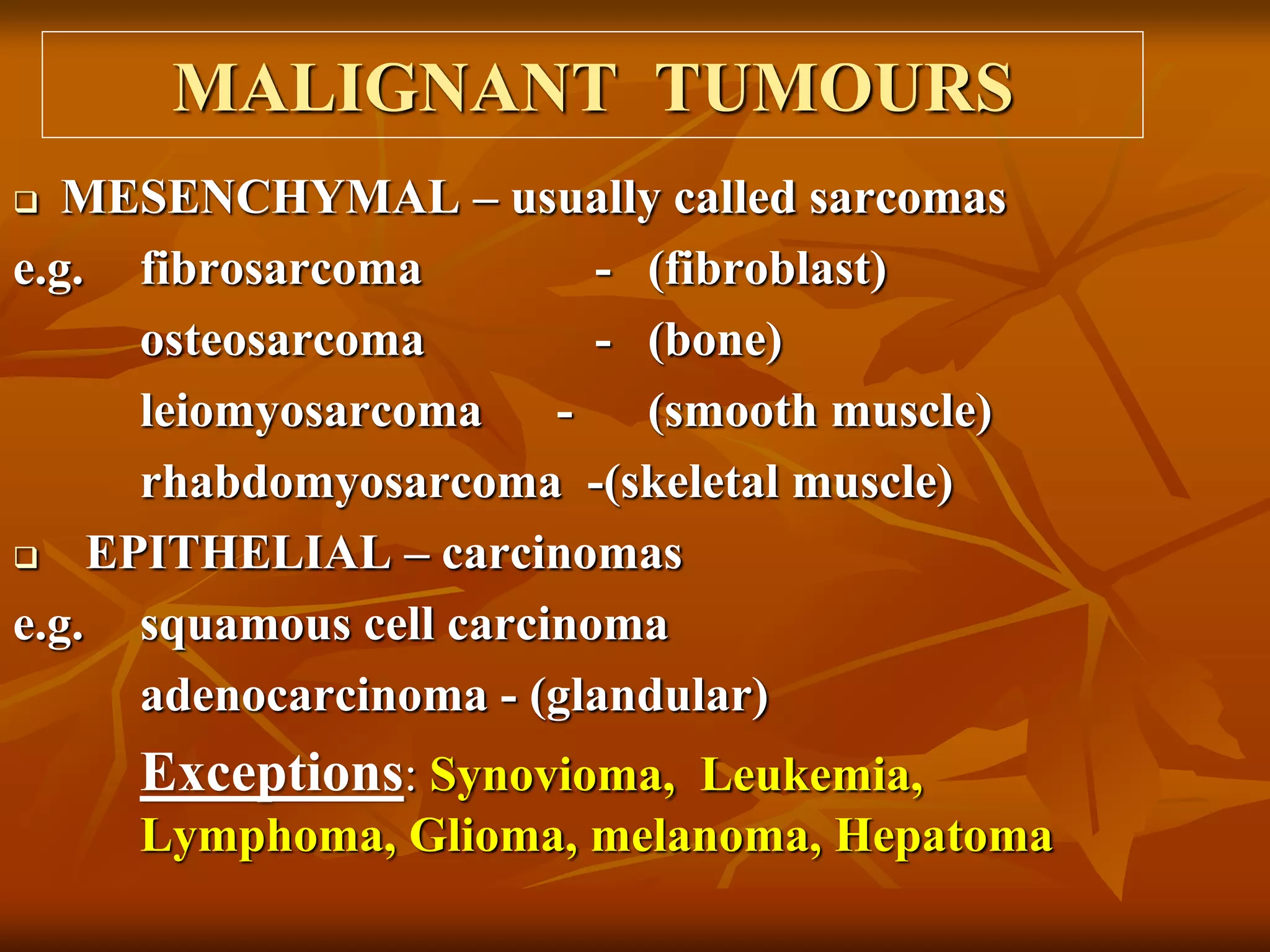
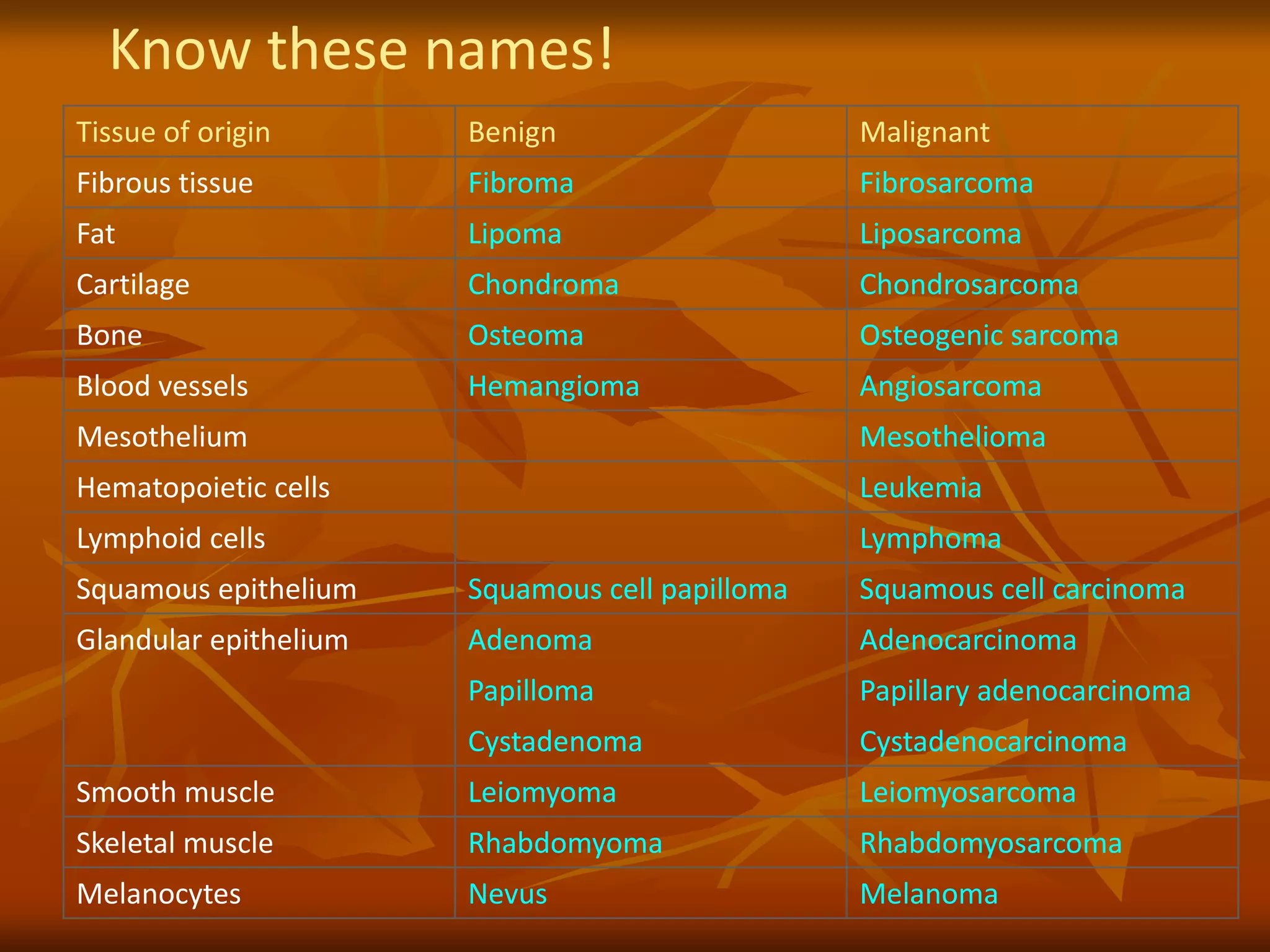
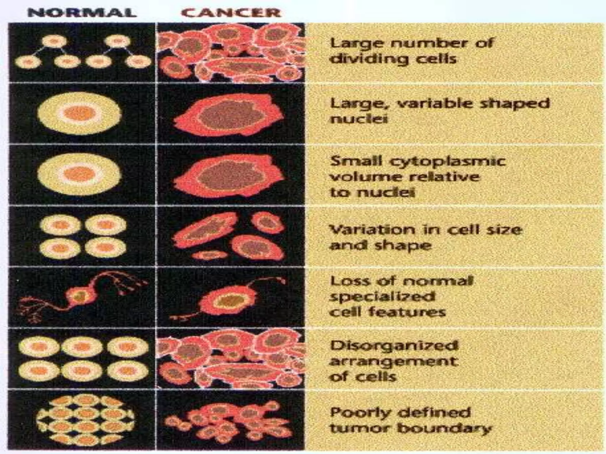
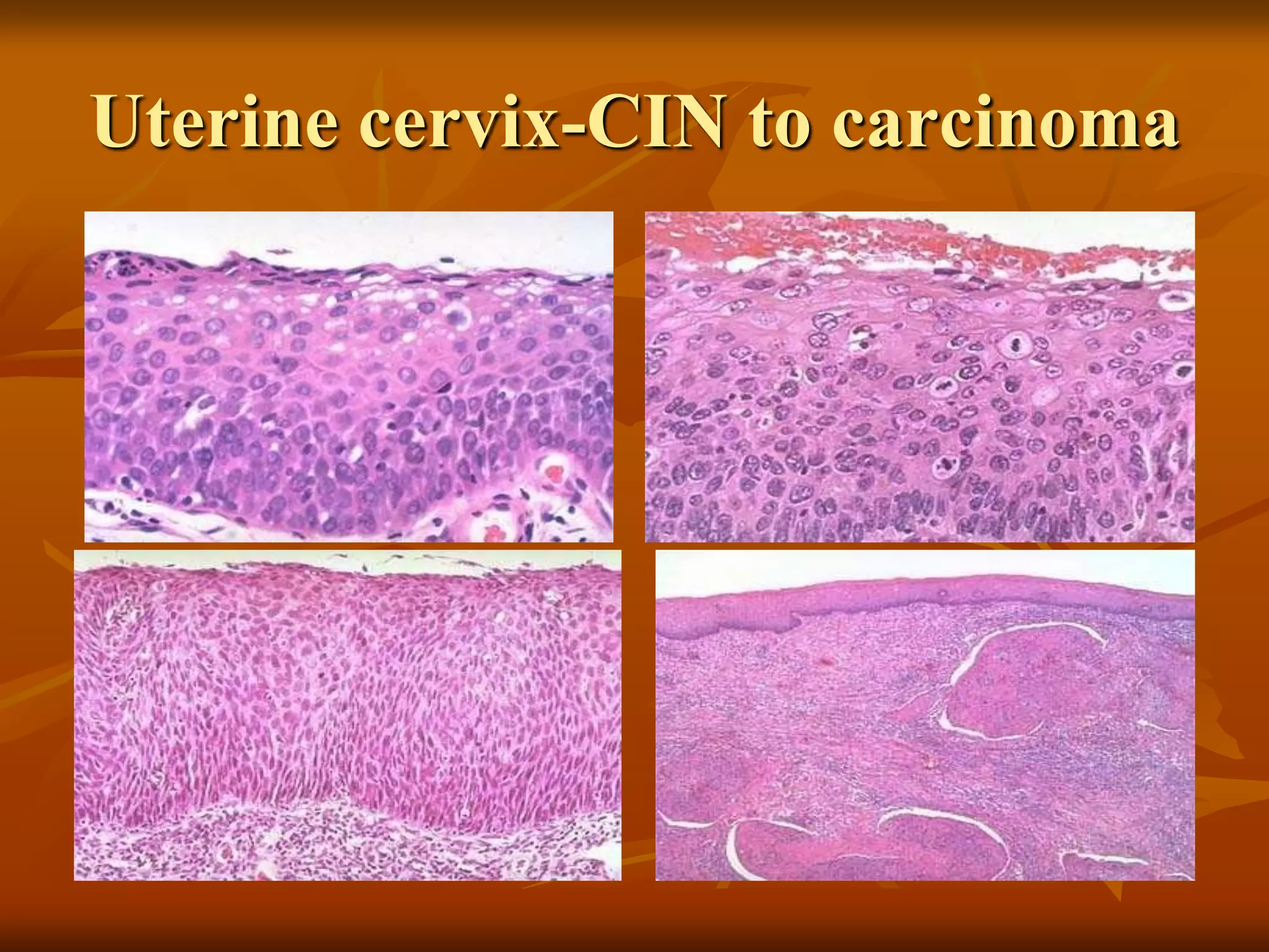
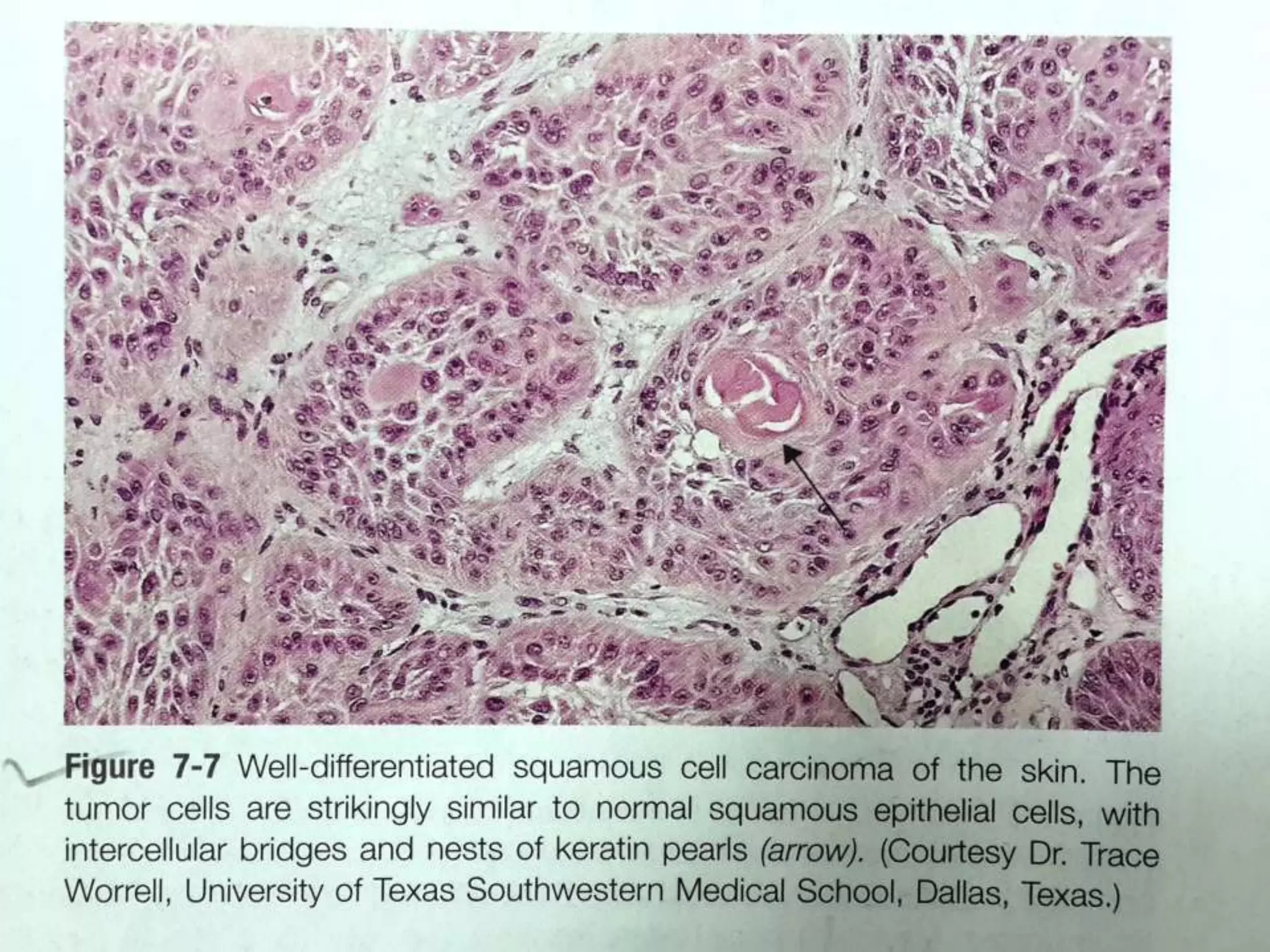
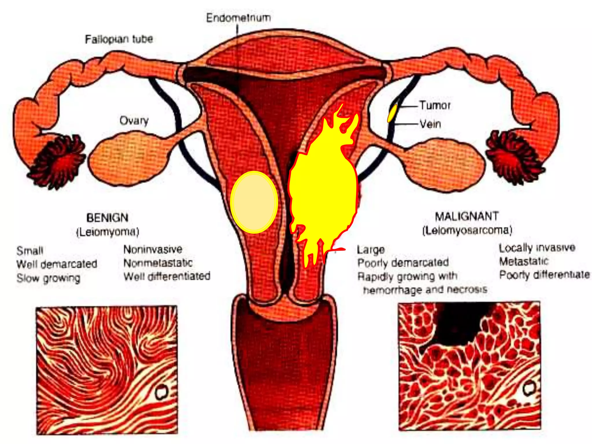
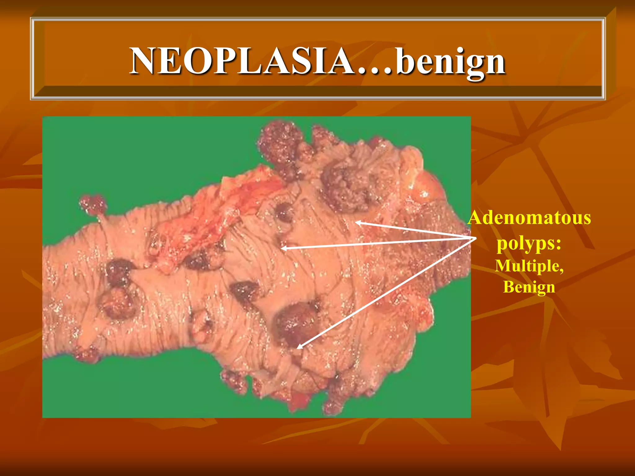
Neoplasia: Tumor
Nomenclature
DR. ROOPAM JAIN
ADDITIONAL DEAN
Cancer = Latin for “crab”
Neoplasia: Terminology
 Neoplasia is “new growth”
 Oncology(Greek oncos –tumor) study of
tumors or neoplasms
 Cancer – common term for all malignant
tumors derives from Latin for crab because a
cancer “adheres to any part that it seizes upon
in an obstinate manner like crab
Willis Definition:
A neoplasm is an abnormal mass of
tissue the growth of which exceeds and
is uncoordinated with that of normal
tissue and persists in the same excessive
manner after cessation of the stimuli
which evoked the change.
Thus neoplasia means:
An abnormal mass of tissue which differs
from the normal in:
 Growth
 Differentiation
 Function
 Organization
Non-Neoplastic Neoplastic
(Polyclonal) (Monoclonal)
Normal Adaptation Benign Malignant
Mechanism of Neoplasia
Neoplasm
Benign Malignant
Carcinoma Sarcoma
Adenoma
Haemangioma
Rhabdomyoma
Nomenclature
Nomenclature: Cell of origin + Suffix
Suffix - oma
 Fibroma
 Osteoma
 Adenoma
 Papilloma
 Chondroma
Carcinoma / Sarcoma
 Fibrosarcoma
 Osteosarcoma
Adenocarcinoma
 Squamous cell
carcinoma
 Chondrosarcoma
N.B. : Teratomas- tumors from totipotent cells containing
parenchymal cells of all the 3 germ layers e.g. dermoid cyst
Mixed tumors
Pleomorphic
Adenoma
e.g. Mixed
tumor of the
salivary gland
origin.
BENIGN TUMOURS
e.g.
 adenoma - (glandular)
 papilloma - (finger-like)
 cystadenoma - (cystic)
 papillary - (combined
cystadenoma features Papille &
cyst)
 polyp - (mucosal projection)
Uterus: leiomyomas
11
Thyroid adenoma
Thyroid adenoma
14
MALIGNANT TUMOURS
 MESENCHYMAL – usually called sarcomas
e.g. fibrosarcoma - (fibroblast)
osteosarcoma - (bone)
leiomyosarcoma - (smooth muscle)
rhabdomyosarcoma -(skeletal muscle)
 EPITHELIAL – carcinomas
e.g. squamous cell carcinoma
adenocarcinoma - (glandular)
Exceptions: Synovioma, Leukemia,
Lymphoma, Glioma, melanoma, Hepatoma
Nomenclature
Hamartoma: not neoplastic, it is rather a
malformation. Aberrant differentiation produce
mass of disorganized but mature of mature (adult-
type) tissue indigenous to particular site e.g.
hemartoma in lung contain island of cartilage,
blood vessels, bronchial type structures &
lymphoid tissue
Choristoma: Heterotrophic rest. It is normal tissue
in abnormal place.
 e.g. rest of adrenal cells under kidney capsule.
pancreatic nodular rest in mucosa of small
intestine
Mixed Tumors
 “Mixed” tumors show divergent differentiation
 Examples
• Pleomorphic adenoma – glands + fibromyxoid
stroma
• Fibroadenoma – glands + fibrous tissue
 Not to be confused with teratomas
 Teratoma: more than one germ-cell layer
 Teratoma contains: bone, epithelium, muscle, fat,
nerve….
Pleomorphic adenoma
19
Tissue of origin Benign Malignant
Fibrous tissue Fibroma Fibrosarcoma
Fat Lipoma Liposarcoma
Cartilage Chondroma Chondrosarcoma
Bone Osteoma Osteogenic sarcoma
Blood vessels Hemangioma Angiosarcoma
Mesothelium Mesothelioma
Hematopoietic cells Leukemia
Lymphoid cells Lymphoma
Squamous epithelium Squamous cell papilloma Squamous cell carcinoma
Glandular epithelium Adenoma Adenocarcinoma
Papilloma Papillary adenocarcinoma
Cystadenoma Cystadenocarcinoma
Smooth muscle Leiomyoma Leiomyosarcoma
Skeletal muscle Rhabdomyoma Rhabdomyosarcoma
Melanocytes Nevus Melanoma
Know these names!
21
Structure of Neoplasm:
Two components
1.Proliferating neoplastic cells
2.Supportive stroma
 Connective tissue
 Blood vessels
Variable amounts of (1) and (2) give tumours their
gross features, e.g. excessive collagenous stroma
(desmoplasia), schirrous tumours.
 Fast growth  less stroma
 Less stroma  more necrosis
TUMOURS
 A spectrum
Benign – Uncertain biologic potential –
Malignant
Benign ? Malignant
Benign and Malignant
 How do we know benign from malignant
tumor?
 Features:
 Differentiation and Anaplasia
 Rate of Growth
 Local Invasion
 Metastasis
Differentiation and Anaplasia
 Differentiation is the extent to which tumor cells
resemble their normal cells morphologically and
functionally
 Generally:
 Benign tumors are well differntiated
 Malignant tumors can be well differentiated, moderately
differentiated or poorly differentiated. They can be
“undifferentiated”
 Anaplasia is lack of differentiation
Differentiation and Anaplasia
 Dysplasia:
 Not neoplstic growth
 Disordered growth and differentiation
 Show mild anaplastic features eg. Nuclear
pleomorphism, hyperchromasia, mitosis….
27
DYSPLASIA AND
CANCER (1)
e.g. Cervical Cancer (HPV)
Mild dysplasia
Moderate
Severe
Carcinoma in situ
- Intact basement membrane
CARCINOMA IN SITU OF THE CERVIX
‘PAP’ smear with atypical cytological
changes of both the nucleus and
cytoplasm
Biopsy shows full thickness
change consistent with C.I.S.
29
Uterine cervix-CIN to carcinoma
CARCINOMA OF THE CERVIX
-41yr old woman with mild discharge
-exophytic friable tumour with positive
cytology
-infiltrating well-differentiated
squamous cell carcinoma
Low power
High power
32
33
Benign Malignant
 Progressive & Slow
growing,
 capsulated,
 Non-invasive
 do not metastasize,
 well differentiated,
structure typical of tissue
of origin. Mitotic figures
rare & normal
 suffix “oma” e.g..
Fibroma.
 Erratic & slow to fast
growing,
 non capsulated,
 Invasive & Infiltrate
 Metastasize.
 Iack of differentiation with
anaplasia, structure often
atypical mitotic figures
numerous & abnormal
 Suffix “Carcinoma” or
“Sarcoma”
NEOPLASIA…benign
Adenomatous
polyps:
Multiple,
Benign
NEOPLASIA…growth
configurations
Malignant
gastric
carcinoma
…ulcer
Rhabdmyosarcoma
 Benign tumours
usually closely
resemble normal cells
(well differentiated).
 Malignant tumours
show a range of
abnormal
differentiation – well
to undifferentiated
(anaplastic)
DIFFERENTIATION…
Cytology – individual cell morphology
 Pleomorphic
- Shape
- Size
 Nuclear/cytoplasmic ratio (N/C)  1:1 (N 1:4 –
1:6)
 Mitoses
- Number
- Normal
- Abnormal
ANAPLASIA
 Anaplasia=move backwards
 Dedifferentiation or… undifferentiation….
 Cytological characteristics of anaplastic cells-
light microscopic as well as ultra structural
 A marker for cancer-Malignancy
CELLULAR ORGANIZATION
Degree of loss of normal organization
 e.g. layered ~ squamous epithelium
- loss of polarity
 e.g. glands
- abnormal size or shape
- loss of ability to form glands
FUNCTIONAL DIFFERENTIATON
Well differentiated tumours  normal product
Poorly differentiated tumours less likely to have
specialized functional activity
Range:
 No product ---Normal---Increased
e.g. insulin, mucin, keratin
 New or ectopic product
e.g. PTH Lung carcinoma
ACTH Lung carcinoma
AFT Liver carcinoma (foetal antigen)
NEOPLASIA…growth
 Size
 May have no bearing on biologic behaviour
BENIGN
OVARIAN
TUMOUR
RATE OF GROWTH
Concept:
Benign tumours grow slowly
Cancers grow rapidly
Exceptions – many
e.g. fast growing benign tumours
hormone dependence
blood supply
RATE OF GROWTH…
Range of malignant tumour progression
 Years to weeks
 Cell cycle time- 3 days
 30 population doubling takes 90 days to
produce 109 cells
 Spontaneous remission, e.g. renal cell
carcinoma
RATE OF GROWTH…
Three Factors:
 Doubling time of tumor cells
 Fraction of tumor cells in replicative pool
 Rate at which cells are shed & lost in the
growing lesion
INVASION
Benign tumours are NOT invasive because of…
 Expansile probing margins
 Localized growth, readily palpable
 Do not have the capacity to infiltrate
 Encapsulation expect Haemangioma,
leiomyoma
Therefore benign tumours…
 If resected do not recur
 If incompletely removed then only local
recurrence occurs.
MECHANISM OF INVASION
 Physical pressure- compression atrophy & destruction
of abutting normal cells.
 Reduced adhesiveness & cohesiveness of cancer
cells.
 Motility of tumor cells-the cells are capable of
locomotion & cytoplasmic processes can be seen
protruding from the cells.
 Loss of contact inhibition- loss of cessation of cell
division and mobility on contact with other cells.
 Release of destructive enzymes- collagenases,
lysosomal enzymes, plasminogen activator.
INVASION
Malignant tumours are INVASIVE because of…
 Progressive growth
 Infiltration ~ poor line of demarcation
 Invasion ( importance of surgical margins)
 Destruction of adjacent tissue
 Metastatic spread
 Death if not treated
Tumor Infiltration
BENIGN VERSUS MALIGNANT
TUMOURS
 Invasion
Distinguish
malignant
tumors
 Metastases
Malignant tumors
Feature Sarcoma Carcinoma
Origin
Arise from mesenchymal
tissue
Arise from epithelial
tissue
Mass size
Usually larger since harder to
detect them in deep
tissue!!!
Usually smaller since
easier to detect on
surface
Time of
diagnosis
Takes longer period to
diagnose
Takes shorter period of
time to diagnose from
onset
Route of
Metastasis
Goes directly to vascular
system/ Blood vessels
Goes through lymphatics
first – longer route to
vascular system
Prognosis
Worse prognosis Good/worse

NEOPLASIA: TUMOR NOMENCLATURE

  • 1.
  • 2.
    Cancer = Latinfor “crab”
  • 3.
    Neoplasia: Terminology  Neoplasiais “new growth”  Oncology(Greek oncos –tumor) study of tumors or neoplasms  Cancer – common term for all malignant tumors derives from Latin for crab because a cancer “adheres to any part that it seizes upon in an obstinate manner like crab
  • 4.
    Willis Definition: A neoplasmis an abnormal mass of tissue the growth of which exceeds and is uncoordinated with that of normal tissue and persists in the same excessive manner after cessation of the stimuli which evoked the change.
  • 5.
    Thus neoplasia means: Anabnormal mass of tissue which differs from the normal in:  Growth  Differentiation  Function  Organization
  • 6.
    Non-Neoplastic Neoplastic (Polyclonal) (Monoclonal) NormalAdaptation Benign Malignant Mechanism of Neoplasia
  • 7.
  • 8.
    Nomenclature: Cell oforigin + Suffix Suffix - oma  Fibroma  Osteoma  Adenoma  Papilloma  Chondroma Carcinoma / Sarcoma  Fibrosarcoma  Osteosarcoma Adenocarcinoma  Squamous cell carcinoma  Chondrosarcoma N.B. : Teratomas- tumors from totipotent cells containing parenchymal cells of all the 3 germ layers e.g. dermoid cyst Mixed tumors Pleomorphic Adenoma e.g. Mixed tumor of the salivary gland origin.
  • 9.
    BENIGN TUMOURS e.g.  adenoma- (glandular)  papilloma - (finger-like)  cystadenoma - (cystic)  papillary - (combined cystadenoma features Papille & cyst)  polyp - (mucosal projection)
  • 10.
  • 11.
  • 12.
  • 13.
  • 14.
  • 15.
    MALIGNANT TUMOURS  MESENCHYMAL– usually called sarcomas e.g. fibrosarcoma - (fibroblast) osteosarcoma - (bone) leiomyosarcoma - (smooth muscle) rhabdomyosarcoma -(skeletal muscle)  EPITHELIAL – carcinomas e.g. squamous cell carcinoma adenocarcinoma - (glandular) Exceptions: Synovioma, Leukemia, Lymphoma, Glioma, melanoma, Hepatoma
  • 16.
    Nomenclature Hamartoma: not neoplastic,it is rather a malformation. Aberrant differentiation produce mass of disorganized but mature of mature (adult- type) tissue indigenous to particular site e.g. hemartoma in lung contain island of cartilage, blood vessels, bronchial type structures & lymphoid tissue Choristoma: Heterotrophic rest. It is normal tissue in abnormal place.  e.g. rest of adrenal cells under kidney capsule. pancreatic nodular rest in mucosa of small intestine
  • 17.
    Mixed Tumors  “Mixed”tumors show divergent differentiation  Examples • Pleomorphic adenoma – glands + fibromyxoid stroma • Fibroadenoma – glands + fibrous tissue  Not to be confused with teratomas  Teratoma: more than one germ-cell layer  Teratoma contains: bone, epithelium, muscle, fat, nerve….
  • 18.
  • 19.
  • 20.
    Tissue of originBenign Malignant Fibrous tissue Fibroma Fibrosarcoma Fat Lipoma Liposarcoma Cartilage Chondroma Chondrosarcoma Bone Osteoma Osteogenic sarcoma Blood vessels Hemangioma Angiosarcoma Mesothelium Mesothelioma Hematopoietic cells Leukemia Lymphoid cells Lymphoma Squamous epithelium Squamous cell papilloma Squamous cell carcinoma Glandular epithelium Adenoma Adenocarcinoma Papilloma Papillary adenocarcinoma Cystadenoma Cystadenocarcinoma Smooth muscle Leiomyoma Leiomyosarcoma Skeletal muscle Rhabdomyoma Rhabdomyosarcoma Melanocytes Nevus Melanoma Know these names!
  • 21.
  • 22.
    Structure of Neoplasm: Twocomponents 1.Proliferating neoplastic cells 2.Supportive stroma  Connective tissue  Blood vessels Variable amounts of (1) and (2) give tumours their gross features, e.g. excessive collagenous stroma (desmoplasia), schirrous tumours.  Fast growth  less stroma  Less stroma  more necrosis
  • 23.
    TUMOURS  A spectrum Benign– Uncertain biologic potential – Malignant Benign ? Malignant
  • 24.
    Benign and Malignant How do we know benign from malignant tumor?  Features:  Differentiation and Anaplasia  Rate of Growth  Local Invasion  Metastasis
  • 25.
    Differentiation and Anaplasia Differentiation is the extent to which tumor cells resemble their normal cells morphologically and functionally  Generally:  Benign tumors are well differntiated  Malignant tumors can be well differentiated, moderately differentiated or poorly differentiated. They can be “undifferentiated”  Anaplasia is lack of differentiation
  • 26.
    Differentiation and Anaplasia Dysplasia:  Not neoplstic growth  Disordered growth and differentiation  Show mild anaplastic features eg. Nuclear pleomorphism, hyperchromasia, mitosis….
  • 27.
  • 28.
    DYSPLASIA AND CANCER (1) e.g.Cervical Cancer (HPV) Mild dysplasia Moderate Severe Carcinoma in situ - Intact basement membrane
  • 29.
    CARCINOMA IN SITUOF THE CERVIX ‘PAP’ smear with atypical cytological changes of both the nucleus and cytoplasm Biopsy shows full thickness change consistent with C.I.S. 29
  • 30.
  • 31.
    CARCINOMA OF THECERVIX -41yr old woman with mild discharge -exophytic friable tumour with positive cytology -infiltrating well-differentiated squamous cell carcinoma Low power High power
  • 32.
  • 33.
  • 34.
    Benign Malignant  Progressive& Slow growing,  capsulated,  Non-invasive  do not metastasize,  well differentiated, structure typical of tissue of origin. Mitotic figures rare & normal  suffix “oma” e.g.. Fibroma.  Erratic & slow to fast growing,  non capsulated,  Invasive & Infiltrate  Metastasize.  Iack of differentiation with anaplasia, structure often atypical mitotic figures numerous & abnormal  Suffix “Carcinoma” or “Sarcoma”
  • 36.
  • 37.
  • 38.
  • 39.
     Benign tumours usuallyclosely resemble normal cells (well differentiated).  Malignant tumours show a range of abnormal differentiation – well to undifferentiated (anaplastic)
  • 40.
    DIFFERENTIATION… Cytology – individualcell morphology  Pleomorphic - Shape - Size  Nuclear/cytoplasmic ratio (N/C)  1:1 (N 1:4 – 1:6)  Mitoses - Number - Normal - Abnormal
  • 41.
    ANAPLASIA  Anaplasia=move backwards Dedifferentiation or… undifferentiation….  Cytological characteristics of anaplastic cells- light microscopic as well as ultra structural  A marker for cancer-Malignancy
  • 42.
    CELLULAR ORGANIZATION Degree ofloss of normal organization  e.g. layered ~ squamous epithelium - loss of polarity  e.g. glands - abnormal size or shape - loss of ability to form glands
  • 43.
    FUNCTIONAL DIFFERENTIATON Well differentiatedtumours  normal product Poorly differentiated tumours less likely to have specialized functional activity Range:  No product ---Normal---Increased e.g. insulin, mucin, keratin  New or ectopic product e.g. PTH Lung carcinoma ACTH Lung carcinoma AFT Liver carcinoma (foetal antigen)
  • 44.
    NEOPLASIA…growth  Size  Mayhave no bearing on biologic behaviour BENIGN OVARIAN TUMOUR
  • 45.
    RATE OF GROWTH Concept: Benigntumours grow slowly Cancers grow rapidly Exceptions – many e.g. fast growing benign tumours hormone dependence blood supply
  • 46.
    RATE OF GROWTH… Rangeof malignant tumour progression  Years to weeks  Cell cycle time- 3 days  30 population doubling takes 90 days to produce 109 cells  Spontaneous remission, e.g. renal cell carcinoma
  • 47.
    RATE OF GROWTH… ThreeFactors:  Doubling time of tumor cells  Fraction of tumor cells in replicative pool  Rate at which cells are shed & lost in the growing lesion
  • 48.
    INVASION Benign tumours areNOT invasive because of…  Expansile probing margins  Localized growth, readily palpable  Do not have the capacity to infiltrate  Encapsulation expect Haemangioma, leiomyoma Therefore benign tumours…  If resected do not recur  If incompletely removed then only local recurrence occurs.
  • 49.
    MECHANISM OF INVASION Physical pressure- compression atrophy & destruction of abutting normal cells.  Reduced adhesiveness & cohesiveness of cancer cells.  Motility of tumor cells-the cells are capable of locomotion & cytoplasmic processes can be seen protruding from the cells.  Loss of contact inhibition- loss of cessation of cell division and mobility on contact with other cells.  Release of destructive enzymes- collagenases, lysosomal enzymes, plasminogen activator.
  • 50.
    INVASION Malignant tumours areINVASIVE because of…  Progressive growth  Infiltration ~ poor line of demarcation  Invasion ( importance of surgical margins)  Destruction of adjacent tissue  Metastatic spread  Death if not treated
  • 51.
  • 52.
    BENIGN VERSUS MALIGNANT TUMOURS Invasion Distinguish malignant tumors  Metastases
  • 53.
    Malignant tumors Feature SarcomaCarcinoma Origin Arise from mesenchymal tissue Arise from epithelial tissue Mass size Usually larger since harder to detect them in deep tissue!!! Usually smaller since easier to detect on surface Time of diagnosis Takes longer period to diagnose Takes shorter period of time to diagnose from onset Route of Metastasis Goes directly to vascular system/ Blood vessels Goes through lymphatics first – longer route to vascular system Prognosis Worse prognosis Good/worse