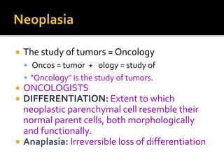
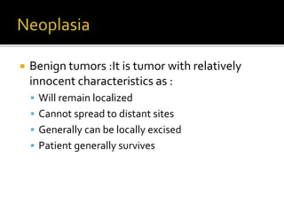
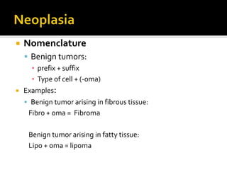
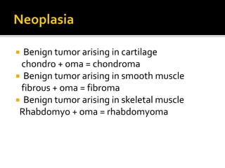
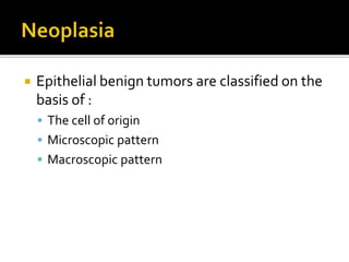
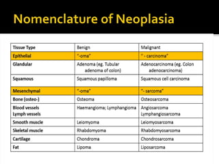
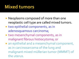
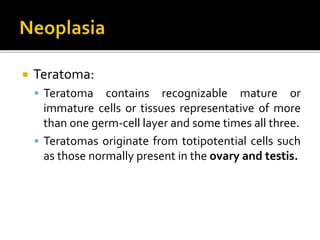
 Cancer is one of the leading causes of death
worldwide.
 Emotional and physical suffering by the
patient.
 Different mortality rate …..
 Some are curable
 Others are fatal
 Neoplasia = latin word (new growth)
 Neoplasm = tumor
 Tumor = swelling
 Neoplasia means new growth and is
characterized by unceasing abnormal and
excessive proliferation of cells.
 Definition:
 is an abnormal mass of tissue,
 the growth of which is uncoordinated with that of
normal tissues,
 and that persists in the same excessive manner after
the cessation of the stimulus which evoked the change
 With the loss of responsiveness to normal growth
controls
Rupert Willis
 The study of tumors = Oncology
 Oncos = tumor + ology = study of
 "Oncology" is the study of tumors.
 ONCOLOGISTS
 DIFFERENTIATION: Extent to which
neoplastic parenchymal cell resemble their
normal parent cells, both morphologically
and functionally.
 Anaplasia: Irreversible loss of differentiation
 Classification
 Benign
 Malignant
 Benign tumors :It is tumor with relatively
innocent characteristics as :
 Will remain localized
 Cannot spread to distant sites
 Generally can be locally excised
 Patient generally survives
Characteristics Benign Malignant
1.Differentiation Well differentiated Ranges from well differentiated to
undifferentiated
2.Anaplasia No anaplasia Certainly present
3. Spread/Infiltration Remains localized Invades and penetrates the surrounding
tissue
4. Metastasis No metastasis Metastasize to regional lymph nodes and
distant organs
5. Rate of growth Usually slow except leiomyoma Usually rapid except cancer of cervix
grows slowly
6. Encapsulation Enclosed within capsule which separates
it from host tissues, except leiomyoma
Capsule never present
7.Gross appearance Degeneration, necrosis ulceration,
hemorrhage less frequent
Degeneration, necrosis ulceration,
hemorrhage more frequent
8. Clinical Effects Do not endanger life until vital organs
involved
Act as parasite and tends to kill the
patient whenever it grows
9.Recurrence Easily local removal-no recurrence Recurrence common
 Components of neoplasms:
1. Parenchyma: made up of proliferating parts of neoplastic
cells
 Determines biological behavior of tumor from which the
tumor derives its name
2.The stroma:
 made up of non-neoplastic, host-derived connective tissue
and blood vessels
 Provides support for the growth of the parenchyma
3. Desmoplasia- the excess of stromal component in tumor
is called desmoplasia and a such tumor is called scirrhous
tumor.
 Tumor is named on the basis of
 Based on the biological behavior :
 Benign and malignant
 Based on the cell or tissue of origin :
 One neoplastic cell type : lipoma,adenocarcinoma
 More than one neoplastic cell type : MIXED like
fibroadenoma
 More than one neoplastic cell type derived from
more than one germ-cell layer: teratoma
 Nomenclature
 Benign tumors:
▪ prefix + suffix
▪ Type of cell + (-oma)
 Examples:
 Benign tumor arising in fibrous tissue:
Fibro + oma = Fibroma
Benign tumor arising in fatty tissue:
Lipo + oma = lipoma
 Benign tumor arising in cartilage
chondro + oma = chondroma
 Benign tumor arising in smooth muscle
fibrous + oma = fibroma
 Benign tumor arising in skeletal muscle
Rhabdomyo + oma = rhabdomyoma
 Epithelial benign tumors are classified on the
basis of :
 The cell of origin
 Microscopic pattern
 Macroscopic pattern
 Adenoma : benign epithelial neoplasms
producing gland pattern….OR … derived from
glands but not necessarily exhibiting gland
pattern
 Papilloma : benign epithelial neoplasms growing
on any surface that produce microscopic or
macroscopic finger-like pattern
 Malignant tumors:
 Malignant tumor arising in mesenchymal tissue :
SARCOMA
▪ From fibrous tissue: Fibrosarcoma
▪ From bone : Osteosarcoma
▪ From cartilage : chondrosarcoma
 Malignant tumors arising from epithelial origin :
CARCINOMA
 Squamous cell carcinoma
 Renal cell adenocarcinoma
 Neoplasms composed of more than one
neoplastic cell type are called mixed tumors.
 two epithelial components, as in
adenosquamous carcinoma;
 two mesenchymal components, as in
malignant fibrous histiocytoma; or
 an epithelial and a mesenchymal component,
as in carcinosarcoma of the lung and
malignant mixed müllerian tumor (MMMT) of
the uterus.
 Teratoma:
 Teratoma contains recognizable mature or
immature cells or tissues representative of more
than one germ-cell layer and some times all three.
 Teratomas originate from totipotential cells such
as those normally present in the ovary and testis.
 MIXEDTUMORS: More than one type of
cells derived from same germ layer
 TERATOMAS
 HAMARTOMAS: Disorganized
overgrowth of mature cells indigenous to
that part
 CHORISTOMA: Presence of well
developed & normally organized tissue of
one organ in another
 Melanoma ( skin )
 Mesothelioma (mesothelium )
 Seminoma ( testis )
 Hepatoma (HCC also called)
 Invasive meningioma
 Leukemia (always malignant)
 Lymphoma ( lymphoid tissue )
1. Gross/Clinical Features
2. Microscopic Features
3. Growth Rate
4. Local Invasion
5. Metastasis
6. Prognosis
 Dysplasia :
 Definiton: a loss in the uniformity of the individual
cells and a loss in their architectural orientation.
 Non-neoplastic and does not necessarily cause
cancer
 Or doesn’t mean cancer and is REVERSIBLE
 Occurs mainly in the epithelia
 Dysplastic cells shows a degree of :
pleomorphism, hyperchromasia,increased mitosis
and loss of polarity.
 Metastasis :
 Definition : the development of secondary implants
discontinuous with the primary tumor, possibly in remote
tissues.
 Cancers have different ability to metastasize
 Approximately 30% patients present with clinically evident
metastases.
 Generally, the more anaplastic and the larger the primary
tumor, the more likely is metastasis
 Metastasis : three pathways
 Lymphatic spread :
 Hematogenous spread :
 Seeding of the body cavities: pleural, peritoneal
cavities and cerebral ventricles
Lymphatic spread :
 favored by carcinomas
 Breast carcinoma  axillary lymph nodes
 Lung carcinomas  bronchial lymph nodes
Hematogenous spread :
 favored by sarcomas
 Also used by carcinomas
 Veins are more commonly invaded
The liver and lungs are the most frequently involved secondary sites
Seeding Body Cavities:
 Malignant neoplasm penetrate into natural cavity (pleural, pericardial,
peritoneal, subarachnoid and joint space)
 This mode of transmission is characteristic of cancers of ovary

Neoplasia,Growth,definition,Differentiation, .ppt

  • 2.
     Cancer isone of the leading causes of death worldwide.  Emotional and physical suffering by the patient.  Different mortality rate …..  Some are curable  Others are fatal
  • 3.
     Neoplasia =latin word (new growth)  Neoplasm = tumor  Tumor = swelling  Neoplasia means new growth and is characterized by unceasing abnormal and excessive proliferation of cells.
  • 4.
     Definition:  isan abnormal mass of tissue,  the growth of which is uncoordinated with that of normal tissues,  and that persists in the same excessive manner after the cessation of the stimulus which evoked the change  With the loss of responsiveness to normal growth controls Rupert Willis
  • 5.
     The studyof tumors = Oncology  Oncos = tumor + ology = study of  "Oncology" is the study of tumors.  ONCOLOGISTS  DIFFERENTIATION: Extent to which neoplastic parenchymal cell resemble their normal parent cells, both morphologically and functionally.  Anaplasia: Irreversible loss of differentiation
  • 6.
  • 7.
     Benign tumors:It is tumor with relatively innocent characteristics as :  Will remain localized  Cannot spread to distant sites  Generally can be locally excised  Patient generally survives
  • 8.
    Characteristics Benign Malignant 1.DifferentiationWell differentiated Ranges from well differentiated to undifferentiated 2.Anaplasia No anaplasia Certainly present 3. Spread/Infiltration Remains localized Invades and penetrates the surrounding tissue 4. Metastasis No metastasis Metastasize to regional lymph nodes and distant organs 5. Rate of growth Usually slow except leiomyoma Usually rapid except cancer of cervix grows slowly 6. Encapsulation Enclosed within capsule which separates it from host tissues, except leiomyoma Capsule never present 7.Gross appearance Degeneration, necrosis ulceration, hemorrhage less frequent Degeneration, necrosis ulceration, hemorrhage more frequent 8. Clinical Effects Do not endanger life until vital organs involved Act as parasite and tends to kill the patient whenever it grows 9.Recurrence Easily local removal-no recurrence Recurrence common
  • 9.
     Components ofneoplasms: 1. Parenchyma: made up of proliferating parts of neoplastic cells  Determines biological behavior of tumor from which the tumor derives its name 2.The stroma:  made up of non-neoplastic, host-derived connective tissue and blood vessels  Provides support for the growth of the parenchyma 3. Desmoplasia- the excess of stromal component in tumor is called desmoplasia and a such tumor is called scirrhous tumor.
  • 10.
     Tumor isnamed on the basis of  Based on the biological behavior :  Benign and malignant  Based on the cell or tissue of origin :  One neoplastic cell type : lipoma,adenocarcinoma  More than one neoplastic cell type : MIXED like fibroadenoma  More than one neoplastic cell type derived from more than one germ-cell layer: teratoma
  • 11.
     Nomenclature  Benigntumors: ▪ prefix + suffix ▪ Type of cell + (-oma)  Examples:  Benign tumor arising in fibrous tissue: Fibro + oma = Fibroma Benign tumor arising in fatty tissue: Lipo + oma = lipoma
  • 12.
     Benign tumorarising in cartilage chondro + oma = chondroma  Benign tumor arising in smooth muscle fibrous + oma = fibroma  Benign tumor arising in skeletal muscle Rhabdomyo + oma = rhabdomyoma
  • 13.
     Epithelial benigntumors are classified on the basis of :  The cell of origin  Microscopic pattern  Macroscopic pattern
  • 14.
     Adenoma :benign epithelial neoplasms producing gland pattern….OR … derived from glands but not necessarily exhibiting gland pattern  Papilloma : benign epithelial neoplasms growing on any surface that produce microscopic or macroscopic finger-like pattern
  • 15.
     Malignant tumors: Malignant tumor arising in mesenchymal tissue : SARCOMA ▪ From fibrous tissue: Fibrosarcoma ▪ From bone : Osteosarcoma ▪ From cartilage : chondrosarcoma  Malignant tumors arising from epithelial origin : CARCINOMA  Squamous cell carcinoma  Renal cell adenocarcinoma
  • 18.
     Neoplasms composedof more than one neoplastic cell type are called mixed tumors.  two epithelial components, as in adenosquamous carcinoma;  two mesenchymal components, as in malignant fibrous histiocytoma; or  an epithelial and a mesenchymal component, as in carcinosarcoma of the lung and malignant mixed müllerian tumor (MMMT) of the uterus.
  • 19.
     Teratoma:  Teratomacontains recognizable mature or immature cells or tissues representative of more than one germ-cell layer and some times all three.  Teratomas originate from totipotential cells such as those normally present in the ovary and testis.
  • 20.
     MIXEDTUMORS: Morethan one type of cells derived from same germ layer  TERATOMAS  HAMARTOMAS: Disorganized overgrowth of mature cells indigenous to that part  CHORISTOMA: Presence of well developed & normally organized tissue of one organ in another
  • 21.
     Melanoma (skin )  Mesothelioma (mesothelium )  Seminoma ( testis )  Hepatoma (HCC also called)  Invasive meningioma  Leukemia (always malignant)  Lymphoma ( lymphoid tissue )
  • 23.
    1. Gross/Clinical Features 2.Microscopic Features 3. Growth Rate 4. Local Invasion 5. Metastasis 6. Prognosis
  • 24.
     Dysplasia : Definiton: a loss in the uniformity of the individual cells and a loss in their architectural orientation.  Non-neoplastic and does not necessarily cause cancer  Or doesn’t mean cancer and is REVERSIBLE  Occurs mainly in the epithelia  Dysplastic cells shows a degree of : pleomorphism, hyperchromasia,increased mitosis and loss of polarity.
  • 25.
     Metastasis : Definition : the development of secondary implants discontinuous with the primary tumor, possibly in remote tissues.  Cancers have different ability to metastasize  Approximately 30% patients present with clinically evident metastases.  Generally, the more anaplastic and the larger the primary tumor, the more likely is metastasis
  • 26.
     Metastasis :three pathways  Lymphatic spread :  Hematogenous spread :  Seeding of the body cavities: pleural, peritoneal cavities and cerebral ventricles
  • 27.
    Lymphatic spread : favored by carcinomas  Breast carcinoma  axillary lymph nodes  Lung carcinomas  bronchial lymph nodes Hematogenous spread :  favored by sarcomas  Also used by carcinomas  Veins are more commonly invaded The liver and lungs are the most frequently involved secondary sites Seeding Body Cavities:  Malignant neoplasm penetrate into natural cavity (pleural, pericardial, peritoneal, subarachnoid and joint space)  This mode of transmission is characteristic of cancers of ovary