Download to read offline

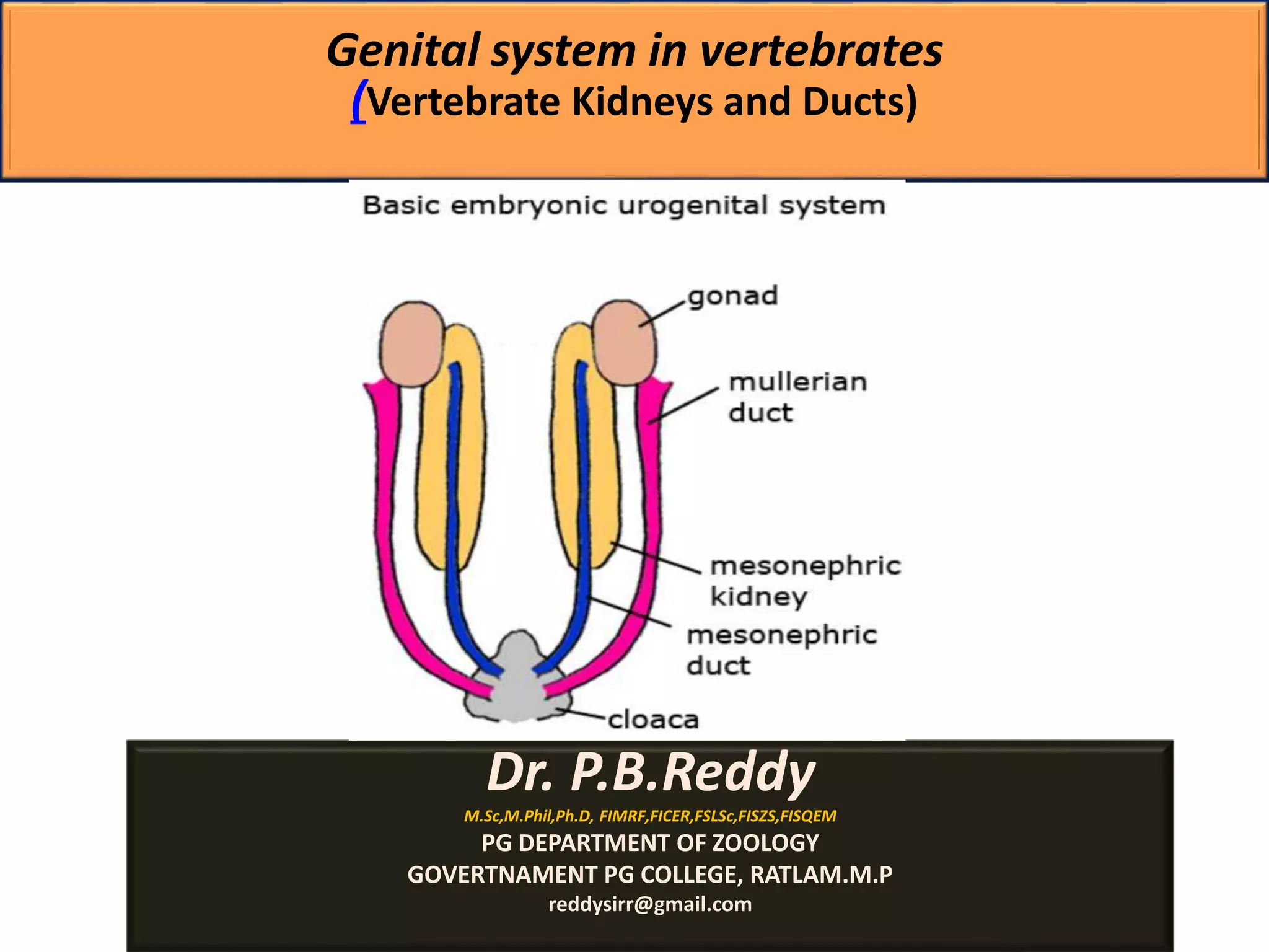
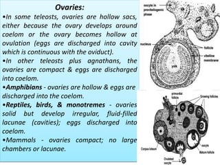
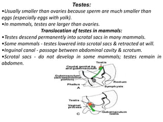
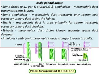

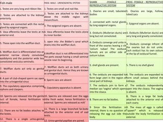
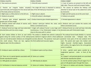


The document discusses the development and anatomy of the genital systems in vertebrates, highlighting the embryonic origins of reproductive organs and the roles of different ducts such as mesonephric and paramesonephric ducts. It details the structure and function of gonads, oviducts, and the male and female genital ducts across various vertebrate classes, including fishes, amphibians, reptiles, birds, and mammals. The text emphasizes the complex differentiation processes influenced by genetic and hormonal factors that lead to the formation of reproductive systems prone to abnormalities.


![Polymer [ बहुलक ] Chemistry Notes PDF - Irfanullah Mehar - JJ Sir Chemistry.pdf](https://cdn.slidesharecdn.com/ss_thumbnails/polymerchemistrynotespdf-irfanullahmehar-jjsirchemistry-260210172118-3f9b37f7-thumbnail.jpg?width=640&height=640&fit=bounds)


