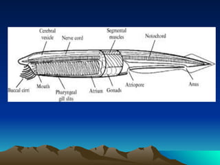
1. Amphioxus is a primitive chordate that undergoes indirect development with a free-swimming ciliated larval stage.
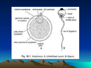
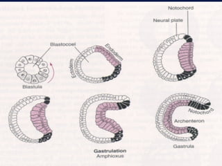
2. Its embryonic development includes external fertilization, equal holoblastic cleavage, blastula formation, and gastrulation by invagination and involution.
3. During gastrulation, the endodermal plate invaginates to form the archenteron while prospective mesoderm and notochord involute inward.


![Human Reproduction [ Reproductive System ] Notes @irfanullah_mehar Irfanullah...](https://cdn.slidesharecdn.com/ss_thumbnails/humanreproductionreproductivesystemnotesirfanullahmeharirfanullahmeharjanantantra-260111172350-56e85778-thumbnail.jpg?width=640&height=640&fit=bounds)

