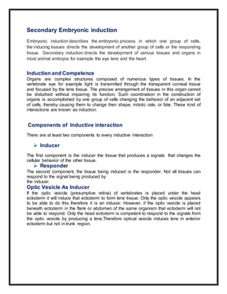
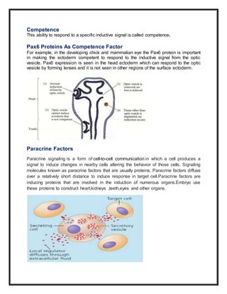
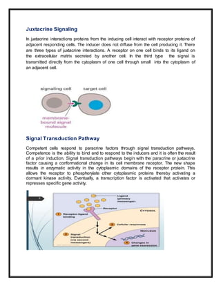
1. Secondary embryonic induction involves one group of cells, known as the inducer, directing the development of another group of cells or responder tissue. For example, the optic vesicle induces the formation of the lens.
2. Induction requires two components - the inducer, which produces signals to change the responder tissue, and the responder tissue which must be competent to receive the signals from the inducer. Competence is conferred by proteins like Pax6.
3. Signaling between tissues can occur through paracrine factors which diffuse short distances, or juxtacrine interactions which involve direct cell contact. This signaling activates gene expression through signal transduction pathways in competent responder tissues.
INTRODUCTION
DEFINATION
GAMETES
STRUCTURE OF GAMETES
SPERM
OVUM
RECOGNITION OF EGG AND SPERM
CAPACITATION
ACROSOME REACTION
SPECIES-SPECIFIC RECOGNITION
GAMETE BINDING AND RECOGNITION
GAMETE FUSION
PREVENTION OF POLYSPERMY
ACTIVATION OF GAMETE METABOLISM
FUSION OF THE GENETIC MATERIAL
SIGNIFICANCE OF FERTILIZATION
CONCLUSIONS
REFERENCES
How 3 germ layers are formed in Chick that are endoderm, mesoderm and ectoderm.As Chick are polylecithal so cell movements are somewhat restricted and gastrulation is modified as compared to frog.
cell commitment and differentiation, stem cell,types of differentiationshallu kotwal
The commitment of cells to specific cell fates and their capacity to differentiate into particular kinds of cells.
Cellular differentiation is the process in which a cell changes from one cell type to another. Usually, the cell changes to a more specialized type. Differentiation occurs numerous times during the development of a multicellular organism as it changes from a simple zygote to a complex system of tissues and cell types. Differentiation continues in adulthood as adult stem cells divide and create fully differentiated daughter cells during tissue repair and during normal cell turnover.
Vittelogenesis is a word developed from Latin vitellus-yolk, and genero-produce
Vitellogenesis (also known as yolk deposition) is the process of yolk formation via nutrients being deposited in the oocyte, or female germ cell involved in reproduction of lecithotrophic organisms. In insects, it starts when the fat body stimulates the release of juvenile hormones and produces vitellogenin protein.
Yolks is the most usual form of food storage in the egg.
Yolks appear in the oocyte in the secondary period of their growth called vittelogenesis.
Thus,the formation and deposition of yolks is known as vittelogenesis
Characteristic
Yolks is a complex variable assembled component.
The principle component are protein,phospholipid and fats in different combination.
Depending upon these component yolks is distinguished into protein yolks and fatty acid
For eg- the avian contain 48.19% water , 16.6 % protein, 32.6% phospholipids and fats and 1% carbohydrates.
A chart showing the fate of each part of an early embryo, in a particular blastula stage is called fate maps. It is done because the correct interpretation of gastrulation is impossible without the knowledge of the position which are the presumptive germinal layers (Ectoderm, Mesoderm and Endoderm) occupy in blastula.
Fate mapping is a method used in developmental biology to study the embryonic origin of various adult tissues and structures. The "fate" of each cell or group of cells is mapped onto the embryo, showing which parts of the embryo will develop into which tissue. When carried out at single-cell resolution, this process is called cell lineage tracing. It is also used to trace the development of tumors.
Polyspermy describes an egg that has been fertilized by more than one sperm. Diploid organisms normally contain two copies of each chromosome, one from each parent. The cell resulting from polyspermy
The first issue that an egg and a sperm of any organism type face in successfully producing an embryo is the possibility of polyspermy. Polyspermy is the fertilization of an egg by multiple sperm, and the results of such unions are lethal.
If multiple sperm fertilize an egg, the embryo inherits multiple paternal centrioles. This causes competition for extra chromosomes and results in the disruption of the creation of the cleavage furrow, thus causing the zygote to die. As an important model organism in the study of fertilization and embryonic development, polyspermy in sea urchins has been studied in detail. The sea urchin’s methods of polyspermy prevention have been broken down into two main pathways. These two primary pathways are known as the fast block and the slow block to polyspermy
After the sperm’s receptors come into contact with the egg’s jelly layer and the acrosomal enzymes are released and break down the jelly layer, the sperm head comes into contact with the vitelline and plasma membranes of the egg. When the two plasma membranes contact one another, signals in the egg are initiated.
First, Na+ channels in the egg open, allowing Na+ to flood into the egg. This causes a depolarization of the egg from it’s normal resting potential of -70 mV.
While depolarization is occurring, the remainder of the jelly layer is dissolving. With the dissolution of the jelly layer and the depolarization of the plasma membrane, the first block to preventing fertilization by multiple sperm is put into place.
These two simple changes are part of the first block to polyspermy, known as the fast block. Within 1/10th of a second of contact, the fast block t
Giving overview of human embryonic development including spermatogenesis, oogenesis, fertilization, gastrulation, cleavage, extraembryonic layers and pregnancy
An embryo has three germ layers - ectoderm, mesoderm and endoderm. T.pdfanubhavnigam2608
An embryo has three germ layers - ectoderm, mesoderm and endoderm. These germ layers
differentiate and form specialized organs for the organism. his process of organ formation from
these germ layers are called Organogenesis. Ectoderm develops into skin, sensory organs, brain,
spinal cord etc. Mesoderm develops into bones,muscle etc. Endoderm forms organs like lungs,
gut, liver etc.
There is a portion in ectoderm at the dorsal side of the germ layer which is destined to form
neural tube, that portion is called neural plate. This neural plate folds and forms neural tube. The
rostral end of the neural tube forms the brain, while the caudal end forms the spinal cord. The
process of folding which creates the neural tube from the neural plate is called Neurulation.
All these developments are done after embryonic induction. During embryonic induction, some
cells of the embryo which have the capability to induce other cells to develop into various
structures.
One example of induction is the formation of the eye lens from epidermis under the effect of eye
cup. An eye cup grows toward the skin from the brain. When eye cup comes in contact with any
nearby epidermis, it transforms that particular area into a lens. The exact nature of the stimulus
for lens induction is not known, although ribonucleic acid (RNA) has been implicated as a
messenger.
Induction for neurulation - There is a signal named bone morphogenetic protein 4 (BMP4) which
is received by ectoderm. So cells which receive it forms epidermis, while which do not receive
forms neural plate. The sending of this signal is blocked by inhibitory signals like - follistatin,
chordin and noggin. These inhibitory signals are produced from Spemanns organizer.
Embryonic inducers must be stable, reach to the responsible cells by diffusion, direct contact or
through gap junctions.
Solution
An embryo has three germ layers - ectoderm, mesoderm and endoderm. These germ layers
differentiate and form specialized organs for the organism. his process of organ formation from
these germ layers are called Organogenesis. Ectoderm develops into skin, sensory organs, brain,
spinal cord etc. Mesoderm develops into bones,muscle etc. Endoderm forms organs like lungs,
gut, liver etc.
There is a portion in ectoderm at the dorsal side of the germ layer which is destined to form
neural tube, that portion is called neural plate. This neural plate folds and forms neural tube. The
rostral end of the neural tube forms the brain, while the caudal end forms the spinal cord. The
process of folding which creates the neural tube from the neural plate is called Neurulation.
All these developments are done after embryonic induction. During embryonic induction, some
cells of the embryo which have the capability to induce other cells to develop into various
structures.
One example of induction is the formation of the eye lens from epidermis under the effect of eye
cup. An eye cup grows toward the skin from the brain. When eye cup comes in.
Operation “Blue Star” is the only event in the history of Independent India where the state went into war with its own people. Even after about 40 years it is not clear if it was culmination of states anger over people of the region, a political game of power or start of dictatorial chapter in the democratic setup.
The people of Punjab felt alienated from main stream due to denial of their just demands during a long democratic struggle since independence. As it happen all over the word, it led to militant struggle with great loss of lives of military, police and civilian personnel. Killing of Indira Gandhi and massacre of innocent Sikhs in Delhi and other India cities was also associated with this movement.
Synthetic Fiber Construction in lab .pptxPavel ( NSTU)
Synthetic fiber production is a fascinating and complex field that blends chemistry, engineering, and environmental science. By understanding these aspects, students can gain a comprehensive view of synthetic fiber production, its impact on society and the environment, and the potential for future innovations. Synthetic fibers play a crucial role in modern society, impacting various aspects of daily life, industry, and the environment. ynthetic fibers are integral to modern life, offering a range of benefits from cost-effectiveness and versatility to innovative applications and performance characteristics. While they pose environmental challenges, ongoing research and development aim to create more sustainable and eco-friendly alternatives. Understanding the importance of synthetic fibers helps in appreciating their role in the economy, industry, and daily life, while also emphasizing the need for sustainable practices and innovation.
Palestine last event orientationfvgnh .pptxRaedMohamed3
An EFL lesson about the current events in Palestine. It is intended to be for intermediate students who wish to increase their listening skills through a short lesson in power point.
Instructions for Submissions thorugh G- Classroom.pptxJheel Barad
This presentation provides a briefing on how to upload submissions and documents in Google Classroom. It was prepared as part of an orientation for new Sainik School in-service teacher trainees. As a training officer, my goal is to ensure that you are comfortable and proficient with this essential tool for managing assignments and fostering student engagement.
We all have good and bad thoughts from time to time and situation to situation. We are bombarded daily with spiraling thoughts(both negative and positive) creating all-consuming feel , making us difficult to manage with associated suffering. Good thoughts are like our Mob Signal (Positive thought) amidst noise(negative thought) in the atmosphere. Negative thoughts like noise outweigh positive thoughts. These thoughts often create unwanted confusion, trouble, stress and frustration in our mind as well as chaos in our physical world. Negative thoughts are also known as “distorted thinking”.
This is a presentation by Dada Robert in a Your Skill Boost masterclass organised by the Excellence Foundation for South Sudan (EFSS) on Saturday, the 25th and Sunday, the 26th of May 2024.
He discussed the concept of quality improvement, emphasizing its applicability to various aspects of life, including personal, project, and program improvements. He defined quality as doing the right thing at the right time in the right way to achieve the best possible results and discussed the concept of the "gap" between what we know and what we do, and how this gap represents the areas we need to improve. He explained the scientific approach to quality improvement, which involves systematic performance analysis, testing and learning, and implementing change ideas. He also highlighted the importance of client focus and a team approach to quality improvement.
How to Make a Field invisible in Odoo 17Celine George
It is possible to hide or invisible some fields in odoo. Commonly using “invisible” attribute in the field definition to invisible the fields. This slide will show how to make a field invisible in odoo 17.
TESDA TM1 REVIEWER FOR NATIONAL ASSESSMENT WRITTEN AND ORAL QUESTIONS WITH A...
Embryonic induction
1. Name Naveen Gul
Topic Secondary Embryonic Induction
Contents
1 Induction and Competence
Inducer
Responder
2 Optic Vesical As inducer
3 Competence
Pax6 protein as competence factor
4 Paracrine and Juxtacrine signaling
5 Signal Transduction pathway
6 Reciprocal and Sequential induction events
7 Rciprocal events
Optic cup formation by lens
Cornea formation from lens
8 Sequential events
Hyaloronidase
Thyroxine signal
9 Instructive Induction
10 Permissive Induction
11 Epithelial Mesenchyma Interaction
12 Regional Specificity of Induction
13 Conclusion
14 References
2. Secondary Embryonic induction
Embryonic induction describes the embryonic process in which one group of cells,
the inducing tissues directs the development of another group of cells or the responding
tissue. Secondary induction directs the development of various tissues and organs in
most animal embryos for example the eye lens and the heart.
Induction and Competence
Organs are complex structures composed of numerous types of tissues. In the
vertebrate eye for example light is transmitted through the transparent corneal tissue
and focused by the lens tissue. The precise arrangement of tissues in this organ cannot
be disturbed without impairing its function. Such coordination in the construction of
organs is accomplished by one group of cells changing the behavior of an adjacent set
of cells, thereby causing them to change their shape, mitotic rate, or fate. These kind of
interactions are known as induction.
Components of Inductive interaction
There are at least two components to every inductive interaction
Inducer
The first component is the inducer the tissue that produces a signals that changes the
cellular behavior of the other tissue.
Responder
The second component, the tissue being induced is the responder. Not all tissues can
respond to the signal being produced by
the inducer.
Optic Vesicle As Inducer
If the optic vesicle (presumptive retina) of vertebrates is placed under the head
ectoderm it will induce that ectoderm to form lens tissue. Only the optic vesicle appears
to be able to do this therefore it is an inducer. However, if the optic vesicle is placed
beneath ectoderm in the flank or abdomen of the same organism that ectoderm will not
be able to respond. Only the head ectoderm is competent to respond to the signals from
the optic vesicle by producing a lens.Therefore optical vesicle induces lens in anterior
ectoderm but not in trunk region.
3. Competence
This ability to respond to a specific inductive signal is called competence.
Pax6 Proteins As Competence Factor
For example, in the developing chick and mammalian eye the Pax6 protein is important
in making the ectoderm competent to respond to the inductive signal from the optic
vesicle. Pax6 expression is seen in the head ectoderm which can respond to the optic
vesicle by forming lenses and it is not seen in other regions of the surface ectoderm.
Paracrine Factors
Paracrine signaling is a form of cell-to-cell communication in which a cell produces a
signal to induce changes in nearby cells altering the behavior of those cells. Signaling
molecules known as paracrine factors that are usually proteins. Paracrine factors diffuse
over a relatively short distance to induce response in target cell.Paracrine factors are
inducing proteins that are involved in the induction of numerous organs.Embryo use
these proteins to construct heart,kidneys ,teeth,eyes and other organs.
4. Juxtacrine Signaling
In juxtacrine interactions proteins from the inducing cell interact with receptor proteins of
adjacent responding cells. The inducer does not diffuse from the cell producing it. There
are three types of juxtacrine interactions. A receptor on one cell binds to its ligand on
the extracellular matrix secreted by another cell. In the third type the signal is
transmitted directly from the cytoplasm of one cell through small into the cytoplasm of
an adjacent cell.
Signal Transduction Pathway
Competent cells respond to paracrine factors through signal transduction pathways.
Competence is the ability to bind and to respond to the inducers and it is often the result
of a prior induction. Signal transduction pathways begin with the paracrine or juxtacrine
factor causing a conformational change in its cell membrane receptor. The new shape
results in enzymatic activity in the cytoplasmic domains of the receptor protein. This
allows the receptor to phosphorylate other cytoplasmic proteins thereby activating a
dormant kinase activity. Eventually, a transcription factor is activated that activates or
represses specific gene activity.
5. Reciprocal and sequential inductive events
Reciprocal Events
Lens induces formation of optic cup
Another feature of induction is the reciprocal nature of many inductive interactions.Once
the lens has formed it can then induce other tissues. One of these responding tissues is
the optic vesicle itself. Now the inducer is being induced under the influence of factors
secreted by the lens the optic vesicle becomes the optic cup and the wall of the optic
cup differentiates into two layers the pigmented retina and the neural retina. Such
interactions are called reciprocal inductions.
Lens induces formation of Cornea
At the same time, the lens is also inducing the ectoderm above it to become the cornea.
Like the lens forming ectoderm the cornea forming ectoderm has achieved a particular
competence to respond to inductive signals and in this case the signals is from the
lens. Under the influence of the lens, the corneal ectodermal cells become columnar
and secrete multiple layers of collagen.
Sequantial Events
Hyaluronidase
Mesenchymal cells from the neural crest use this collagen matrix to enter the area and
secrete a set of proteins including the enzyme hyaluronidase that further differentiate
the cornea.
Thyroxin Signal
Another signal the hormone thyroxine dehydrates the tissue and makes it transparent.
Thus, there are sequential inductive events and multiple causes for each induction.
6. Instructive interaction
Howard Holtzer (1968) distinguished two major modes of inductive interaction.In
instructive interaction a signal from the inducing cell is necessary for initiating new gene
expression in the responding cell. Without the inducing cell the responding cell would
not be capable of differentiating in that particular way.For example when the optic
vesicle is experimentally placed under a new region of the head ectoderm and causes
that region of the ectoderm to form a lens that is an instructive interaction.
Permissive Interaction
The second type of induction is permissive interaction. Here the responding tissue
contains all the potentials that are to be expressed and needs only an environment that
allows the expression of these traits. For instance, many tissues need a solid substrate
containing fibronectin or laminin in order to develop. The fibronectin or laminin does not
alter the type of cell. They help in the expresssion of traits.
Epithelial-mesenchymal interactions
Induction that are invovled in the interactions of sheets of epithelial cells with adjacent
mesenchymal cells.These interactions are called epithelial mesenchymal
interactions.Epithelia are sheets or tubes of connected cells they can originate from any
germ layer. Mesenchyme refers to loosely packed unconnected cells. Mesenchymal
cells are derived from the mesoderm or neural crest. All organs consist of an epithelium
and an associated mesenchyme so epithelial-mesenchymal interactions are among the
most important phenomena in nature.
8. Regionalspecificity of induction
The first property of epithelial-mesenchyma interaction is regional specificity of
induction. Skin is composed of two main tissues an outer epidermis an epithelial tissue
derived from ectoderm and a dermis a mesenchymal tissue derived from mesoderm.
mesenchyme is responsible for the regional specificity of induction in the competent
epidermal epithelium. The same type of epithelium develops cutaneous structures
according to the region from which the mesenchyme was taken.
Here, the mesenchyme plays an instructive role.
Wing dermal mesenchyma and wing epidermal epithelium give rise to wing
epithelium.
Thigh dermal mesenchyma and wing epiderml epithelium give rise to thigh
feather.
Foot dermal mesenchyma and wing epidermal epithelium give rise to scales and
claws.
Regionally specific inductions can generate different structures from the same tissue.
Conclusion
Interaction among different cells occurs through paracrine and juxtacrine
induction.Formation of heart and lens are secondary embryonic induction whereas
tissue interaction that that generate neural tube is known as primary embryonic
induction.