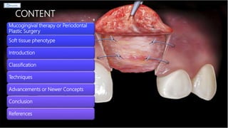
RIDGE AUGMENTATION: II
SOFT TISSUE AUGMENTATION
DR. RINISHA SINHA
MDS III
DEPARTMENT OF PERIODONTOLOGY
CONTENT
Mucogingival therapy or Periodontal
Plastic Surgery
Soft tissue phenotype
Introduction
Classification
Techniques
Advancements or Newer Concepts
Conclusion
References
MUCOGINGIVAL THERAPY
Friedman, 1957
• “Surgical procedures designed to preserve gingiva, remove aberrant frenulum or
muscle attachments, and increase the depth of the vestibule.”
American Academy of Periodontology, 2001
• General term to describe the periodontal treatment involving procedures for the
correction of defects in morphology, position, and/or amount of soft tissue and
underlying bone support at teeth and implants.
Described for all procedures that involved both the gingiva and the alveolar mucosa
PERIODONTAL PLASTIC SURGERY
Miller, 1993
Proceedings of the 1996 World Workshop in Periodontics, 1996
• “Surgical procedures performed to prevent or correct the anatomic, developmental,
traumatic or disease-induced defects of the gingiva, alveolar mucosa or bone.”
Included treatment procedures are:
• Gingival augmentation
• Root coverage
• Correction of mucosal defects at implants
• Crown lengthening
• Gingival preservation at ectopic tooth eruption
• Removal of aberrant frenulum
• Prevention of ridge collapse associated with tooth extraction
• Augmentation of the edentulous ridges
PERIODONTAL PHENOTYPE
= GINGIVAL BIOTYPE
 TISSUE THICKNESS has been reported to be an important factor that influences implant
success. References: Wang et al. 2007; Jung, Hammerle. 2007
CLASSIFICATION AUTHORS
Scalloped and Thin
Flat and Thick
Oschenbien and Ross; 1969
Thin Gingival Tissues as <1.5 mm
Thick Gingival Tissues as ≥ 2.0 mm
Clafey N, Shanley D; 1986
Thin – Scalloped
Thick – Flat
Siebert and Lindhe; 1989
Flat = 2.1 mm
Scalloped = 2.8 mm
Pronounced scalloped gingiva = 4.1mm
Becker at al. 1997
References: Muller, Eger 1997
References: Jepsen et al. 2018
METHODS TO ASSESS THICKNESS OF SOFT
TISSUES
Direct method
Ultrasonic devices
Soft tissue Cone beam computed tomography (CBCT)
Factors affecting soft tissue deficiencies
Soft-tissue
deficiencies prior
to implant
placement
Tooth loss
Periodontal disease
Systemic diseases
Soft-tissue deficiencies prior to implant placement encompass the following
situations:
Improve
esthetics
Enhance
pontic
adaptation
the available amount of soft tissue does not
1) easily allow soft-tissue coverage of bone volume augmentations;
2) allow tension free primary coverage of the site of implant placement; or
3) allow tension free adaptation of the keratinized soft-tissue flap around the neck of
the placed implant
Does the thickness of the soft tissues influences the behavior of the crestal
bone during tissue integration of implants?
References: Linkevicius T, Apse P, Grybauskas S, Puisys A. The influence of
soft tissue thickness on crestal bone changes around implants: a 1-year
prospective controlled clinical trial. Int J Oral Maxillofac Implants.
2009;24:712–719
References: Kaminaka A, Nakano T, Ono S, Kato T, Yatani H. Conebeam
computed tomography evaluation of horizontal and vertical dimensional
changes in buccal peri-implant alveolar bone and soft tissue: a 1-year
prospective clinical study. Clin Implant Dent Relat Res. 2015;17(Suppl.
2):e576–585.
THIN SOFT TISSUES LEAD TO INCREASED MARGINAL BONE LOSS
INTRODUCTION
 Tissue deficiencies at implant sites are common clinical findings
 Soft-tissue defects include volume and quality deficiencies, i.e. lack of keratinized tissue.
References: Acharya A, Hao J, Mattheos N, Chau A, Shirke P, Lang NP. Residual ridge dimensions at edentulous maxillary first molar sites and periodontal bone loss
among two ethnic cohorts seeking tooth replacement. Clin Oral Implants Res. 2014;25:1386–1394.
References: Thoma DS, Buranawat B, Hammerle CH, Held U, Jung RE. Efficacy of soft tissue
augmentation around dental implants and in partially edentulous areas: a systematic review. J Clin
Periodontol. 2014;41(Suppl. 15):S77–91.
PLASTIC
PERIODONTAL
PROCEDURES
To augment keratinized
tissue
Apically positioned flap
Vestibuloplasty
To increase the soft tissue
volume
Free gingival grafts
Sub-epithelial connective tissue grafts
Roll and Pedicle grafts
References:
Cairo
et
al.
2008;
Thoma
et
al.
2009
References: Thoma et al. 2009
References: Seibert 1983a; Studer
et al. 2000
References: Langer B 1980; Abrams L
1980; Rosenberg ES 1982
CORRECTION OF SOFT TISSUE RIDGE DEFECTS
SIEBERT;
1983
Class I:
Loss of bucco-
lingual width but
normal apico-
coronal height
Class II:
Loss of apico-
coronal height
but normal
bucco-lingual
width
Class III:
A combination
of loss of both
height and width
of the ridge
ALLEN’s
1985
Type A:
Apico-coronal
loss of tissue
Type B:
Bucco-lingual
loss of tissue
Type C:
Combination
…
MILD:
Less than 3 mm
reduction
MODERATE:
Between 3 to 6
mm reduction
SEVERE:
More than 6
mm reduction
Factors to be determined prior to initiation of soft
tissue augmentation therapy
Volume of tissue
required to eliminate
the ridge deformity
Type of graft
procedure to be used
Timing of various
treatment
procedures
Design of the
provisional
Provisional problems
tissue discolorations and
matching tissue color
SOFT TISSUE AUGMENTATION
PROCEDURES
Pedicle graft procedures
Roll flap
procedure
Free graft procedures
Pouch graft
procedures
Interpositional graft
procedures
Onlay graft
procedures
References: Studer et al. 1997 References: Studer et al. 2000
PEDICLE GRAFT PROCEDURES
 Studer et al. 1997
 For correction of a single tooth ridge defect
 With minor horizontal and vertical loss
 Esthetic results
 Difficult to perform
 Donor site might not be satisfactory due to anatomic considerations and finite
tissue thickness
ROLL FLAP PROCEDURE
 Abrams 1980
 Preparation of a de-epithelialized connective tissue pedicle graft subsequently placed in a sub-
epithelial pouch
 Indications:
 Treatment of small to moderate class I ridge defects
 primarily in cases with a single-tooth space
 most useful in restoring defects in the apico-coronal dimension but requires sufficient soft
tissue thickness directly over and palatal to the residual ridge crest.
 Enables
 to augment tissue apically and labially to the cervical area of a pontic
 To give recipient site the appearance of a normal tooth-gingiva interface
 Converts a bucco-lingual ridge concavity into a ridge convexity resembling the eminence
produced by the roots of the adjacent teeth
References: Lindhe, J., Lang, N. P., & Karring, T. (2008). Clinical
periodontology and implant dentistry. Oxford: Blackwell
Munksgaard.
References: Abrams L. Augmentation of the deformed residual
edentulous ridge for fixed prosthesis. Compend Contin Educ
Dent 1980;1:205.
Technique References: Lindhe, J., Lang, N. P., & Karring, T. (2008). Clinical
periodontology and implant dentistry. Oxford: Blackwell
Munksgaard.
Adjustment of pontic contours
 Common to all soft tissue ridge augmentation procedures in patients with
fixed bridgework
 A light contact is maintained between the pedicle graft and the tissue surface
of the pontics
 Post-operative swelling conforms to the shape of the pontic
 Enables to shape the soft tissue into a form that is intended for the
augmented site
References: Lindhe, J., Lang, N. P., & Karring, T. (2008). Clinical
periodontology and implant dentistry. Oxford: Blackwell
Munksgaard.
Post-operative care
Periodontal dressing is placed over the donor site; to be changed at weekly intervals and maintained until
wound healing has progressed to a point where the tissue is no longer tender to touch.
No dressing to be placed over the facial (labial) surface of the grafted area where the swelling will occur
References: Lindhe, J., Lang, N. P., & Karring, T. (2008). Clinical
periodontology and implant dentistry. Oxford: Blackwell
Munksgaard.
FREE GRAFT PROCEDURES
Studer et al. 2000
Submerged free connective tissue graft procedure for larger defects
Onlay full-thickness graft procedure for ridge augmentation in presence of
additional mucogingival problems such as insufficient gingival width, high
frenum, gingival scarring or tattoo
References: Lindhe, J., Lang, N. P., & Karring, T. (2008). Clinical
periodontology and implant dentistry. Oxford: Blackwell
Munksgaard.
SUBEPITHELIAL CONNECTIVE TISSUE GRAFT
 Garber & Rosenberg 1981
 Preferred over free gingival grafts, especially in areas with high esthetic impact
 Advantages:
 More gain in tissue volume
 Better color and texture match
 Disadvantages:
 Significant shrinkage of grafts following the augmentation procedure
References: Edel et al. 1974; Nemcovsky et al. 2000
References: Mormann et al. 1981; Studer et al. 2000; Thoma et al.
2010.
 Kaldahl et al. 1982; Seibert 1983; Allen et al. 1985; Miller 1986; Cohen 1994
 Subepithelial pouch is prepared in the area of the ridge deformity, into which a free graft of
connective tissue is placed and molded to create the desired contour of the ridge
POUCH GRAFT PROCEDURES
 Indications:
 To correct class I defects
 If large-volume defects may have thin palatal tissues
 insufficient to provide the volume of the donor tissue necessary to fill the deformity
 hard tissue augmentation procedures to be selected.
Entrance incision and the plane of dissection may
be made in different ways:
1. CORONAL-APICALLY
the horizontal incision is made on the palatal or
lingual side of the defect and the plane of
dissection carried in an apical direction
References: Lindhe, J., Lang, N. P., & Karring, T. (2008). Clinical
periodontology and implant dentistry. Oxford: Blackwell
Munksgaard.
2. APICAL-CORONALLY
the horizontal incision is made high in the
vestibule near the mucobuccal fold and the
plane of dissection is carried coronally to
the crest of the ridge
3. LATERALLY
one or tow vertical entrance incisions are
started from either side of the defect. The
plane of dissection is made laterally across
the span of the deformity. (80)
References: Lindhe, J., Lang, N. P., & Karring, T. (2008). Clinical
periodontology and implant dentistry. Oxford: Blackwell
Munksgaard.
HEALING OF FREE SOFT TISSUE GRAFT
References: Oliver, Loe, Karring et al. 1968; Janson et al. 1969
 Seibert 1991, 1993 a,b
 Not completely submerged and covered in the manner that a subepithelial connective tissue
graft is placed
 No need to remove the epithelium from the surface of the donor tissue
 Indications:
 When augmentation is required in both bucco-lingual and apico-coronal direction, a
position of the graft must be positioned above the surface of the tissue surrounding the
recipient site
 To correct class I as well as small to moderate class II defects
 Thus, a certain amount of the grafted connective tissue will be exposed in the oral cavity
 Large amount of donor tissue
INTERPOSITIONAL GRAFT PROCEDURES
a. Pre-treatment view: Class III ridge defect –
A two-stage procedure used
b. A pouch was prepared to receive an
interpositional graft; epithelium removed
from the borders of the recipient site to
permit some graft to be placed above the
level of surrounding tissue (gaining apico-
coronal augmentation)
c. Wedge-shaped graft – 10 mm thick at the
center
d. Interpositional graft is both displacing the
labial surface of the pouch in the labial
directions as well as adding height to the
ridge
e. 2 months post-op – Additional
augmentation needed
f. A second-stage onlay graft used to create
a papilla and fill the dark triangle between
the pontics
 Meltzer 1979; Seibert 1983
 Epithelized free grafts
 Indications:
 To augment ridge defects in apico-coronal plane i.e. to gain ridge height
 In treatment of large class II and III defects
 Not suitable in areas where the blood supply at the recipient site has been compromised
by scar tissue formation from previous wound healing
 Following placement, receive their nutrition from the de-epithelialized connective tissue of the
recipient site
 Amount of apico-coronal augmentation is related to the initial thickness of the graft, the
events of the wound healing processes, and the amount of grafts tissue that survives
 If necessary, can be repeated at 2-month intervals to gradually increase the ridge height
ONLAY GRAFT PROCEDURES
Selection of donor site
Require large amount of donor tissue
Palatal vault region of premolars and first molars, midway between the gingival
margin and the midline raphae is the only area in the maxilla that contains
necessary volume of tissue required to augment large ridge defects
Tissue to be probed with a 30-gauge syringe needle to ensure that an
acceptable volume of tissue can be obtained at the time of surgery
Major palatine artery emerges from the posterior palatine foramen located
adjacent to the distal surface of the maxillary second molar, midway between
the gingival margin and the midline raphae
Artery passes in an anterior direction close to the surface of the palatal bone
Important therefore that the second and third molar regions are not used as
donor site for large volume grafts
References: Lindhe, J., Lang, N. P., & Karring, T.
(2008). Clinical periodontology and implant
dentistry. Oxford: Blackwell Munksgaard.
Dissection of donor tissue
Base of the graft should be V- or U- shaped to match the shape of the defect in the ridge
Therefore the different planes of incision in the palate must converge towards an area under the
center or toward one edge of the donor site
Comparatively easy with the use of a scalpel to dissect in an antero-posterior or from an area
high in the palate in a lateral direction towards the teeth
Difficult to dissect in an anterior direction from the distal edge of the donor site
Variety of blade holders available which permit the scalpel blade to be positioned at different
angles to the holder and which enables the surgeon to cut with a back-action
After removal of the donor tissue, it must be stored in pieces of surgical gauze moistened in
isotonic saline at all times
Treatment of the donor site
Difficult to anchor and maintain a periodontal dressing at the donor site in the palatal vault,
an acrylic stent should be fabricated prior to surgery
Stent to be made with wrought wire clasps on each side to add retention and to aid the
patient in removing and inserting the device
Donor site must be inspected carefully for signs of arterial bleeding
If any small vessel bleeding is observed, a circumferential suture must be placed around the
vessel distal to the bleeding point
Immediately thereafter the void at the donor site should be packed with a suitable
hemostatic agent and the edges of the wound be brought closer together with sutures
Then put stent into position
Try-in and stabilization of graft
Graft is transferred with tissue forceps to the recipient site for a try-in.
Graft is trimmed to the proper shape and adjusted to fit the connective tissue
surface of the prepared ridge
A series of parallel cuts may be made deep into the exposed lamina propria
of the recipient site to sever large blood vessels immediately before suturing
Series of interrupted sutures is placed along the borders of the graft
Assistant stabilizes the onlay graft against the surface of the recipient site,
while placement of sutures is done
References: Lindhe, J., Lang, N. P., & Karring, T. (2008). Clinical
periodontology and implant dentistry. Oxford: Blackwell
Munksgaard.
Wound healing in the recipient site
Considerable post-operative swelling often occurs during the first week after pouch and onlay
augmentation procedures
The epithelium of the graft will slough to form a white film on the surface of the graft
Patients should rinse two-four times per day with an antimicrobial mouthwash during the first
week after surgery
Refrain from mechanical cleaning measures in the area until a new epithelial covering has formed
over the graft, which will not occur until a functional capillary circulation has been re-established
in the graft (4-7 days after the surgery)
Grafted tissue will assume a normal color as the epithelium thickens via stratification
Stable tissue form after 3 months but further shrinkage may occur over a period of several
months
Final restorative measures should not be initiated until after 6 months
Wound healing in the donor site
Granulation tissue will gradually fill the donor site
Initial healing – complete within 3-4 weeks after the removal of a 4-5 mm
thick graft
Patient to wear surgical stent for about 2 weeks to protect the healing
wound
Palate returns to its pre-surgical contour after about 3 months
g. 2 months after the first surgical
procedure, ridge was de-epitheliliazed
and cuts were made into the connective
tissue prior to placing the second-stage
onlay graft into position
h. The onlay graft was sutured into
position
i. The pontics were adjjusted and brought
into light contact with the graft
j. Marked swelling occurred within the
graft at 14 days post-surgery
k. 2 months following the surgical
procedure, gingivoplasty was done
l. Post-treatment view 1 year after the final
surgical procedure
 Seibert & Louis 1996
 Indications:
 Class III : major challenge;
 to be augmented in both vertical and horizontal dimensions
COMBINED ONLAY-INTERPOSITIONAL GRAFT
PROCEDURES
Advantages
Submerged connective
tissue section of the
interpositional graft aids in
the revascularisation of the
onlay section of the graft,
thereby gaining a greater
percentage of take of the
overall graft
A smaller post-operative
open wound in the palate
donor site
Faster healing in the palate
donor site with less patient
discomfort
Greater latitude or ability to
control the degree of
bucco-lingual and apico-
coronal augmentation
within a single procedure
Vestibular depth is not
decreased and the muco
gingival junction is not
moved coronally, thereby
eliminating the need for
follow-up corrective
procedures
a. & b. The right maxillary lateral and central
incisors were lost due to trauma. These views
show the horizontal and vertical loss of ridge
tissue 10 months after the extractions.
c. A partial-thickness path of incision was extended labially and apically
to create a pouch. The amount of space created within the pouch and
the degree of relaxation of the flap was tested with a periosteal elevator.
d. The epithelialized section of the graft.
e. The premolar area, maxillary right side, was used as a donor area.
The area of exposed connective tissue corresponds to the onlay section
of the graft. The incisions were extended another 5–7 mm towards the
midline on a long bevel to obtain the interpositional segment of the
graft.
f. The graft was tucked into the labial pouch and sutured first along its
palatal border. The labial flap was then sutured along the epithelial
connective border of the graft. The residual labial socket defect in the
flap created a soft tissue discontinuity defect along the labial margin of
the flap.
g. At 6 weeks post surgery, further augmentation would be required to
gain additional soft tissue in both the vertical and horizontal planes.
Second-stage procedure was done at this time.
h. An incision 1.5 mm in depth utilized to de-epithelialize the crestal
surface of the ridge. papillae were not included within the surgical field.
The mesial and distal borders of the onlay section of the recipient site
were then extended apically to create vertical releasing incisions. The
overall recipient site was to be trapezoidal in shape. A labial flap to
create the pouch section of the recipient site was made using partial-
thickness dissection.
i. The left maxillary premolar area was used as the donor site for the
second-stage surgery.
j. This side view clearly shows the epithelialized onlay section of the
graft and the de-epithelialized connective tissue section of the graft, as
well as tissue thickness.
k. The graft was sutured first along the fixed
palatal border to gain initial stabilization. Then
the connective tissue interpositional section was
sutured along the lateral borders. The flap was
then sutured over the interpositional section of
the graft at the epithelialized edge of the onlay
section of the graft and along the vertical
incisions.
l. At 6 weeks post surgery, the provisional
prosthesis was modified to bring the tissue
surface of the pontics into contact with the
healing ridge.
m. At 2 months post surgery, tooth form was
further modified on the provisional prosthesis
and gingivoplasty was done to sculpt the tissues
to final form and smooth out surface
irregularities.
n. The final ceramo-metal prosthesis was
inserted 4 months later. The life-like
reconstruction of the soft tissues and dentition
restored dentofacial esthetics for the patient.
ADVANCEMENTS / NEWER
CONCEPTS
ACELLULAR DERMAL MATRIX
 An acellular dermal matrix designed to serve as a biologic scaffold for normal tissue
remodeling. Derived from donated human skin.
 ADM contains both the structure and the biochemical information to direct normal
revascularization and cell repopulation because blood vessels, collagens, proteoglycans,
and elastin are preserved.
 This extracellular matrix contains the blood vessel channels that serve as conduits for
revascularization
 Collagens, proteoglycans, and elastin provide structure and information for cell
repopulation.
 Most importantly, preserved proteoglycans and proteins direct the patient’s own cells to
initiate revascularization and cell repopulation.
 Grafted ADMs maintain their ultrastructural acellular matrix integrity and do not provoke a
rejection or inflammatory response in host tissues.
 Does not have the limitations in availability that palatal donor tissue does.
References: Wainwright et al. 1996; Eppley 2000
INDICATIONS
Skin burn management
For correction of gingival recession
Bone regeneration
Soft tissue ridge augmentation
Minimise soft tissue fenestration prior to block
grafting
References: Wainwright et al. 1996
References: Henderson et al. 2001
References: Fowler et al. 2000
References: Harris et al. 2003; Batista and Batista
2001
References: AlGhamdi and Buhite, 2008
Implant site preparation with osetotome Folding the ADM and
stabilization with sutures
Flap closure
10 days follow-up 8 weeks healing
VASCULARIZED INTERPOSITIONAL PERIOSTEAL
CONNECTIVE TISSUE FLAP
 A novel pedicle autograft
 ADVANTAGES:
 Excellent blood supply,
 Less morbidity,
 Primary closure of donor and recipient bed
 Does not alter the color of the area
 More agreeable to the patient because it involves a single surgical site
References: Agarwal, et al.: Ridge augmentation using VIP-CT flap
References: Sclar A. Vascularized interpositional periosteal-connective tissue (VIP-CT) flap. In: Sclar A, editor. Soft Tissue and Esthetic Considerations in Implant Dentistry. Chicago:
Quintessence Publishing; 2003. p. 163-87.
Kim CS, Jang YJ, Choi SH, Cho KS. Long-term results from soft and hard tissue augmentation by a modified vascularized interpositional periosteal-connective tissue technique in
the maxillary anterior region. J Oral Maxillofac Surg 2012;70:484-91.
TRANSMUCOSAL PERIOSTEAL RELEASING
INCISION: THE “BUTTONHOLE TECHNIQUE”
References: Aranda J et al. Clinical Advances in Periodontics, Vol. 5, No. 2, May 2015
THE DOME TECHNIQUE
References: Irinakis T, Aldahlawi S. The dome technique: a new surgical technique to enhance soft-tissue margins and emergence profiles around implants placed in the esthetic zone.
Clin Cosmet Investig Dent. 2018;10:1-7
SELF-INFLATING
SOFT
TISSUE
EXPANDERS
References:
Dhadse
P
et
al.
J
Indian
Soc
Periodontol,
2014
Jul-Aug;
18(4):
433-
440.
CONCLUSION
The reconstruction with
autografts can be broadly
divided into 3 major groups:
• free gingival grafts,
• free buccal mucosa grafts,
• and buccal fat pad grafts.
Require large amounts of
tissue which cannot always
be easily harvested in the
oral cavity; therefore, new
materials are being
developed, that is, from
allogeneic, xenogeneic, and
synthetic origin or alloplastic
materials.
Self-inflating soft tissue
hydrogel expanders
successfully used to acquire
surplus amounts of soft
tissue to cover bone grafts.
Advantages - their high
biocompatibility, low
complication rates, non-
genotoxic and non-immune
reactivity, material pureness,
and safety which
subsequently ensures a low
risk of infection.
Drawbacks, such as patient
discomfort during expansion
and waiting time of 6 to 10
weeks; a second surgery
required to remove the
expander and
simultaneously perform a
bone augmentation, only a
limited amount of tissue can
be expanded in comparison
to alloplastic materials
Collagen matrix, resembles
the extracellular matrix,
widely used for soft tissue
augmentation.
Disadvantages such as low
mechanical stiffness and
rapid biodegradation, lack
important ECM proteins.
Therefore, acellularized
xenogenic tissues - a
promising alternative
material for soft tissue
regeneration because they
maintain their 3D structure
and subsequently offer good
mechanical properties.
Recently, tissue-engineered
three-dimensional (3D) oral
mucosal grafts have been
tested and could offer a
promising alternative to
conventional grafts.
Newman, Takei, Klokkevold, Carranza:
Carrazanza’s Clinical Periodontology,
Saunders, 10th edition.
Jan Wolff, DDS, Dr Med Dent, Elisabet
Farré-Guasch; Soft Tissue Augmentation
Techniques and Materials Used in the Oral
Cavity: An Overview; Implant Dentistry;
Volume 25; Number 3; 2016
Lindhe, Lang, Karring: Clinical
Periodontology and Implant Dentistry.
Blackwell Munksgaard, 5th edition.
Urban IA, Montero E, Monje A, Sanz-
Sánchez I. Effectiveness of vertical ridge
augmentation interventions: A systematic
review and meta-analysis. J Clin
Periodontol. 2019 Jun;46 Suppl 21:319-339.
Goyal M, Mittal N, Gupta GK, Singhal M.
Ridge augmentation in implant dentistry. J
Int Clin Dent Res Organ 2015;7:94-112.
Linkevicius T, Apse P
, Grybauskas S, Puisys
A. The influence of soft tissue thickness on
crestal bone changes around implants: a 1-
year prospective controlled clinical trial. Int
J Oral Maxillofac Implants. 2009;24:712–719
Dhadse P et al. J Indian Soc Periodontol,
2014 Jul-Aug; 18(4): 433-440.
Thoma DS, Buranawat B, Hammerle CH,
Held U, Jung RE. Efficacy of soft tissue
augmentation around dental implants and
in partially edentulous areas: a systematic
review. Periodontol 2000. 2014;41(Suppl.
15):S77–91.
Ridge Augmentation II.pptx

Ridge Augmentation II.pptx

  • 1.
    RIDGE AUGMENTATION: II SOFTTISSUE AUGMENTATION DR. RINISHA SINHA MDS III DEPARTMENT OF PERIODONTOLOGY
  • 2.
    CONTENT Mucogingival therapy orPeriodontal Plastic Surgery Soft tissue phenotype Introduction Classification Techniques Advancements or Newer Concepts Conclusion References
  • 3.
    MUCOGINGIVAL THERAPY Friedman, 1957 •“Surgical procedures designed to preserve gingiva, remove aberrant frenulum or muscle attachments, and increase the depth of the vestibule.” American Academy of Periodontology, 2001 • General term to describe the periodontal treatment involving procedures for the correction of defects in morphology, position, and/or amount of soft tissue and underlying bone support at teeth and implants. Described for all procedures that involved both the gingiva and the alveolar mucosa
  • 4.
    PERIODONTAL PLASTIC SURGERY Miller,1993 Proceedings of the 1996 World Workshop in Periodontics, 1996 • “Surgical procedures performed to prevent or correct the anatomic, developmental, traumatic or disease-induced defects of the gingiva, alveolar mucosa or bone.” Included treatment procedures are: • Gingival augmentation • Root coverage • Correction of mucosal defects at implants • Crown lengthening • Gingival preservation at ectopic tooth eruption • Removal of aberrant frenulum • Prevention of ridge collapse associated with tooth extraction • Augmentation of the edentulous ridges
  • 5.
    PERIODONTAL PHENOTYPE = GINGIVALBIOTYPE  TISSUE THICKNESS has been reported to be an important factor that influences implant success. References: Wang et al. 2007; Jung, Hammerle. 2007 CLASSIFICATION AUTHORS Scalloped and Thin Flat and Thick Oschenbien and Ross; 1969 Thin Gingival Tissues as <1.5 mm Thick Gingival Tissues as ≥ 2.0 mm Clafey N, Shanley D; 1986 Thin – Scalloped Thick – Flat Siebert and Lindhe; 1989 Flat = 2.1 mm Scalloped = 2.8 mm Pronounced scalloped gingiva = 4.1mm Becker at al. 1997 References: Muller, Eger 1997 References: Jepsen et al. 2018
  • 6.
    METHODS TO ASSESSTHICKNESS OF SOFT TISSUES Direct method Ultrasonic devices Soft tissue Cone beam computed tomography (CBCT)
  • 7.
    Factors affecting softtissue deficiencies Soft-tissue deficiencies prior to implant placement Tooth loss Periodontal disease Systemic diseases
  • 8.
    Soft-tissue deficiencies priorto implant placement encompass the following situations: Improve esthetics Enhance pontic adaptation the available amount of soft tissue does not 1) easily allow soft-tissue coverage of bone volume augmentations; 2) allow tension free primary coverage of the site of implant placement; or 3) allow tension free adaptation of the keratinized soft-tissue flap around the neck of the placed implant
  • 9.
    Does the thicknessof the soft tissues influences the behavior of the crestal bone during tissue integration of implants? References: Linkevicius T, Apse P, Grybauskas S, Puisys A. The influence of soft tissue thickness on crestal bone changes around implants: a 1-year prospective controlled clinical trial. Int J Oral Maxillofac Implants. 2009;24:712–719 References: Kaminaka A, Nakano T, Ono S, Kato T, Yatani H. Conebeam computed tomography evaluation of horizontal and vertical dimensional changes in buccal peri-implant alveolar bone and soft tissue: a 1-year prospective clinical study. Clin Implant Dent Relat Res. 2015;17(Suppl. 2):e576–585. THIN SOFT TISSUES LEAD TO INCREASED MARGINAL BONE LOSS
  • 10.
    INTRODUCTION  Tissue deficienciesat implant sites are common clinical findings  Soft-tissue defects include volume and quality deficiencies, i.e. lack of keratinized tissue. References: Acharya A, Hao J, Mattheos N, Chau A, Shirke P, Lang NP. Residual ridge dimensions at edentulous maxillary first molar sites and periodontal bone loss among two ethnic cohorts seeking tooth replacement. Clin Oral Implants Res. 2014;25:1386–1394. References: Thoma DS, Buranawat B, Hammerle CH, Held U, Jung RE. Efficacy of soft tissue augmentation around dental implants and in partially edentulous areas: a systematic review. J Clin Periodontol. 2014;41(Suppl. 15):S77–91. PLASTIC PERIODONTAL PROCEDURES To augment keratinized tissue Apically positioned flap Vestibuloplasty To increase the soft tissue volume Free gingival grafts Sub-epithelial connective tissue grafts Roll and Pedicle grafts References: Cairo et al. 2008; Thoma et al. 2009 References: Thoma et al. 2009 References: Seibert 1983a; Studer et al. 2000 References: Langer B 1980; Abrams L 1980; Rosenberg ES 1982
  • 11.
    CORRECTION OF SOFTTISSUE RIDGE DEFECTS SIEBERT; 1983 Class I: Loss of bucco- lingual width but normal apico- coronal height Class II: Loss of apico- coronal height but normal bucco-lingual width Class III: A combination of loss of both height and width of the ridge ALLEN’s 1985 Type A: Apico-coronal loss of tissue Type B: Bucco-lingual loss of tissue Type C: Combination … MILD: Less than 3 mm reduction MODERATE: Between 3 to 6 mm reduction SEVERE: More than 6 mm reduction
  • 12.
    Factors to bedetermined prior to initiation of soft tissue augmentation therapy Volume of tissue required to eliminate the ridge deformity Type of graft procedure to be used Timing of various treatment procedures Design of the provisional Provisional problems tissue discolorations and matching tissue color
  • 13.
    SOFT TISSUE AUGMENTATION PROCEDURES Pediclegraft procedures Roll flap procedure Free graft procedures Pouch graft procedures Interpositional graft procedures Onlay graft procedures References: Studer et al. 1997 References: Studer et al. 2000
  • 14.
    PEDICLE GRAFT PROCEDURES Studer et al. 1997  For correction of a single tooth ridge defect  With minor horizontal and vertical loss  Esthetic results  Difficult to perform  Donor site might not be satisfactory due to anatomic considerations and finite tissue thickness
  • 15.
    ROLL FLAP PROCEDURE Abrams 1980  Preparation of a de-epithelialized connective tissue pedicle graft subsequently placed in a sub- epithelial pouch  Indications:  Treatment of small to moderate class I ridge defects  primarily in cases with a single-tooth space  most useful in restoring defects in the apico-coronal dimension but requires sufficient soft tissue thickness directly over and palatal to the residual ridge crest.  Enables  to augment tissue apically and labially to the cervical area of a pontic  To give recipient site the appearance of a normal tooth-gingiva interface  Converts a bucco-lingual ridge concavity into a ridge convexity resembling the eminence produced by the roots of the adjacent teeth References: Lindhe, J., Lang, N. P., & Karring, T. (2008). Clinical periodontology and implant dentistry. Oxford: Blackwell Munksgaard. References: Abrams L. Augmentation of the deformed residual edentulous ridge for fixed prosthesis. Compend Contin Educ Dent 1980;1:205.
  • 16.
    Technique References: Lindhe,J., Lang, N. P., & Karring, T. (2008). Clinical periodontology and implant dentistry. Oxford: Blackwell Munksgaard.
  • 18.
    Adjustment of ponticcontours  Common to all soft tissue ridge augmentation procedures in patients with fixed bridgework  A light contact is maintained between the pedicle graft and the tissue surface of the pontics  Post-operative swelling conforms to the shape of the pontic  Enables to shape the soft tissue into a form that is intended for the augmented site References: Lindhe, J., Lang, N. P., & Karring, T. (2008). Clinical periodontology and implant dentistry. Oxford: Blackwell Munksgaard.
  • 19.
    Post-operative care Periodontal dressingis placed over the donor site; to be changed at weekly intervals and maintained until wound healing has progressed to a point where the tissue is no longer tender to touch. No dressing to be placed over the facial (labial) surface of the grafted area where the swelling will occur References: Lindhe, J., Lang, N. P., & Karring, T. (2008). Clinical periodontology and implant dentistry. Oxford: Blackwell Munksgaard.
  • 20.
    FREE GRAFT PROCEDURES Studeret al. 2000 Submerged free connective tissue graft procedure for larger defects Onlay full-thickness graft procedure for ridge augmentation in presence of additional mucogingival problems such as insufficient gingival width, high frenum, gingival scarring or tattoo References: Lindhe, J., Lang, N. P., & Karring, T. (2008). Clinical periodontology and implant dentistry. Oxford: Blackwell Munksgaard.
  • 21.
    SUBEPITHELIAL CONNECTIVE TISSUEGRAFT  Garber & Rosenberg 1981  Preferred over free gingival grafts, especially in areas with high esthetic impact  Advantages:  More gain in tissue volume  Better color and texture match  Disadvantages:  Significant shrinkage of grafts following the augmentation procedure References: Edel et al. 1974; Nemcovsky et al. 2000 References: Mormann et al. 1981; Studer et al. 2000; Thoma et al. 2010.
  • 22.
     Kaldahl etal. 1982; Seibert 1983; Allen et al. 1985; Miller 1986; Cohen 1994  Subepithelial pouch is prepared in the area of the ridge deformity, into which a free graft of connective tissue is placed and molded to create the desired contour of the ridge POUCH GRAFT PROCEDURES  Indications:  To correct class I defects  If large-volume defects may have thin palatal tissues  insufficient to provide the volume of the donor tissue necessary to fill the deformity  hard tissue augmentation procedures to be selected.
  • 23.
    Entrance incision andthe plane of dissection may be made in different ways: 1. CORONAL-APICALLY the horizontal incision is made on the palatal or lingual side of the defect and the plane of dissection carried in an apical direction References: Lindhe, J., Lang, N. P., & Karring, T. (2008). Clinical periodontology and implant dentistry. Oxford: Blackwell Munksgaard.
  • 24.
    2. APICAL-CORONALLY the horizontalincision is made high in the vestibule near the mucobuccal fold and the plane of dissection is carried coronally to the crest of the ridge 3. LATERALLY one or tow vertical entrance incisions are started from either side of the defect. The plane of dissection is made laterally across the span of the deformity. (80) References: Lindhe, J., Lang, N. P., & Karring, T. (2008). Clinical periodontology and implant dentistry. Oxford: Blackwell Munksgaard.
  • 25.
    HEALING OF FREESOFT TISSUE GRAFT References: Oliver, Loe, Karring et al. 1968; Janson et al. 1969
  • 26.
     Seibert 1991,1993 a,b  Not completely submerged and covered in the manner that a subepithelial connective tissue graft is placed  No need to remove the epithelium from the surface of the donor tissue  Indications:  When augmentation is required in both bucco-lingual and apico-coronal direction, a position of the graft must be positioned above the surface of the tissue surrounding the recipient site  To correct class I as well as small to moderate class II defects  Thus, a certain amount of the grafted connective tissue will be exposed in the oral cavity  Large amount of donor tissue INTERPOSITIONAL GRAFT PROCEDURES
  • 28.
    a. Pre-treatment view:Class III ridge defect – A two-stage procedure used b. A pouch was prepared to receive an interpositional graft; epithelium removed from the borders of the recipient site to permit some graft to be placed above the level of surrounding tissue (gaining apico- coronal augmentation) c. Wedge-shaped graft – 10 mm thick at the center d. Interpositional graft is both displacing the labial surface of the pouch in the labial directions as well as adding height to the ridge e. 2 months post-op – Additional augmentation needed f. A second-stage onlay graft used to create a papilla and fill the dark triangle between the pontics
  • 29.
     Meltzer 1979;Seibert 1983  Epithelized free grafts  Indications:  To augment ridge defects in apico-coronal plane i.e. to gain ridge height  In treatment of large class II and III defects  Not suitable in areas where the blood supply at the recipient site has been compromised by scar tissue formation from previous wound healing  Following placement, receive their nutrition from the de-epithelialized connective tissue of the recipient site  Amount of apico-coronal augmentation is related to the initial thickness of the graft, the events of the wound healing processes, and the amount of grafts tissue that survives  If necessary, can be repeated at 2-month intervals to gradually increase the ridge height ONLAY GRAFT PROCEDURES
  • 30.
    Selection of donorsite Require large amount of donor tissue Palatal vault region of premolars and first molars, midway between the gingival margin and the midline raphae is the only area in the maxilla that contains necessary volume of tissue required to augment large ridge defects Tissue to be probed with a 30-gauge syringe needle to ensure that an acceptable volume of tissue can be obtained at the time of surgery Major palatine artery emerges from the posterior palatine foramen located adjacent to the distal surface of the maxillary second molar, midway between the gingival margin and the midline raphae Artery passes in an anterior direction close to the surface of the palatal bone Important therefore that the second and third molar regions are not used as donor site for large volume grafts References: Lindhe, J., Lang, N. P., & Karring, T. (2008). Clinical periodontology and implant dentistry. Oxford: Blackwell Munksgaard.
  • 31.
    Dissection of donortissue Base of the graft should be V- or U- shaped to match the shape of the defect in the ridge Therefore the different planes of incision in the palate must converge towards an area under the center or toward one edge of the donor site Comparatively easy with the use of a scalpel to dissect in an antero-posterior or from an area high in the palate in a lateral direction towards the teeth Difficult to dissect in an anterior direction from the distal edge of the donor site Variety of blade holders available which permit the scalpel blade to be positioned at different angles to the holder and which enables the surgeon to cut with a back-action After removal of the donor tissue, it must be stored in pieces of surgical gauze moistened in isotonic saline at all times
  • 32.
    Treatment of thedonor site Difficult to anchor and maintain a periodontal dressing at the donor site in the palatal vault, an acrylic stent should be fabricated prior to surgery Stent to be made with wrought wire clasps on each side to add retention and to aid the patient in removing and inserting the device Donor site must be inspected carefully for signs of arterial bleeding If any small vessel bleeding is observed, a circumferential suture must be placed around the vessel distal to the bleeding point Immediately thereafter the void at the donor site should be packed with a suitable hemostatic agent and the edges of the wound be brought closer together with sutures Then put stent into position
  • 33.
    Try-in and stabilizationof graft Graft is transferred with tissue forceps to the recipient site for a try-in. Graft is trimmed to the proper shape and adjusted to fit the connective tissue surface of the prepared ridge A series of parallel cuts may be made deep into the exposed lamina propria of the recipient site to sever large blood vessels immediately before suturing Series of interrupted sutures is placed along the borders of the graft Assistant stabilizes the onlay graft against the surface of the recipient site, while placement of sutures is done References: Lindhe, J., Lang, N. P., & Karring, T. (2008). Clinical periodontology and implant dentistry. Oxford: Blackwell Munksgaard.
  • 34.
    Wound healing inthe recipient site Considerable post-operative swelling often occurs during the first week after pouch and onlay augmentation procedures The epithelium of the graft will slough to form a white film on the surface of the graft Patients should rinse two-four times per day with an antimicrobial mouthwash during the first week after surgery Refrain from mechanical cleaning measures in the area until a new epithelial covering has formed over the graft, which will not occur until a functional capillary circulation has been re-established in the graft (4-7 days after the surgery) Grafted tissue will assume a normal color as the epithelium thickens via stratification Stable tissue form after 3 months but further shrinkage may occur over a period of several months Final restorative measures should not be initiated until after 6 months
  • 35.
    Wound healing inthe donor site Granulation tissue will gradually fill the donor site Initial healing – complete within 3-4 weeks after the removal of a 4-5 mm thick graft Patient to wear surgical stent for about 2 weeks to protect the healing wound Palate returns to its pre-surgical contour after about 3 months
  • 36.
    g. 2 monthsafter the first surgical procedure, ridge was de-epitheliliazed and cuts were made into the connective tissue prior to placing the second-stage onlay graft into position h. The onlay graft was sutured into position i. The pontics were adjjusted and brought into light contact with the graft j. Marked swelling occurred within the graft at 14 days post-surgery k. 2 months following the surgical procedure, gingivoplasty was done l. Post-treatment view 1 year after the final surgical procedure
  • 37.
     Seibert &Louis 1996  Indications:  Class III : major challenge;  to be augmented in both vertical and horizontal dimensions COMBINED ONLAY-INTERPOSITIONAL GRAFT PROCEDURES
  • 38.
    Advantages Submerged connective tissue sectionof the interpositional graft aids in the revascularisation of the onlay section of the graft, thereby gaining a greater percentage of take of the overall graft A smaller post-operative open wound in the palate donor site Faster healing in the palate donor site with less patient discomfort Greater latitude or ability to control the degree of bucco-lingual and apico- coronal augmentation within a single procedure Vestibular depth is not decreased and the muco gingival junction is not moved coronally, thereby eliminating the need for follow-up corrective procedures
  • 40.
    a. & b.The right maxillary lateral and central incisors were lost due to trauma. These views show the horizontal and vertical loss of ridge tissue 10 months after the extractions. c. A partial-thickness path of incision was extended labially and apically to create a pouch. The amount of space created within the pouch and the degree of relaxation of the flap was tested with a periosteal elevator. d. The epithelialized section of the graft. e. The premolar area, maxillary right side, was used as a donor area. The area of exposed connective tissue corresponds to the onlay section of the graft. The incisions were extended another 5–7 mm towards the midline on a long bevel to obtain the interpositional segment of the graft. f. The graft was tucked into the labial pouch and sutured first along its palatal border. The labial flap was then sutured along the epithelial connective border of the graft. The residual labial socket defect in the flap created a soft tissue discontinuity defect along the labial margin of the flap. g. At 6 weeks post surgery, further augmentation would be required to gain additional soft tissue in both the vertical and horizontal planes. Second-stage procedure was done at this time. h. An incision 1.5 mm in depth utilized to de-epithelialize the crestal surface of the ridge. papillae were not included within the surgical field. The mesial and distal borders of the onlay section of the recipient site were then extended apically to create vertical releasing incisions. The overall recipient site was to be trapezoidal in shape. A labial flap to create the pouch section of the recipient site was made using partial- thickness dissection. i. The left maxillary premolar area was used as the donor site for the second-stage surgery. j. This side view clearly shows the epithelialized onlay section of the graft and the de-epithelialized connective tissue section of the graft, as well as tissue thickness.
  • 41.
    k. The graftwas sutured first along the fixed palatal border to gain initial stabilization. Then the connective tissue interpositional section was sutured along the lateral borders. The flap was then sutured over the interpositional section of the graft at the epithelialized edge of the onlay section of the graft and along the vertical incisions. l. At 6 weeks post surgery, the provisional prosthesis was modified to bring the tissue surface of the pontics into contact with the healing ridge. m. At 2 months post surgery, tooth form was further modified on the provisional prosthesis and gingivoplasty was done to sculpt the tissues to final form and smooth out surface irregularities. n. The final ceramo-metal prosthesis was inserted 4 months later. The life-like reconstruction of the soft tissues and dentition restored dentofacial esthetics for the patient.
  • 42.
  • 43.
    ACELLULAR DERMAL MATRIX An acellular dermal matrix designed to serve as a biologic scaffold for normal tissue remodeling. Derived from donated human skin.  ADM contains both the structure and the biochemical information to direct normal revascularization and cell repopulation because blood vessels, collagens, proteoglycans, and elastin are preserved.  This extracellular matrix contains the blood vessel channels that serve as conduits for revascularization  Collagens, proteoglycans, and elastin provide structure and information for cell repopulation.  Most importantly, preserved proteoglycans and proteins direct the patient’s own cells to initiate revascularization and cell repopulation.  Grafted ADMs maintain their ultrastructural acellular matrix integrity and do not provoke a rejection or inflammatory response in host tissues.  Does not have the limitations in availability that palatal donor tissue does. References: Wainwright et al. 1996; Eppley 2000
  • 44.
    INDICATIONS Skin burn management Forcorrection of gingival recession Bone regeneration Soft tissue ridge augmentation Minimise soft tissue fenestration prior to block grafting References: Wainwright et al. 1996 References: Henderson et al. 2001 References: Fowler et al. 2000 References: Harris et al. 2003; Batista and Batista 2001 References: AlGhamdi and Buhite, 2008
  • 45.
    Implant site preparationwith osetotome Folding the ADM and stabilization with sutures Flap closure 10 days follow-up 8 weeks healing
  • 46.
    VASCULARIZED INTERPOSITIONAL PERIOSTEAL CONNECTIVETISSUE FLAP  A novel pedicle autograft  ADVANTAGES:  Excellent blood supply,  Less morbidity,  Primary closure of donor and recipient bed  Does not alter the color of the area  More agreeable to the patient because it involves a single surgical site References: Agarwal, et al.: Ridge augmentation using VIP-CT flap References: Sclar A. Vascularized interpositional periosteal-connective tissue (VIP-CT) flap. In: Sclar A, editor. Soft Tissue and Esthetic Considerations in Implant Dentistry. Chicago: Quintessence Publishing; 2003. p. 163-87. Kim CS, Jang YJ, Choi SH, Cho KS. Long-term results from soft and hard tissue augmentation by a modified vascularized interpositional periosteal-connective tissue technique in the maxillary anterior region. J Oral Maxillofac Surg 2012;70:484-91.
  • 48.
    TRANSMUCOSAL PERIOSTEAL RELEASING INCISION:THE “BUTTONHOLE TECHNIQUE” References: Aranda J et al. Clinical Advances in Periodontics, Vol. 5, No. 2, May 2015
  • 49.
    References: Aranda Jet al. Clinical Advances in Periodontics, Vol. 5, No. 2, May 2015
  • 50.
    THE DOME TECHNIQUE References:Irinakis T, Aldahlawi S. The dome technique: a new surgical technique to enhance soft-tissue margins and emergence profiles around implants placed in the esthetic zone. Clin Cosmet Investig Dent. 2018;10:1-7
  • 51.
  • 52.
    CONCLUSION The reconstruction with autograftscan be broadly divided into 3 major groups: • free gingival grafts, • free buccal mucosa grafts, • and buccal fat pad grafts. Require large amounts of tissue which cannot always be easily harvested in the oral cavity; therefore, new materials are being developed, that is, from allogeneic, xenogeneic, and synthetic origin or alloplastic materials. Self-inflating soft tissue hydrogel expanders successfully used to acquire surplus amounts of soft tissue to cover bone grafts. Advantages - their high biocompatibility, low complication rates, non- genotoxic and non-immune reactivity, material pureness, and safety which subsequently ensures a low risk of infection. Drawbacks, such as patient discomfort during expansion and waiting time of 6 to 10 weeks; a second surgery required to remove the expander and simultaneously perform a bone augmentation, only a limited amount of tissue can be expanded in comparison to alloplastic materials Collagen matrix, resembles the extracellular matrix, widely used for soft tissue augmentation. Disadvantages such as low mechanical stiffness and rapid biodegradation, lack important ECM proteins. Therefore, acellularized xenogenic tissues - a promising alternative material for soft tissue regeneration because they maintain their 3D structure and subsequently offer good mechanical properties. Recently, tissue-engineered three-dimensional (3D) oral mucosal grafts have been tested and could offer a promising alternative to conventional grafts.
  • 53.
    Newman, Takei, Klokkevold,Carranza: Carrazanza’s Clinical Periodontology, Saunders, 10th edition. Jan Wolff, DDS, Dr Med Dent, Elisabet Farré-Guasch; Soft Tissue Augmentation Techniques and Materials Used in the Oral Cavity: An Overview; Implant Dentistry; Volume 25; Number 3; 2016 Lindhe, Lang, Karring: Clinical Periodontology and Implant Dentistry. Blackwell Munksgaard, 5th edition. Urban IA, Montero E, Monje A, Sanz- Sánchez I. Effectiveness of vertical ridge augmentation interventions: A systematic review and meta-analysis. J Clin Periodontol. 2019 Jun;46 Suppl 21:319-339. Goyal M, Mittal N, Gupta GK, Singhal M. Ridge augmentation in implant dentistry. J Int Clin Dent Res Organ 2015;7:94-112. Linkevicius T, Apse P , Grybauskas S, Puisys A. The influence of soft tissue thickness on crestal bone changes around implants: a 1- year prospective controlled clinical trial. Int J Oral Maxillofac Implants. 2009;24:712–719 Dhadse P et al. J Indian Soc Periodontol, 2014 Jul-Aug; 18(4): 433-440. Thoma DS, Buranawat B, Hammerle CH, Held U, Jung RE. Efficacy of soft tissue augmentation around dental implants and in partially edentulous areas: a systematic review. Periodontol 2000. 2014;41(Suppl. 15):S77–91.