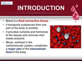
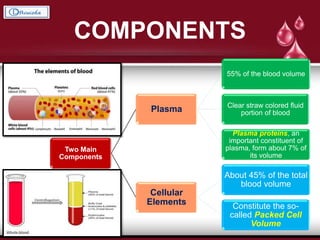
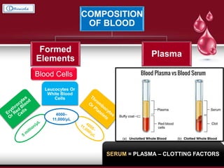
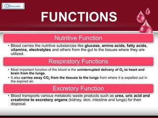
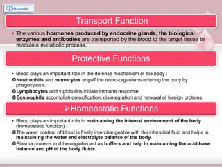
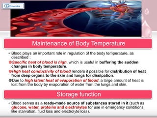
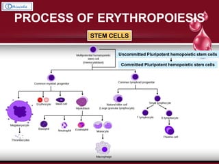
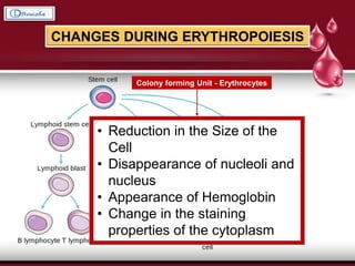
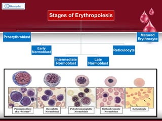
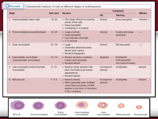
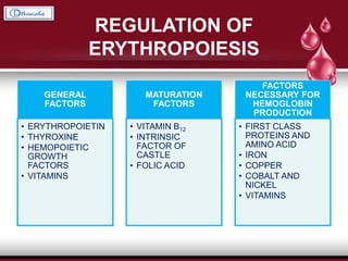
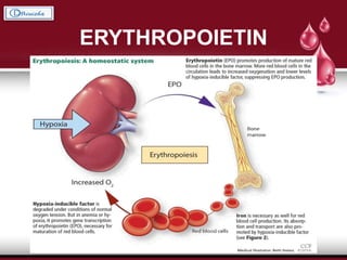
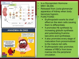
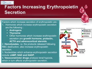
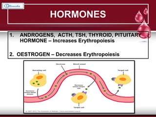
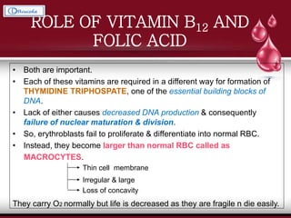
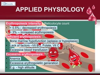
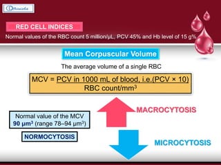
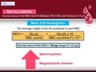
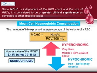
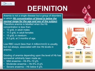
Blood – Part I
Dr. Rinisha Sinha
MDS Part I
Introduction
Characteristics of Blood
Components of Blood
Function of Blood
Red Blood Cells
Erythropoiesis
Hemoglobin
Hematocrit Values
Anemia
INTRODUCTION
• Blood is a fluid connective tissue.
• It transports substances from one
part of the body to another.
• It provides nutrients and hormones
to the tissues and removes their
waste products.
• Blood, confined in the
cardiovascular system, constitutes
a major part of the extracellular
fluid of the body.
CHARACTERISTICS
Color of the blood : Opaque Red due to the pigment hemoglobin in the red
blood cells (RBC).
Arterial blood : Bright red ; Venous blood : Dark red in color.
Volume of blood in an average adult is about 5–6 Liters (8% of the body
weight or 80 mL/kg body weight).
Viscosity of blood is five times more than that of water.
Specific gravity of blood is 1.050–1.060.
Specific gravity of RBC is greater (1.090) than that of plasma (1.030).
pH of blood is about 7.4 (ranges from 7.38 to 7.42), i.e. it is alkaline in nature.
In acidosis, pH of blood falls below 7.38 and in alkalosis, pH is more than 7.42.
COMPONENTS
Two Main
Components
Plasma
55% of the blood volume
Clear straw colored fluid
portion of blood
Plasma proteins, an
important constituent of
plasma, form about 7% of
its volume
Cellular
Elements
About 45% of the total
blood volume
Constitute the so-
called Packed Cell
Volume
COMPOSITION
OF BLOOD
Formed
Elements
Plasma
Blood Cells Liquid Portion
Leucocytes Or
White Blood
Cells
4000–
11,000/μL
SOLIDS
WATER
GASES
7-8%
92-93%
• Oxygen
• Carbon
Dioxide
• Nitrogen
SERUM = PLASMA – CLOTTING FACTORS
FUNCTIONS
Nutritive Function
• Blood carries the nutritive substances like glucose, amino acids, fatty acids,
vitamins, electrolytes and others from the gut to the tissues where they are
utilized.
Respiratory Functions
• Most important function of the blood is the uninterrupted delivery of O2 to heart and
brain from the lungs.
• It also carries away CO2 from the tissues to the lungs from where it is expelled out in
the expired air.
Excretory Function
• Blood transports various metabolic waste products such as urea, uric acid and
creatinine to excretory organs (kidney, skin, intestine and lungs) for their
disposal.
Transport Function
• The various hormones produced by endocrine glands, the biological
enzymes and antibodies are transported by the blood to the target tissue to
modulate metabolic process.
Protective Functions
• Blood plays an important role in the defense mechanism of the body :
Neutrophils and monocytes engulf the micro-organisms entering the body by
phagocytosis.
Lymphocytes and γ globulins initiate immune response.
Eosinophils accomplish detoxification, disintegration and removal of foreign proteins.
Homeostatic Functions
• Blood plays an important role in maintaining the internal environment of the body
(homeostatic function) :
The water content of blood is freely interchangeable with the interstitial fluid and helps in
maintaining the water and electrolyte balance of the body.
Plasma proteins and hemoglobin act as buffers and help in maintaining the acid-base
balance and pH of the body fluids.
Maintenance of Body Temperature
• Blood plays an important role in regulation of the body temperature, as
described :
Specific heat of blood is high, which is useful in buffering the sudden
changes in body temperature.
High heat conductivity of blood renders it possible for distribution of heat
from deep organs to the skin and lungs for dissipation.
Due to high latent heat of evaporation of blood, a large amount of heat is
lost from the body by evaporation of water from the lungs and skin.
Storage function
• Blood serves as a ready-made source of substances stored in it (such as
glucose, water, proteins and electrolytes for use in emergency conditions
like starvation, fluid loss and electrolyte loss).
RED BLOOD CELLS
CHARACTERISTIC FEATURES
OF RED BLOOD CELLS
INTRODUCTION
• Non – nucleated formed elements in the blood.
• Also known as “Erythrocytes”. { Erythros = Red }
• The color Red is due to the presence of the coloring pigment called hemoglobin.
• Vital role in the transport of respiratory gases.
• Largest in number compared to other blood cells.
NORMAL VALUES 4 – 5.5 millions per cubic mm of blood
5 – 6.5 millions per cubic mm of blood
4.5 – 5.5 millions per cubic mm of blood
6 - 7 millions per cubic mm of blood
FUNCTIONAL MORPHOLOGY
Bounded by a cell membrane but
is non-nucleated and lacks the
usual cell organelles.
Normal size
Diameter = 7.2 μm (range 6.9–7.4 μm)
Thickness (in the periphery) = 2 - 2.5 μm
(in the center) = 1 μm
Surface area = 120–140 μm2
Volume = 80 μm3 (range 78–86 μm3)
ADVANTAGES OF BICONCAVE SHAPE
• It renders FLEXIBILITY to the red cells so that
they can pass through the capillaries whose
minimum diameter is 3.5 μm.
• Greater surface area provided as compared to
volume, which allows considerable alterations in
the cell volume. Thus, the RBCs can withstand
changes of osmotic pressure. In this way, the
RBCs can resist hemolysis to certain extent
when placed in the hypotonic solution.
• Greater surface area allows EASY EXCHANGE
OF O2 and CO2, and RAPID DIFFUSION of other
substances.
VARIATIONS IN COUNTS OF RBCs
PHYSIOLOGICAL
POLYCYTHAEMIA
PATHOLOGICAL
POLYCYTHAEMIA
PHYSIOLOGICAL
DECREASE
ANAEMIA
AGE
SEX
HIGH
ALTITUDE
EXCESSIVE
EXERCISE
EMOTIONAL
CONDITION
INCREASE IN
ATMOSPHERIC
PRESSURE
AFTER
MEALS
PHYSIOLOGICAL
POLYCYTHAEMIA
PATHOLOGICAL
POLYCYTHAEMIA
Primary polycythaemia or
polycythaemia vera
occurs in Myeloproliferative disorder-
like malignancies of the bone
marrow. The RBC count is
persistently above 14 million/mm3
and is always associated with high
white blood cell (WBC) count (above
24,000/mm3).
Secondary polycythaemia
occurs due to certain conditions
producing a state of chronic
hypoxia in the body such as:
Congenital heart disease
•Chronic respiratory disorders like
emphysema
•Ayerza’s Disease.
Pathological increase in the RBC
count (above 7 million/mm3)
Physiological Decrease
in RBC count
At HIGH Barometric Pressure
RBC count is decreased slightly due
to high O2 tension of the blood.
After Sleep and
Immedicately after
getting up from sleep
RBC count is
decreased slightly
In Pregnancy
relative reduction in RBC
count due to
hemodilution caused by
increase in the plasma
volume
ANEMIA
VARIATIONS IN SHAPE OF
RED BLOOD CELLS
ROULEAUX FORMATION
The tendency of the RBCs to pile one over the other like a pile of coins.
Major role in Rouleaux formation :
DISCOID SHAPE + PROTEIN
COATING of the RED CELLS
Reversible Phenomenon
• It promotes sedimentation
of the RBCs.
• Does not occur in the
normal circulation under
physiological conditions.
Irreversible
Phenomenon
Albumin
Fibrinogen, Globulin
ERYTHROCYTE SEDIMENTATION
RATE
• The rate at which the RBCs sediment (settle down) when the blood
containing an anticoagulant is allowed to stand in a vertically placed tube.
• It is expressed in millimeter at the end of first hour.
3 – 7 mm 5 – 9 mmBy Westergren’s method
• Increased ESR : indicative of chronic inflammatory conditions in the body.
• Estimation of ESR is more useful as a prognostic test, i.e. to judge the progress of
the disease in patients under treatment.
Also called
• Sedimentation rate
• Sed rate
• BIERNACKI REACTION
Wintrobe’s method
 Rouleaux formation.
Increased tendency of the rouleaux formation RAISES the
ESR. Fibrinogen and the proteins favor rouleaux formation
and thus increase the ESR.
 Size of the RBCs.
Increase in the size of the RBCs (Macrocytosis) RAISES
the ESR.
 Number of RBCs.
INVERSELY PROPORTIONAL
In Anemia, ESR is Increased.
 Viscosity of blood.
ESR is INCREASED when the viscosity of blood is
decreased and vice versa.
 Sex.
ESR is greater in the females (5–9 mm) than the males (3–
7 mm).
 Menstruation.
ESR is slightly raised during menstruation in the females.
 Pregnancy.
ESR is raised in pregnancy from third month to parturition,
and returns to normal after 3–4 weeks of delivery.
Factors affecting ESR
•1. Tuberculosis
2. Malignant
diseases
3. Collagen diseases
4. All anemias
(except sickle cell
anemia)
5. Chronic infections
1. Polycythemia
2. Decreased
fibrinogen levels
3. Sickle cell anemia
4. Allergic conditions
IncreaseinESR
DecreaseinESR
Pathological variations in ESR
HEMOPOIESIS
ERYTHROPOIESIS
LEUCOPOIESIS
THROMBOPOIESIS or
MEGAKARYOCYTOPOIESIS
ERYTHROPOESIS
The process of the origin, development and maturation of erythrocytes.
Definition
In Fetal Life, the erythropoiesis occurs in three stages :
Site of Erythropoiesis
MESOBLASTIC
STAGE
During the first
two months,
the RBCs are
produced from
Mesenchyme of
the yolk sac
HEPATIC STAGE
From the third
month of
Intrauterine life,
Liver is the main
organ that
produces RBCs
Spleen and
Lymphoid organs
are also involved.
MYELOID
STAGE
During the last
three months of
intrauterine life,
the RBCs are
produced from
the Red Bone
Marrow and
Liver.
In Newborn babies, growing Children and Adults :
Only RED BONE MARROW produces the Red Blood Cells
• Up to the age of 20 years : Red Bone Marrow of all bones (Long Bones and all
the Flat Bones.
• After the age of 20 years : Red Bone Marrow of Membranous Bones like
vertebrae, sternum, ribs, scapula, iliac bones and skull bones and from the long
ends of long bones.
If the bone marrow is destroyed or fibrosed, then, LIVER and SPLEEN may
produced the red blood cells in adults.
PROCESS OF ERYTHROPOIESIS
STEM CELLS
Uncommitted Pluripotent hemopoietic stem cells
Committed Pluripotent hemopoietic stem cells
CHANGES DURING ERYTHROPOIESIS
Colony forming Unit - Erythrocytes
• Reduction in the Size of the
Cell
• Disappearance of nucleoli and
nucleus
• Appearance of Hemoglobin
• Change in the staining
properties of the cytoplasm
Stages of Erythropoiesis
Proerythroblast
Early
Normoblast
Intermediate
Normoblast
Late
Normoblast
Reticulocyte
Matured
Erythrocyte
REGULATION OF
ERYTHROPOIESIS
GENERAL
FACTORS
• ERYTHROPOIETIN
• THYROXINE
• HEMOPOIETIC
GROWTH
FACTORS
• VITAMINS
MATURATION
FACTORS
• VITAMIN B12
• INTRINSIC
FACTOR OF
CASTLE
• FOLIC ACID
FACTORS
NECESSARY FOR
HEMOGLOBIN
PRODUCTION
• FIRST CLASS
PROTEINS AND
AMINO ACID
• IRON
• COPPER
• COBALT AND
NICKEL
• VITAMINS
ERYTHROPOIETIN
Factors which increase secretion of erythropoietin are :
1. Hormones which increase erythropoietin secretion
are the following :
 Androgens
 Thyroxine
 Other hormones which increase erythropoietin
secretion are growth hormone, prolactin,
ACTH and adrenocortical steroids.
2. Hemolysates, i.e. the products released following
RBC destruction, also increase erythropoietin
secretion.
3. Nucleotides which enhance erythropoietin secretion
include cAMP, NAD and NADP.
4. Vasoconstrictor drugs produce renal hypoxia,
which in turn affects erythropoietin secretion.
Factors Increasing Erythropoietin
Secretion
HORMONES
1. ANDROGENS, ACTH, TSH, THYROID, PITUITARY
HORMONE – Increases Erythropoiesis
2. OESTROGEN – Decreases Erythropoiesis
ROLE OF VITAMIN B12 AND
FOLIC ACID
• Both are important.
• Each of these vitamins are required in a different way for formation of
THYMIDINE TRIPHOSPATE, one of the essential building blocks of
DNA.
• Lack of either causes decreased DNA production & consequently
failure of nuclear maturation & division.
• So, erythroblasts fail to proliferate & differentiate into normal RBC.
• Instead, they become larger than normal RBC called as
MACROCYTES.
They carry O2 normally but life is decreased as they are fragile n die easily.
Thin cell membrane
Irregular & large
Loss of concavity
APPLIED PHYSIOLOGY
Erythropoiesis intensity – Reticulocyte count
• < 0.5%-- decreased erythropoiesis
• > 3% -- increased erythropoiesis
Deficient erythropoiesis
• Bone marrow hypofunction (aplasia or hypoplasia)
• Lack of factors –Vit B12, Folate, Vit C
• Excessive blood transfusion
Excessive erythropoiesis
• Anemia
• Excessive erythropoietin generation
• E.g. - high altitude.
HEMOGLOBIN
• The cytoplasm of erythrocytes (RBCs)
contains an oxygen binding protein
called hemoglobin.
• Erythrocyte precursors synthesize
hemoglobin.
• Iron containing coloring matter of Red
Blood Cells (RBCs).
• Chromoprotein ; forms 95% of dry
weight of RBC and 30-34% of wet
weight.
15 g/dL 14.5 g/dL
At Birth
After 3 months
After 1 year
From Puberty
onwards
25 g/dL
20 g/dL
17 g/dL
14-16
g/dL
Average Hemoglobin content in blood
14 to 16 g/dL
4 Pyrrole rings
CH4
CH4
CH4
CH4
Structure of Hemoglobin
Protein
GLOBIN
Iron –
containing
pigment
HAEM
4 Polypeptide chains
Iron–porphyrin complex called
Iron– protoporphyrin IX, i.e. it
consists of
• a porphyrin nucleus
• the iron
4 Pyrrole rings
Synthesis of
Hemoglobin
Adult hemoglobin - HbA : globin contains 2 α and 2 β chains
Fetal hemoglobin - HbF : 2 α and 2 γ chains instead of β chains
MITOCHONDRIA
CYTOPLASM
UROPORPHOBILINOGEN I UROPORPHOBILINOGEN III
COPROPORPHYRINOGEN III
MITOCHONDRIAMITOCHONDRIA
Uroporphobilinogen I
Synthase
Porphobilinogen III
co-Synthase
Uroporphobilinogen
decarboxylase
DERIVATIVES OF HAEMOGLOBIN
(REACTIONS OF HAEMOGLOBIN)
Oxyhaemoglobin
Hb reacts readily with oxygen
An unstable and reversible
compound, i.e. oxygen can be
released from this compound.
In this compound iron remains in
the ferrous state.
Reduced haemoglobin
Deoxygenated Hb is formed
when oxygen is released from the
oxyhaemoglobin.
HbO2 → Hb + O 2
Carbaminohaemoglobin
compound of Hb with carbon
dioxide.
HbNH2 + CO2 → HbNHCOOH
Carboxyhaemoglobin
Carbon monoxyhaemoglobin is
a compound of Hb with carbon
monoxide (CO):
Hb + CO → COHb
The affinity of Hb for CO is much
more (200–250 times) than its
affinity for oxygen.
Because of this, the CO displaces
oxygen from Hb, thereby reducing
the oxygen carrying capacity of
the blood.
Methaemoglobin
When reduced or oxygenated Hb
is treated with an oxidizing agent,
e.g. potassium ferricyanide, the
ferrous Fe2+, is oxidized to ferric
(Fe3+); the sixth bond is attached
to OH to form the compound
methaemoglobin.
Methaemoglobin is represented
as HbOH.
Disadvantages : It cannot unite
reversibly with gaseous oxygen;
the O2 of the attached OH is not
given off in a vacuum
Glycosylated haemoglobin
Derivative of HbA present in very
small amount, e.g. haemoglobin
A1C (HbA1C), in which glucose is
attached to terminal valine in the
β chains. The level of
glycosylated Hb in the blood
increases in poorly controlled
patients of diabetes mellitus
LIFE SPAN AND FATE OF RED BLOOD CELLS
Normally, the average life span of RBCs is 120 days
Spleen  Graveyard of Red blood cells.
Daily 10% red blood cells, which are senile, get destroyed in normal young
healthy adults.
BILIRUBIN AND JAUNDICE
The normal serum bilirubin level
ranges from 0.3 to 1.0 mg/100 mL.
Van den Bergh test
Performed using the diazo reagent
(mixture of sulphanilic acid, hydrochloric
acid and sodium nitrite).
• Direct Van den Bergh reaction.
When diazo reagent is added to the serum
containing conjugated bilirubin (water
soluble) a reddish brown coloration is
obtained within 30 s.
• Indirect Van den Bergh reaction.
When diazo reagent is added to the serum
containing mainly unconjugated bilirubin
(water insoluble), no color is obtained.
However, if some solvent like alcohol (which
dissolves the unconjugated bilirubin) is
added, the reddish brown coloration is
obtained.
CLINICAL IMPORTANCE
 Many drugs e.g.
METHANDROSTENOLONE
competes with bilirubin for
conjugating with glucuronides. So
free bilirubin is accumulated in
plasma leading to jaundice.
 CRIGLER- NAJJAR DISEASE :
GLUCURONYL TRANSFERASE
is absent, leading to jaundice.
 PHENOBARBITONE, increases
activity of glucuronyltransferase. So
hyperbilirubinemia & kernicterus
in neonates can be treated.
Due to gene mutation, structural variation in polypeptide chains
occurs.
• Hemoglobin S  Sickle cell anemia
α chains normal & β chains abnormal.
(Glutamic acid is replaced by valine at 6th position).
• Thalassemia, polypeptide chains are decreased, absent or
abnormal.
 In α thalassemia, α chains are decreased, absent or abnormal.
 In β thalassemia, β chains are decreased, absent or abnormal.
ABNORMAL HEMOGLOBIN
PORPHYRIAS - Is defective synthesis of heme
• Hb level lets us know if our body is making enough red blood cells. It is
important to know this before surgery because we may lose some blood
during or after surgery. Too few red blood cells, indicates anemia. This
increases risk of needing transfusions.
If found to be low there are ways to increase it depending on cause of
anaemia.
 If we lack certain nutrients, we may need to take supplements.
 A newer method is to give Epoetin alfa (PROCRIT).
• PROCRIT helps increase the body's natural supply of
erythropoietin.
– stimulates bone marrow to produce "progenitor" cells.
– helps increase the number of red blood cells.
– may reduce the need for blood transfusions.
Why Is Knowing Hb Level Before Surgery so Important ?
HEMATOCRIT VALUES
PACKED CELL VOLUME
The percentage of the cellular elements (RBCs, WBCs and platelets) in
the whole blood.
40 - 45 % 38 - 42 %
“PCV” EQUIVALENT TO
HEMATOCRIT VOLUME
Since, Volume of WBCs and
Platelets is very less
SIGNIFICANCE OF DETERMINING PCV
• Diagnosis and treatment planning of anemia &
polycythemia (viscosity)
• Recovery from dehydration after treatment.
VARIATIONS IN PCV
• PCV increases in polycythemia and
dehydration.
• PCV decreases in anemia.
RED CELL INDICES
Normal values of the RBC count 5 million/μL, PCV 45% and Hb level of 15 g%
Mean Corpuscular Volume
The average volume of a single RBC
MCV = PCV in 1000 mL of blood, i.e.(PCV × 10)
RBC count/mm3
Normal value of the MCV
90 μm3 (range 78–94 μm3)
NORMOCYTOSIS
MACROCYTOSIS
MICROCYTOSIS
RED CELL INDICES
Normal values of the RBC count 5 million/μL, PCV 45% and Hb level of 15 g%
Mean Cell Hemoglobin
The average weight of the Hb contained in each RBC
MCH = Hb g/L___ = Hb g% x 10____
RBC count/L RBC count/mm3 x 1012
Normal value of the MCH = 30 pg (range 27–33 pg)
Spherocytosis
Megaloblastic Anemia
Since MCHC is independent of the RBC count and the size of
RBCs, it is considered to be of greater clinical significance as
compared to other absolute values.
Mean Cell Haemoglobin Concentration
The amount of Hb expressed as a percentage of the volume of a RBC
MCHC = Hb g%___
PCV/100 mL
Normal value of the MCHC
33.3% (range 30–38%)
NORMOCHROMIC
HYPERCHROMIC
Very Rare
MCHC > 38% cannot
occur
HYPOCHROMIC
Iron – Deficiency
Anemia
DEFINITION
Anemia is not a single disease but a group of disorders
in which Hb concentration of blood is below the
normal range for the age and sex of the subject.
Therefore anemia is labelled when the Hb
concentration is less than:
• 13 g/dL in adult males
• 11.5 g/dL in adult females
• 15 g/dL in newborn
• 9.5 g/dL at 3 months of age.
Low RBC count (less than 4 million/mm3) is usually,
but not always, associated with low Hb levels in
anemia.
Grading of anemia depending upon the level of Hb has
somewhat arbitrarily been made as :
• Mild anaemia – Hb 8%–10 g%
• Moderate anaemia – Hb 6%–8 g%
• Severe anaemia – Hb below 6 g%
CLASSIFICATION
Etiological
(Whitby’s)
Classification
Deficiency anemias: Iron-deficiency anemia;
Megaloblastic anemia (pernicious anemia) due to deficiency of vitamin B12
Megaloblastic anemia due to deficiency of folic acid
Protein and vitamin C deficiency can also cause anaemia.
Blood loss anaemias or haemorrhagic anaemias are commonly known and can be:
•Acute post-hemorrhagic anemia as in accidents
•Chronic post-hemorrhagic anemia
Hemolytic anemias
Hereditary hemolytic anemias, e.g. Thalassemia, Sickle cell anemia, Hereditary
spherocytosis and Glucose 6-phosphate dehydrogenase (G6PD) deficiency.
Acquired hemolytic anemias such as Immunohaemolytic anemia (due to antibodies
against RBCs): Hemolytic anemia due to direct toxic effects (e.g. in malaria, snake
venom, toxic effects of drugs and chemicals, etc.); Hemolytic anemia in
splenomegaly and Hemolytic anemia in paroxysmal nocturnal hemoglobinuria (PNH).
Aplastic anemia. It occurs due to the failure of bone marrow to produce RBCs.
Anemia due to chronic diseases. It is seen in tuberculosis, chronic infections,
malignancies, chronic lung diseases, etc.
Morphological
(Wintrobe’s)
classification
Normocytic normochromic anemias.
These are characterized by normal MCV (78–94 μm3 or 78–94 μL) and
normal MCHC (30%–38%). Such a morphological picture is seen in:
Acute post-hemorrhagic anemia, Hemolytic anemias and Aplastic
anemias.
Microcytic hypochromic anemias.
These are characterized by reduced MCV (< 78 μm3) and reduced
MCHC (< 30%). Examples of such anemias are:
Iron-deficiency anemia, Chronic post-hemorrhagic anaemia and
Thalassemia.
Macrocytic normochromic anaemia.
It is characterized by increased MCV (> 94 μm3) and normal MCHC
(30–38%). Examples are:
Megaloblastic anemia (pernicious anemia) due to deficiency of vitamin
B12 and Megaloblastic anemia due to deficiency of folic acid
INTRODUCTION
Iron-deficiency anemia is the commonest nutritional deficiency disorder present
throughout the world, but its prevalence is higher in the developing countries.
In India, iron-deficiency is the commonest cause of anemia. It is much more
common :
• In women between 20–45 years than in men,
• At periods of active growth in infancy, childhood and adolescence.
Only 10% of the dietary intake of iron is absorbed.
to compensate menstrual loss
Pregnant and lactating women require about
Daily requirement
5–10 mg/day 20 mg/day
40 mg of iron per day
Causes of Iron – Deficiency Anemia Characteristic Features
Laboratory Findings
Blood picture and red cell indices
 Hb concentration is decreased.
 RBCs are hypochromic (deficient in Hb) and microcytic (smaller in
size). They show anisocytosis and poikilocytosis.
Red cell indices like MCV, MCH and MCHC are decreased.
Bone marrow findings
 Marrow cellularity: Erythroid hyperplasia
 Erythropoiesis: Normoblastic
 Marrow iron: Deficient
Biochemical findings
 Serum iron decreases, often under 50 mg% (normal 60%–160
mg%).
 Serum bilirubin is less than 0.4 mg%.
 Serum ferritin is very low, indicating poor tissue iron stores.
 Total iron binding capacity (TIBC) is increased.
Treatment of Iron – Deficiency anemia
• Oral administration of Fe2+ salts
• Correction of causative factor, if possible.
INTRODUCTION
Megaloblastic anemias are characterized by the abnormally large cells of
erythrocyte series.
These are caused by defective DNA synthesis due to deficiency of
vitamin B12 and/or folic acid (folate).
Megaloblastic anaemia due to vitamin B12 deficiency
Megaloblastic anemia due to folate deficiency
White Blood Cells
Leukopoiesis
Neutrophils
Leukemia
Platelets
Thrombopoiesis
Hemostasis
Coagulation
Anti-hemostatic Mechanism and Anticoagulants
Procoagulants
Bleeding Disorders
Laboratory Investigations
Conclusion and References
CHARACTERISTIC FEATURES
OF WHITE BLOOD CELLS
INTRODUCTION
• Nucleated Mobile Units of the body’s protective systems.
• Also known as “LEUCOCYTES” ; they are colorless in contrast to the RBCs.
• Partial formation in the Bone Marrow and partial in the Lymph Tissues.
• Granulocytes
• Monocytes
• Few Lymphocytes
• Lymphocytes
• Plasma Cells
After formation, transported to areas of serious infection and inflammation,
thereby providing a rapid and potent defense against infectious agents.
NORMAL VALUES 4000–11,000/mm3 of blood
5000–15,000/mm3 of blood
4500–13,500/mm3 of blood
10,000–25,000/mm3 of blood
6000–16,000/mm3 of blood
62.0% 2.3%
0.4%
5.3%
30.0%
VARIATIONS IN WHITE
BLOOD CELL COUNT
Increase in total WBC count
above 11,000/mm3
Physiological causes
1. Age
2. Exercise
3. After food intake
4. Mental stress
5. Pregnancy
6. Exposure to low temperature also
causes leukocytosis
Pathological causes
1. Acute bacterial infections
especially by the pyogenic organisms
2. Acute hemorrhage
3. Burns
4. Postoperative period
5. Tuberculosis
6. Glandular fever
Decrease in the total WBC count
below 4000/mm3
Causes of leucopenia
1. Infections by the non-pyogenic
bacteria, especially typhoid fever and
paratyphoid fever.
2. Viral infections, such as influenza,
smallpox, mumps, etc.
3. Protozoal infections.
4. Starvation and malnutrition.
5. Disorders of Spleen.
6. Aplasia of bone marrow and its
depression due to :
o Drugs such as chloromycetin and
cytotoxic drugs used in malignant
diseases.
 Repeated exposure to X-rays or
radiations.
 Chemical poisons like arsenic,
dinitrophenol and antimony.
7. Pernicious Anemia
Leukocytosis
Leucopenia
Granulocytosis
The abnormal increase in the
number of granulocytes.
Agranulocytosis
The acute pathological condition
characterized by absolute lack of
granulocytes.
Granulocytopenia
The abnormal reduction in the
number of granulocytes.
Leukemia
The condition which is
characterized by
abnormal and
uncontrolled increase in
leukocyte count more
than 1,000,000/cubic mm.
It is also called Blood
Cancer.
Pathological variations in different
types of WBCs
LEUKOPOIESIS
The development and maturation of Leukocytes
formedonlyinthe
BONEMARROW
The BONE MARROW and
THYMUS form the Primary
Lymphopoietic Organs.
Secondary or Reactive lymphoid tissue
• Lymph Node
• Spleen
• Gut-associated Lymphoid Tissue (GALT)
Two major lineages of white blood cells are formed
The Myelocytic lineages The Lymphocytic lineages
Myeloblast
Promyelocyte
Neutrophil
Myelocyte
Young
Neutrophil
Metamyelocyte
“Band”
Neutrophil
Metamyelocyte
Polymorphonuclear
Neutrophil
Eosinophil
Myelocyte
Eosinophil
Metamyelocyte
Polymorphonuclear
Eosinophil
Basophil
Myelocyte
Polymorphonuclear
Basophil
StagesofMonocyteformation
PLATELETS Megakaryocyte
Lymphoblast
Prolymphocyte
Lymphocytes
REGULATION OF LEUCOPOIESIS
During tissue injury and inflammation, bacterial toxins, products of injury, etc. cause a great
increase in the rate of production and release of leucocytes.
The products of dead and dying white cells themselves control leucopoiesis.
Granulopenia or dead granulocytes and monocytes
G-CSF
M-CSF
GM-CSF
Interleukins
Bone marrow
Granulocytes
Monocytes/macrophages
Release
Stimulate
Increased formationNormal counts inhibit
Colony Stimulating Factors (CSFs)
The cytokines controlling the formation
of different types of granulocytes.
They are glycoproteins formed by
monocytes and T lymphocytes.
Interleukins
The cytokines that control lymphocyte
formation e.g. IL-I, IL-3, etc.
They are formed by monocytes,
macrophages and endothelial cells.
LIFESPAN OF WHITE
BLOOD CELLS
• Not constant.
• It depends upon the demand in the body and their
function.
• May be as short as half a day or it may be as long as 3
to 6 months.
PROPERTIES OF WHITE
BLOOD CELLS
DIAPEDESIS
The process by which
the Leukocytes
squeeze through the
narrow blood vessels.
AMEBOID MOVEMENT
Neutrophils, monocytes and
lymphocytes show amebic
movement, characterized by
protrusion of the cytoplasm
and change in the shape.
PHAGOCYTOSIS
Neutrophils and monocytes
engulf the foreign bodies by
means of phagocytosis.
CHEMOTAXIS
The attraction of WBCs
towards the injured tissues by
the chemical substances
released at the site of injury.
Phagocytosis
Engulfment of the foreign particles or
bacteria, their digestion and
ultimately death.
Reaction of inflammation
Neutrophils release leukotrienes,
prostaglandins, thromboxane, etc.
that bring about the reactions of
inflammation like vasodilatation and
edema.
Febrile response
Neutrophils contain a fever producing
substance called endogenous
pyrogen, which is an important
mediator of febrile response to the
bacterial pyrogens.
NEUTROPHILS
Diameter = 10 – 14 μm
Nucleus
• A single horseshoe shaped nucleus,
becomes lobed as the cell grows.
• Purple in color
• Multilobed (2–6 lobes); called
polymorphonuclear leucocytes.
• The lobes are connected by the
chromatin filaments, seen clearly
through the cytoplasm.
Cytoplasm
• Pale bluish in color.
• Full of fine (pinpoint) granules.
• Granules take both acidic and basic
stain, and look violet-pink in color.
First Line of Defense
Neutrophils + Monocytes
PHAGOCYTOSIS
Substances secreted by
WBCs and their Functions
LEUKEMIA
• A group of malignant diseases of
the blood in which there occurs an
increase in the total WBC count
associated with presence of
immature WBCs in the peripheral
blood.
• Can be caused by cancerous
mutation of a myelogenous or
lymphogenous cell.
4% of all cancer deaths
LEUKEMIAS
MYELOID
LYMPHOID
LEUKEMIAS
ACUTE
CHRONIC
Effects of Leukemia on the Body
Important effect of leukemia on the body is excessive use of metabolic substrates by the
growing cancerous cells. The leukemic tissues reproduce new cells so rapidly that
tremendous demands are made on the body reserves for foodstuffs, specific amino acids, and
vitamins. Consequently, the energy of the patient is greatly depleted, and excessive
utilization of amino acids by the leukemic cells causes rapid deterioration of the normal
protein tissues of the body. Thus, while the leukemic tissues grow, other tissues become
debilitated. After metabolic starvation has continued long enough, this alone is sufficient to
cause death.
PLATELETS
THROMBOCYTES
Thrombo = clot; Cytes = cells
STRUCTURE AND COMPOSITION
Size
The smallest, colorless, non-nucleated and moderately refractive bodies.
Diameter : 2.5 µm (2 to 4 µm) Volume : 7.5 cu µm (7 to 8 cu µm)
Leishman staining shows a platelet consisting of faint bluish cytoplasm
containing reddish purple granules.
Platelet is constituted by:
Cell
membrane or
surface
membrane
Microtubules Cytoplasm
NORMAL VALUES
2,50,000 mm3 of blood
Range = 2,00,000 and 4,00,000
mm3 of blood
CELL MEMBRANE
Extensive invagination of cell membrane forms an open canalicular system.
Glycoproteins
• Prevent the adherence of platelets to
normal endothelium.
• But accelerate the adherence to
collagen and damaged endothelium
in ruptured blood vessels.
• Form the receptors for adenosine
diphosphate (ADP) and thrombin.
Phospholipids
• Accelerate the clotting reactions.
• Form the precursors of thromboxane
A2 .
MICROTUBULES
 Form a ring around cytoplasm below
the cell membrane.
 Made up of polymerized proteins
called tubulin.
 These provide structural support for
the inactivated platelets to maintain
the disk like shape.
CYTOPLASM
 Contains the cellular organelles, Golgi apparatus, endoplasmic reticulum,
mitochondria, microtubule, micro vessels, filaments and granules.
Proteins
Enzymes
Adenosine triphosphatase
Enzymes necessary for
synthesis of prostaglandins
Hormonal Substances
Adrenaline
5-hydroxytryptamine
Histamine
Other Chemical
Substances
Glycogen
Substances like blood group
antigens
Platelet
Granules
1. Contractile proteins
i. Actin and myosin
ii. Thrombosthenin
2. von Willebrand factor
3. Fibrin-stabilizing factor
4. Platelet-derived growth
factor (PDGF)
5. Platelet-activating factor
(PAF)
6. Vitronectin (serum
spreading factor)
7. Thrombospondin
LIFESPAN AND FATE OF
PLATELETS
Average lifespan = 10 days
(varies between 8 and 11 days)
They are destroyed by tissue
macrophage system in spleen.
Splenomegaly platelet count
Splenectomy platelet count
PROPERTIES
OF
PLATELETS
ADHESIVENESS
AGGREGATION
(GROUPING OF
PLATELETS)
AGGLUTINATION
PHYSIOLOGICAL
VARIATIONS
• Age: Platelets are less in infants (1,50,000 to 2,00,000/cu
mm) and reaches normal level at 3rd month after birth
• Sex: There is no difference in the platelet count between
males and females. In females, it is reduced during
menstruation
• High altitude: Platelet count increases
• After meals: After taking food, the platelet count increases.
APPLIED
PHYSIOLOGY –
PLATELET
DISORDERS
• Thrombocytopenia
• Thrombocytosis
• Thrombocythemia
• Glanzmann’s thrombasthenia
Thrombocytopenia
• Acute infections
• Acute leukemia
• Aplastic and
pernicious
anemia
• Chickenpox
• Smallpox
• Splenomegaly
• Scarlet fever
• Typhoid
• Tuberculosis
• Purpura
• Gaucher’s
disease
Thrombocytosis
• Allergic
conditions
• Asphyxia
• Hemorrhage
• Bone fractures
• Surgical
operations
• Splenectomy
• Rheumatic fever
• Trauma (wound
or injury or
damage caused
by external force)
Thrombocythemia
• Carcinoma i
• Chronic leukemia
• Hodgkin’s
disease
Glanzmann’s
thrombasthenia
• An inherited
hemorrhagic
disorder, caused
by structural or
functional
abnormality of
platelets.
• Normal Platelet
Count
• Normal clotting
time, normal or
prolonged
bleeding time but
defective clot
retraction
THROMBOPOIESIS
Platelets are formed from bone marrow.
Cytoplasm has
Pseudopodium
FACTORS AFFECTING
THROMBOPOIESIS
Thrombopoiesis seems to be regulated by following humoral
factors:
• Thrombopoietin
• Megakaryocyte colony stimulating activity (Meg-CSA)
Vascular
Constriction
Formation of a
Platelet Plug
Formation of a
blood Clot as a
result of blood
coagulation,
Eventual growth of
fibrous tissue into
the blood clot to
close the hole in the
vessel permanently.
Hemostasis refers to the spontaneous arrest or prevention of bleeding
from the injured/damaged vessels by the physiological process.
DEFINITION
Coagulation or Clotting is defined as the process in which blood loses its fluidity
and becomes a jelly-like mass few minutes after it is shed from the blood vessels or
collected in a container.
Coagulation of blood occurs through a series of reactions due to the activation of
a group of substances.
Factor I
Factor II
Factor III
Factor IV
Factor V
Factor VI
Factor VII
Factor VIII
Factor IX
Factor X
Factor XI
Factor XII
Factor XIII
Fibrinogen
Prothrombin
Thromboplastin (Tissue factor)
Calcium
Labile factor (Proaccelerin or accelerator globulin)
Presence has not been proved
Stable factor
Antihemophilic factor (Antihemophilic globulin)
Christmas factor
Stuart-Prower factor
Plasma thromboplastin antecedent
Hageman factor (Contact factor)
Fibrin-stabilizing factor (Fibrinase)
THIRTEEN CLOTTING FACTORS
Stages of Blood Clotting
Formation of
prothrombin
activator
Conversion
of
prothrombin
into
thrombin
Conversion
of fibrinogen
into fibrin
BLOOD CLOT
The mass of coagulated blood which contains RBCs, WBCs and
platelets entrapped in fibrin meshwork.
CLOT RETRACTION
After the formation,
the blood clot starts
contracting. And after
about 30 to 45
minutes, the straw-
colored serum oozes
out of the clot. The
process involving the
contraction of blood
clot and oozing of
serum is called Clot
Retraction.
FIBRINOLYSISLysis of blood clot inside the blood
vessel is called fibrinolysis. It
helps to remove the clot from lumen
of the blood vessel. This process
requires a substance called plasmin
or fibrinolysin.
PROCOAGULANTS
Procoagulants or hemostatic agents
are the substances which accelerate
the process of blood coagulation.
THROMBIN
SNAKE VENOM
EXTRACTS OF LUNGS
AND THYMUS
SODIUM OR CALCIUM
ALGINATE
OXIDIZED
CELLULOSE
ANTIHEMOSTATIC
MECHANISMS
The factors which balance the tendency of the blood to clot in vivo constitute the
anti-hemostatic factors.
Plasmin or
fibrinolysin
Present in the
blood in an
inactive form
called
plasminogen or
profibrinolysin
Factors causing
fibrinolysis (fibrinolytic
mechanism).
Heparin
Antithrombin III
or heparin co-
factor II
Protein C
Factors preventing
coagulation (circulatory
anticoagulants)
Prostacyclin
An endogenous
factor which
prevents platelet
aggregation by
inhibiting the
thromboxane A2
formation (which
promotes
platelet
aggregation).
Factors preventing
platelet aggregation
Heparin is a naturally
produced anticoagulant
in the body. It is
produced by mast cells
which are the wandering
cells.
Protein C is a plasma protein synthesized in liver.
It, along with thrombomodulin and protein S,
constitutes an important negative feedback pathway
that keeps the coagulatory process under control.
Substances which prevent or postpone coagulation of blood are called Anticoagulants.
Anticoagulants used to
prevent blood clotting inside
the body, i.e. in vivo.
Anticoagulants used to
prevent clotting of blood that
is collected from the body,
i.e. in vitro.
Anticoagulants used to
prevent blood clotting both in
vivo and in vitro.
Endogenous anticoagulants
Those which are present
inside the blood naturally :
• Heparin
• Antithrombin III
• Protein C
Exogenous anticoagulants
Administered from outside or
are used in vitro :
• Heparin
• Calcium sequesters
• Vitamin K antagonist
• Defibration substances
PHYSICAL METHODS TO PREVENT BLOOD CLOTTING
COLD
Reducing the
temperature to about 5°C
postpones the
coagulation of blood.
COLLECTING BLOOD
IN A CONTAINER WITH
SMOOTH SURFACE
Collecting the blood in a container with smooth
surface like a silicon-coated container prevents
clotting. The smooth surface inhibits the
activation of factor XII and platelets. So, the
formation of prothrombin activator is prevented.
Hemophilia Purpura
von Willebrand
disease
• It is Inherited disorders. X-linked recessive.
• Females are carriers and males suffer from the disease.
• Factor VIII deficiency : Hemophilia A
• Factor IX deficiency : Hemophilia B (Christmas disease)
Purpura
• Faulty Primary hemostatic mechanism
• Prolonged BT but normal CT, PT, APTT
• Spontaneous bleeding from capillaries-- small tiny
hemorrhagic spots in body -- purpuric spots – PURPURA
Blood coagulation tests
 Bleeding time: -
o 1-6 minutes. It is usually determined by Duke’s method.
o Bleeding occurs from the skin when it is pricked with a needle, which
normally stops of its own within a few minutes. The time lapse between
the skin prick and the arrest of bleeding is called bleeding time (BT).
o Bleeding time is normal in hemophilia but prolonged in purpura.
 Clotting time:-
o By modified Lee and White’s method.
o Normal values are 6 - 10 mins.
o Clotting time is prolonged in hemophilia but normal in purpura.
 Prothrombin time :-
o Normal value is 10-14 secs.
o It is prolonged in deficiency of factor VII,X,V.
 Activated Partial Prothrombin time:- 30-40 secs
REFERENCES
• Guyton and Hall textbook of
Medical Physiology – 12th
edition
• Essentials of Medical
Physiology – K.
Sembulingam (5th edition)
• Principles of Physiology – J.
Tortora (8th edition)
• Textbook of Human
Physiology – Indu khurrana
(2nd edition)

Blood

  • 1.
    Blood – PartI Dr. Rinisha Sinha MDS Part I
  • 2.
    Introduction Characteristics of Blood Componentsof Blood Function of Blood Red Blood Cells Erythropoiesis Hemoglobin Hematocrit Values Anemia
  • 3.
    INTRODUCTION • Blood isa fluid connective tissue. • It transports substances from one part of the body to another. • It provides nutrients and hormones to the tissues and removes their waste products. • Blood, confined in the cardiovascular system, constitutes a major part of the extracellular fluid of the body.
  • 4.
    CHARACTERISTICS Color of theblood : Opaque Red due to the pigment hemoglobin in the red blood cells (RBC). Arterial blood : Bright red ; Venous blood : Dark red in color. Volume of blood in an average adult is about 5–6 Liters (8% of the body weight or 80 mL/kg body weight). Viscosity of blood is five times more than that of water. Specific gravity of blood is 1.050–1.060. Specific gravity of RBC is greater (1.090) than that of plasma (1.030). pH of blood is about 7.4 (ranges from 7.38 to 7.42), i.e. it is alkaline in nature. In acidosis, pH of blood falls below 7.38 and in alkalosis, pH is more than 7.42.
  • 5.
    COMPONENTS Two Main Components Plasma 55% ofthe blood volume Clear straw colored fluid portion of blood Plasma proteins, an important constituent of plasma, form about 7% of its volume Cellular Elements About 45% of the total blood volume Constitute the so- called Packed Cell Volume
  • 6.
    COMPOSITION OF BLOOD Formed Elements Plasma Blood CellsLiquid Portion Leucocytes Or White Blood Cells 4000– 11,000/μL SOLIDS WATER GASES 7-8% 92-93% • Oxygen • Carbon Dioxide • Nitrogen SERUM = PLASMA – CLOTTING FACTORS
  • 7.
    FUNCTIONS Nutritive Function • Bloodcarries the nutritive substances like glucose, amino acids, fatty acids, vitamins, electrolytes and others from the gut to the tissues where they are utilized. Respiratory Functions • Most important function of the blood is the uninterrupted delivery of O2 to heart and brain from the lungs. • It also carries away CO2 from the tissues to the lungs from where it is expelled out in the expired air. Excretory Function • Blood transports various metabolic waste products such as urea, uric acid and creatinine to excretory organs (kidney, skin, intestine and lungs) for their disposal.
  • 8.
    Transport Function • Thevarious hormones produced by endocrine glands, the biological enzymes and antibodies are transported by the blood to the target tissue to modulate metabolic process. Protective Functions • Blood plays an important role in the defense mechanism of the body : Neutrophils and monocytes engulf the micro-organisms entering the body by phagocytosis. Lymphocytes and γ globulins initiate immune response. Eosinophils accomplish detoxification, disintegration and removal of foreign proteins. Homeostatic Functions • Blood plays an important role in maintaining the internal environment of the body (homeostatic function) : The water content of blood is freely interchangeable with the interstitial fluid and helps in maintaining the water and electrolyte balance of the body. Plasma proteins and hemoglobin act as buffers and help in maintaining the acid-base balance and pH of the body fluids.
  • 9.
    Maintenance of BodyTemperature • Blood plays an important role in regulation of the body temperature, as described : Specific heat of blood is high, which is useful in buffering the sudden changes in body temperature. High heat conductivity of blood renders it possible for distribution of heat from deep organs to the skin and lungs for dissipation. Due to high latent heat of evaporation of blood, a large amount of heat is lost from the body by evaporation of water from the lungs and skin. Storage function • Blood serves as a ready-made source of substances stored in it (such as glucose, water, proteins and electrolytes for use in emergency conditions like starvation, fluid loss and electrolyte loss).
  • 10.
  • 11.
    CHARACTERISTIC FEATURES OF REDBLOOD CELLS INTRODUCTION • Non – nucleated formed elements in the blood. • Also known as “Erythrocytes”. { Erythros = Red } • The color Red is due to the presence of the coloring pigment called hemoglobin. • Vital role in the transport of respiratory gases. • Largest in number compared to other blood cells. NORMAL VALUES 4 – 5.5 millions per cubic mm of blood 5 – 6.5 millions per cubic mm of blood 4.5 – 5.5 millions per cubic mm of blood 6 - 7 millions per cubic mm of blood
  • 12.
    FUNCTIONAL MORPHOLOGY Bounded bya cell membrane but is non-nucleated and lacks the usual cell organelles. Normal size Diameter = 7.2 μm (range 6.9–7.4 μm) Thickness (in the periphery) = 2 - 2.5 μm (in the center) = 1 μm Surface area = 120–140 μm2 Volume = 80 μm3 (range 78–86 μm3) ADVANTAGES OF BICONCAVE SHAPE • It renders FLEXIBILITY to the red cells so that they can pass through the capillaries whose minimum diameter is 3.5 μm. • Greater surface area provided as compared to volume, which allows considerable alterations in the cell volume. Thus, the RBCs can withstand changes of osmotic pressure. In this way, the RBCs can resist hemolysis to certain extent when placed in the hypotonic solution. • Greater surface area allows EASY EXCHANGE OF O2 and CO2, and RAPID DIFFUSION of other substances.
  • 13.
    VARIATIONS IN COUNTSOF RBCs PHYSIOLOGICAL POLYCYTHAEMIA PATHOLOGICAL POLYCYTHAEMIA PHYSIOLOGICAL DECREASE ANAEMIA AGE SEX HIGH ALTITUDE EXCESSIVE EXERCISE EMOTIONAL CONDITION INCREASE IN ATMOSPHERIC PRESSURE AFTER MEALS PHYSIOLOGICAL POLYCYTHAEMIA
  • 14.
    PATHOLOGICAL POLYCYTHAEMIA Primary polycythaemia or polycythaemiavera occurs in Myeloproliferative disorder- like malignancies of the bone marrow. The RBC count is persistently above 14 million/mm3 and is always associated with high white blood cell (WBC) count (above 24,000/mm3). Secondary polycythaemia occurs due to certain conditions producing a state of chronic hypoxia in the body such as: Congenital heart disease •Chronic respiratory disorders like emphysema •Ayerza’s Disease. Pathological increase in the RBC count (above 7 million/mm3)
  • 15.
    Physiological Decrease in RBCcount At HIGH Barometric Pressure RBC count is decreased slightly due to high O2 tension of the blood. After Sleep and Immedicately after getting up from sleep RBC count is decreased slightly In Pregnancy relative reduction in RBC count due to hemodilution caused by increase in the plasma volume ANEMIA
  • 16.
    VARIATIONS IN SHAPEOF RED BLOOD CELLS
  • 17.
    ROULEAUX FORMATION The tendencyof the RBCs to pile one over the other like a pile of coins. Major role in Rouleaux formation : DISCOID SHAPE + PROTEIN COATING of the RED CELLS Reversible Phenomenon • It promotes sedimentation of the RBCs. • Does not occur in the normal circulation under physiological conditions. Irreversible Phenomenon Albumin Fibrinogen, Globulin
  • 18.
    ERYTHROCYTE SEDIMENTATION RATE • Therate at which the RBCs sediment (settle down) when the blood containing an anticoagulant is allowed to stand in a vertically placed tube. • It is expressed in millimeter at the end of first hour. 3 – 7 mm 5 – 9 mmBy Westergren’s method • Increased ESR : indicative of chronic inflammatory conditions in the body. • Estimation of ESR is more useful as a prognostic test, i.e. to judge the progress of the disease in patients under treatment. Also called • Sedimentation rate • Sed rate • BIERNACKI REACTION Wintrobe’s method
  • 19.
     Rouleaux formation. Increasedtendency of the rouleaux formation RAISES the ESR. Fibrinogen and the proteins favor rouleaux formation and thus increase the ESR.  Size of the RBCs. Increase in the size of the RBCs (Macrocytosis) RAISES the ESR.  Number of RBCs. INVERSELY PROPORTIONAL In Anemia, ESR is Increased.  Viscosity of blood. ESR is INCREASED when the viscosity of blood is decreased and vice versa.  Sex. ESR is greater in the females (5–9 mm) than the males (3– 7 mm).  Menstruation. ESR is slightly raised during menstruation in the females.  Pregnancy. ESR is raised in pregnancy from third month to parturition, and returns to normal after 3–4 weeks of delivery. Factors affecting ESR
  • 20.
    •1. Tuberculosis 2. Malignant diseases 3.Collagen diseases 4. All anemias (except sickle cell anemia) 5. Chronic infections 1. Polycythemia 2. Decreased fibrinogen levels 3. Sickle cell anemia 4. Allergic conditions IncreaseinESR DecreaseinESR Pathological variations in ESR
  • 21.
  • 22.
    ERYTHROPOESIS The process ofthe origin, development and maturation of erythrocytes. Definition In Fetal Life, the erythropoiesis occurs in three stages : Site of Erythropoiesis MESOBLASTIC STAGE During the first two months, the RBCs are produced from Mesenchyme of the yolk sac HEPATIC STAGE From the third month of Intrauterine life, Liver is the main organ that produces RBCs Spleen and Lymphoid organs are also involved. MYELOID STAGE During the last three months of intrauterine life, the RBCs are produced from the Red Bone Marrow and Liver.
  • 23.
    In Newborn babies,growing Children and Adults : Only RED BONE MARROW produces the Red Blood Cells • Up to the age of 20 years : Red Bone Marrow of all bones (Long Bones and all the Flat Bones. • After the age of 20 years : Red Bone Marrow of Membranous Bones like vertebrae, sternum, ribs, scapula, iliac bones and skull bones and from the long ends of long bones. If the bone marrow is destroyed or fibrosed, then, LIVER and SPLEEN may produced the red blood cells in adults.
  • 24.
    PROCESS OF ERYTHROPOIESIS STEMCELLS Uncommitted Pluripotent hemopoietic stem cells Committed Pluripotent hemopoietic stem cells
  • 25.
    CHANGES DURING ERYTHROPOIESIS Colonyforming Unit - Erythrocytes • Reduction in the Size of the Cell • Disappearance of nucleoli and nucleus • Appearance of Hemoglobin • Change in the staining properties of the cytoplasm
  • 26.
  • 28.
    REGULATION OF ERYTHROPOIESIS GENERAL FACTORS • ERYTHROPOIETIN •THYROXINE • HEMOPOIETIC GROWTH FACTORS • VITAMINS MATURATION FACTORS • VITAMIN B12 • INTRINSIC FACTOR OF CASTLE • FOLIC ACID FACTORS NECESSARY FOR HEMOGLOBIN PRODUCTION • FIRST CLASS PROTEINS AND AMINO ACID • IRON • COPPER • COBALT AND NICKEL • VITAMINS
  • 30.
  • 32.
    Factors which increasesecretion of erythropoietin are : 1. Hormones which increase erythropoietin secretion are the following :  Androgens  Thyroxine  Other hormones which increase erythropoietin secretion are growth hormone, prolactin, ACTH and adrenocortical steroids. 2. Hemolysates, i.e. the products released following RBC destruction, also increase erythropoietin secretion. 3. Nucleotides which enhance erythropoietin secretion include cAMP, NAD and NADP. 4. Vasoconstrictor drugs produce renal hypoxia, which in turn affects erythropoietin secretion. Factors Increasing Erythropoietin Secretion
  • 33.
    HORMONES 1. ANDROGENS, ACTH,TSH, THYROID, PITUITARY HORMONE – Increases Erythropoiesis 2. OESTROGEN – Decreases Erythropoiesis
  • 34.
    ROLE OF VITAMINB12 AND FOLIC ACID • Both are important. • Each of these vitamins are required in a different way for formation of THYMIDINE TRIPHOSPATE, one of the essential building blocks of DNA. • Lack of either causes decreased DNA production & consequently failure of nuclear maturation & division. • So, erythroblasts fail to proliferate & differentiate into normal RBC. • Instead, they become larger than normal RBC called as MACROCYTES. They carry O2 normally but life is decreased as they are fragile n die easily. Thin cell membrane Irregular & large Loss of concavity
  • 35.
    APPLIED PHYSIOLOGY Erythropoiesis intensity– Reticulocyte count • < 0.5%-- decreased erythropoiesis • > 3% -- increased erythropoiesis Deficient erythropoiesis • Bone marrow hypofunction (aplasia or hypoplasia) • Lack of factors –Vit B12, Folate, Vit C • Excessive blood transfusion Excessive erythropoiesis • Anemia • Excessive erythropoietin generation • E.g. - high altitude.
  • 36.
  • 37.
    • The cytoplasmof erythrocytes (RBCs) contains an oxygen binding protein called hemoglobin. • Erythrocyte precursors synthesize hemoglobin. • Iron containing coloring matter of Red Blood Cells (RBCs). • Chromoprotein ; forms 95% of dry weight of RBC and 30-34% of wet weight. 15 g/dL 14.5 g/dL At Birth After 3 months After 1 year From Puberty onwards 25 g/dL 20 g/dL 17 g/dL 14-16 g/dL Average Hemoglobin content in blood 14 to 16 g/dL
  • 38.
    4 Pyrrole rings CH4 CH4 CH4 CH4 Structureof Hemoglobin Protein GLOBIN Iron – containing pigment HAEM 4 Polypeptide chains Iron–porphyrin complex called Iron– protoporphyrin IX, i.e. it consists of • a porphyrin nucleus • the iron 4 Pyrrole rings
  • 39.
    Synthesis of Hemoglobin Adult hemoglobin- HbA : globin contains 2 α and 2 β chains Fetal hemoglobin - HbF : 2 α and 2 γ chains instead of β chains MITOCHONDRIA CYTOPLASM UROPORPHOBILINOGEN I UROPORPHOBILINOGEN III COPROPORPHYRINOGEN III MITOCHONDRIAMITOCHONDRIA Uroporphobilinogen I Synthase Porphobilinogen III co-Synthase Uroporphobilinogen decarboxylase
  • 40.
    DERIVATIVES OF HAEMOGLOBIN (REACTIONSOF HAEMOGLOBIN) Oxyhaemoglobin Hb reacts readily with oxygen An unstable and reversible compound, i.e. oxygen can be released from this compound. In this compound iron remains in the ferrous state. Reduced haemoglobin Deoxygenated Hb is formed when oxygen is released from the oxyhaemoglobin. HbO2 → Hb + O 2 Carbaminohaemoglobin compound of Hb with carbon dioxide. HbNH2 + CO2 → HbNHCOOH Carboxyhaemoglobin Carbon monoxyhaemoglobin is a compound of Hb with carbon monoxide (CO): Hb + CO → COHb The affinity of Hb for CO is much more (200–250 times) than its affinity for oxygen. Because of this, the CO displaces oxygen from Hb, thereby reducing the oxygen carrying capacity of the blood. Methaemoglobin When reduced or oxygenated Hb is treated with an oxidizing agent, e.g. potassium ferricyanide, the ferrous Fe2+, is oxidized to ferric (Fe3+); the sixth bond is attached to OH to form the compound methaemoglobin. Methaemoglobin is represented as HbOH. Disadvantages : It cannot unite reversibly with gaseous oxygen; the O2 of the attached OH is not given off in a vacuum Glycosylated haemoglobin Derivative of HbA present in very small amount, e.g. haemoglobin A1C (HbA1C), in which glucose is attached to terminal valine in the β chains. The level of glycosylated Hb in the blood increases in poorly controlled patients of diabetes mellitus
  • 41.
    LIFE SPAN ANDFATE OF RED BLOOD CELLS Normally, the average life span of RBCs is 120 days Spleen  Graveyard of Red blood cells. Daily 10% red blood cells, which are senile, get destroyed in normal young healthy adults.
  • 42.
    BILIRUBIN AND JAUNDICE Thenormal serum bilirubin level ranges from 0.3 to 1.0 mg/100 mL. Van den Bergh test Performed using the diazo reagent (mixture of sulphanilic acid, hydrochloric acid and sodium nitrite). • Direct Van den Bergh reaction. When diazo reagent is added to the serum containing conjugated bilirubin (water soluble) a reddish brown coloration is obtained within 30 s. • Indirect Van den Bergh reaction. When diazo reagent is added to the serum containing mainly unconjugated bilirubin (water insoluble), no color is obtained. However, if some solvent like alcohol (which dissolves the unconjugated bilirubin) is added, the reddish brown coloration is obtained.
  • 43.
    CLINICAL IMPORTANCE  Manydrugs e.g. METHANDROSTENOLONE competes with bilirubin for conjugating with glucuronides. So free bilirubin is accumulated in plasma leading to jaundice.  CRIGLER- NAJJAR DISEASE : GLUCURONYL TRANSFERASE is absent, leading to jaundice.  PHENOBARBITONE, increases activity of glucuronyltransferase. So hyperbilirubinemia & kernicterus in neonates can be treated.
  • 44.
    Due to genemutation, structural variation in polypeptide chains occurs. • Hemoglobin S  Sickle cell anemia α chains normal & β chains abnormal. (Glutamic acid is replaced by valine at 6th position). • Thalassemia, polypeptide chains are decreased, absent or abnormal.  In α thalassemia, α chains are decreased, absent or abnormal.  In β thalassemia, β chains are decreased, absent or abnormal. ABNORMAL HEMOGLOBIN PORPHYRIAS - Is defective synthesis of heme
  • 45.
    • Hb levellets us know if our body is making enough red blood cells. It is important to know this before surgery because we may lose some blood during or after surgery. Too few red blood cells, indicates anemia. This increases risk of needing transfusions. If found to be low there are ways to increase it depending on cause of anaemia.  If we lack certain nutrients, we may need to take supplements.  A newer method is to give Epoetin alfa (PROCRIT). • PROCRIT helps increase the body's natural supply of erythropoietin. – stimulates bone marrow to produce "progenitor" cells. – helps increase the number of red blood cells. – may reduce the need for blood transfusions. Why Is Knowing Hb Level Before Surgery so Important ?
  • 46.
  • 47.
    PACKED CELL VOLUME Thepercentage of the cellular elements (RBCs, WBCs and platelets) in the whole blood. 40 - 45 % 38 - 42 % “PCV” EQUIVALENT TO HEMATOCRIT VOLUME Since, Volume of WBCs and Platelets is very less SIGNIFICANCE OF DETERMINING PCV • Diagnosis and treatment planning of anemia & polycythemia (viscosity) • Recovery from dehydration after treatment. VARIATIONS IN PCV • PCV increases in polycythemia and dehydration. • PCV decreases in anemia.
  • 48.
    RED CELL INDICES Normalvalues of the RBC count 5 million/μL, PCV 45% and Hb level of 15 g% Mean Corpuscular Volume The average volume of a single RBC MCV = PCV in 1000 mL of blood, i.e.(PCV × 10) RBC count/mm3 Normal value of the MCV 90 μm3 (range 78–94 μm3) NORMOCYTOSIS MACROCYTOSIS MICROCYTOSIS
  • 49.
    RED CELL INDICES Normalvalues of the RBC count 5 million/μL, PCV 45% and Hb level of 15 g% Mean Cell Hemoglobin The average weight of the Hb contained in each RBC MCH = Hb g/L___ = Hb g% x 10____ RBC count/L RBC count/mm3 x 1012 Normal value of the MCH = 30 pg (range 27–33 pg) Spherocytosis Megaloblastic Anemia
  • 50.
    Since MCHC isindependent of the RBC count and the size of RBCs, it is considered to be of greater clinical significance as compared to other absolute values. Mean Cell Haemoglobin Concentration The amount of Hb expressed as a percentage of the volume of a RBC MCHC = Hb g%___ PCV/100 mL Normal value of the MCHC 33.3% (range 30–38%) NORMOCHROMIC HYPERCHROMIC Very Rare MCHC > 38% cannot occur HYPOCHROMIC Iron – Deficiency Anemia
  • 52.
    DEFINITION Anemia is nota single disease but a group of disorders in which Hb concentration of blood is below the normal range for the age and sex of the subject. Therefore anemia is labelled when the Hb concentration is less than: • 13 g/dL in adult males • 11.5 g/dL in adult females • 15 g/dL in newborn • 9.5 g/dL at 3 months of age. Low RBC count (less than 4 million/mm3) is usually, but not always, associated with low Hb levels in anemia. Grading of anemia depending upon the level of Hb has somewhat arbitrarily been made as : • Mild anaemia – Hb 8%–10 g% • Moderate anaemia – Hb 6%–8 g% • Severe anaemia – Hb below 6 g%
  • 53.
    CLASSIFICATION Etiological (Whitby’s) Classification Deficiency anemias: Iron-deficiencyanemia; Megaloblastic anemia (pernicious anemia) due to deficiency of vitamin B12 Megaloblastic anemia due to deficiency of folic acid Protein and vitamin C deficiency can also cause anaemia. Blood loss anaemias or haemorrhagic anaemias are commonly known and can be: •Acute post-hemorrhagic anemia as in accidents •Chronic post-hemorrhagic anemia Hemolytic anemias Hereditary hemolytic anemias, e.g. Thalassemia, Sickle cell anemia, Hereditary spherocytosis and Glucose 6-phosphate dehydrogenase (G6PD) deficiency. Acquired hemolytic anemias such as Immunohaemolytic anemia (due to antibodies against RBCs): Hemolytic anemia due to direct toxic effects (e.g. in malaria, snake venom, toxic effects of drugs and chemicals, etc.); Hemolytic anemia in splenomegaly and Hemolytic anemia in paroxysmal nocturnal hemoglobinuria (PNH). Aplastic anemia. It occurs due to the failure of bone marrow to produce RBCs. Anemia due to chronic diseases. It is seen in tuberculosis, chronic infections, malignancies, chronic lung diseases, etc.
  • 54.
    Morphological (Wintrobe’s) classification Normocytic normochromic anemias. Theseare characterized by normal MCV (78–94 μm3 or 78–94 μL) and normal MCHC (30%–38%). Such a morphological picture is seen in: Acute post-hemorrhagic anemia, Hemolytic anemias and Aplastic anemias. Microcytic hypochromic anemias. These are characterized by reduced MCV (< 78 μm3) and reduced MCHC (< 30%). Examples of such anemias are: Iron-deficiency anemia, Chronic post-hemorrhagic anaemia and Thalassemia. Macrocytic normochromic anaemia. It is characterized by increased MCV (> 94 μm3) and normal MCHC (30–38%). Examples are: Megaloblastic anemia (pernicious anemia) due to deficiency of vitamin B12 and Megaloblastic anemia due to deficiency of folic acid
  • 57.
    INTRODUCTION Iron-deficiency anemia isthe commonest nutritional deficiency disorder present throughout the world, but its prevalence is higher in the developing countries. In India, iron-deficiency is the commonest cause of anemia. It is much more common : • In women between 20–45 years than in men, • At periods of active growth in infancy, childhood and adolescence.
  • 58.
    Only 10% ofthe dietary intake of iron is absorbed. to compensate menstrual loss Pregnant and lactating women require about Daily requirement 5–10 mg/day 20 mg/day 40 mg of iron per day Causes of Iron – Deficiency Anemia Characteristic Features
  • 59.
    Laboratory Findings Blood pictureand red cell indices  Hb concentration is decreased.  RBCs are hypochromic (deficient in Hb) and microcytic (smaller in size). They show anisocytosis and poikilocytosis. Red cell indices like MCV, MCH and MCHC are decreased. Bone marrow findings  Marrow cellularity: Erythroid hyperplasia  Erythropoiesis: Normoblastic  Marrow iron: Deficient Biochemical findings  Serum iron decreases, often under 50 mg% (normal 60%–160 mg%).  Serum bilirubin is less than 0.4 mg%.  Serum ferritin is very low, indicating poor tissue iron stores.  Total iron binding capacity (TIBC) is increased.
  • 60.
    Treatment of Iron– Deficiency anemia • Oral administration of Fe2+ salts • Correction of causative factor, if possible.
  • 62.
    INTRODUCTION Megaloblastic anemias arecharacterized by the abnormally large cells of erythrocyte series. These are caused by defective DNA synthesis due to deficiency of vitamin B12 and/or folic acid (folate).
  • 63.
    Megaloblastic anaemia dueto vitamin B12 deficiency
  • 64.
    Megaloblastic anemia dueto folate deficiency
  • 66.
    White Blood Cells Leukopoiesis Neutrophils Leukemia Platelets Thrombopoiesis Hemostasis Coagulation Anti-hemostaticMechanism and Anticoagulants Procoagulants Bleeding Disorders Laboratory Investigations Conclusion and References
  • 67.
    CHARACTERISTIC FEATURES OF WHITEBLOOD CELLS INTRODUCTION • Nucleated Mobile Units of the body’s protective systems. • Also known as “LEUCOCYTES” ; they are colorless in contrast to the RBCs. • Partial formation in the Bone Marrow and partial in the Lymph Tissues. • Granulocytes • Monocytes • Few Lymphocytes • Lymphocytes • Plasma Cells After formation, transported to areas of serious infection and inflammation, thereby providing a rapid and potent defense against infectious agents.
  • 69.
    NORMAL VALUES 4000–11,000/mm3of blood 5000–15,000/mm3 of blood 4500–13,500/mm3 of blood 10,000–25,000/mm3 of blood 6000–16,000/mm3 of blood
  • 70.
  • 71.
    VARIATIONS IN WHITE BLOODCELL COUNT Increase in total WBC count above 11,000/mm3 Physiological causes 1. Age 2. Exercise 3. After food intake 4. Mental stress 5. Pregnancy 6. Exposure to low temperature also causes leukocytosis Pathological causes 1. Acute bacterial infections especially by the pyogenic organisms 2. Acute hemorrhage 3. Burns 4. Postoperative period 5. Tuberculosis 6. Glandular fever Decrease in the total WBC count below 4000/mm3 Causes of leucopenia 1. Infections by the non-pyogenic bacteria, especially typhoid fever and paratyphoid fever. 2. Viral infections, such as influenza, smallpox, mumps, etc. 3. Protozoal infections. 4. Starvation and malnutrition. 5. Disorders of Spleen. 6. Aplasia of bone marrow and its depression due to : o Drugs such as chloromycetin and cytotoxic drugs used in malignant diseases.  Repeated exposure to X-rays or radiations.  Chemical poisons like arsenic, dinitrophenol and antimony. 7. Pernicious Anemia Leukocytosis Leucopenia
  • 72.
    Granulocytosis The abnormal increasein the number of granulocytes. Agranulocytosis The acute pathological condition characterized by absolute lack of granulocytes. Granulocytopenia The abnormal reduction in the number of granulocytes. Leukemia The condition which is characterized by abnormal and uncontrolled increase in leukocyte count more than 1,000,000/cubic mm. It is also called Blood Cancer.
  • 73.
    Pathological variations indifferent types of WBCs
  • 74.
    LEUKOPOIESIS The development andmaturation of Leukocytes formedonlyinthe BONEMARROW The BONE MARROW and THYMUS form the Primary Lymphopoietic Organs. Secondary or Reactive lymphoid tissue • Lymph Node • Spleen • Gut-associated Lymphoid Tissue (GALT)
  • 75.
    Two major lineagesof white blood cells are formed The Myelocytic lineages The Lymphocytic lineages Myeloblast Promyelocyte Neutrophil Myelocyte Young Neutrophil Metamyelocyte “Band” Neutrophil Metamyelocyte Polymorphonuclear Neutrophil Eosinophil Myelocyte Eosinophil Metamyelocyte Polymorphonuclear Eosinophil Basophil Myelocyte Polymorphonuclear Basophil StagesofMonocyteformation PLATELETS Megakaryocyte Lymphoblast Prolymphocyte Lymphocytes
  • 77.
    REGULATION OF LEUCOPOIESIS Duringtissue injury and inflammation, bacterial toxins, products of injury, etc. cause a great increase in the rate of production and release of leucocytes. The products of dead and dying white cells themselves control leucopoiesis. Granulopenia or dead granulocytes and monocytes G-CSF M-CSF GM-CSF Interleukins Bone marrow Granulocytes Monocytes/macrophages Release Stimulate Increased formationNormal counts inhibit Colony Stimulating Factors (CSFs) The cytokines controlling the formation of different types of granulocytes. They are glycoproteins formed by monocytes and T lymphocytes. Interleukins The cytokines that control lymphocyte formation e.g. IL-I, IL-3, etc. They are formed by monocytes, macrophages and endothelial cells.
  • 78.
    LIFESPAN OF WHITE BLOODCELLS • Not constant. • It depends upon the demand in the body and their function. • May be as short as half a day or it may be as long as 3 to 6 months.
  • 79.
    PROPERTIES OF WHITE BLOODCELLS DIAPEDESIS The process by which the Leukocytes squeeze through the narrow blood vessels. AMEBOID MOVEMENT Neutrophils, monocytes and lymphocytes show amebic movement, characterized by protrusion of the cytoplasm and change in the shape. PHAGOCYTOSIS Neutrophils and monocytes engulf the foreign bodies by means of phagocytosis. CHEMOTAXIS The attraction of WBCs towards the injured tissues by the chemical substances released at the site of injury.
  • 80.
    Phagocytosis Engulfment of theforeign particles or bacteria, their digestion and ultimately death. Reaction of inflammation Neutrophils release leukotrienes, prostaglandins, thromboxane, etc. that bring about the reactions of inflammation like vasodilatation and edema. Febrile response Neutrophils contain a fever producing substance called endogenous pyrogen, which is an important mediator of febrile response to the bacterial pyrogens. NEUTROPHILS Diameter = 10 – 14 μm Nucleus • A single horseshoe shaped nucleus, becomes lobed as the cell grows. • Purple in color • Multilobed (2–6 lobes); called polymorphonuclear leucocytes. • The lobes are connected by the chromatin filaments, seen clearly through the cytoplasm. Cytoplasm • Pale bluish in color. • Full of fine (pinpoint) granules. • Granules take both acidic and basic stain, and look violet-pink in color. First Line of Defense Neutrophils + Monocytes
  • 82.
  • 84.
    Substances secreted by WBCsand their Functions
  • 86.
    LEUKEMIA • A groupof malignant diseases of the blood in which there occurs an increase in the total WBC count associated with presence of immature WBCs in the peripheral blood. • Can be caused by cancerous mutation of a myelogenous or lymphogenous cell. 4% of all cancer deaths LEUKEMIAS MYELOID LYMPHOID LEUKEMIAS ACUTE CHRONIC
  • 88.
    Effects of Leukemiaon the Body Important effect of leukemia on the body is excessive use of metabolic substrates by the growing cancerous cells. The leukemic tissues reproduce new cells so rapidly that tremendous demands are made on the body reserves for foodstuffs, specific amino acids, and vitamins. Consequently, the energy of the patient is greatly depleted, and excessive utilization of amino acids by the leukemic cells causes rapid deterioration of the normal protein tissues of the body. Thus, while the leukemic tissues grow, other tissues become debilitated. After metabolic starvation has continued long enough, this alone is sufficient to cause death.
  • 89.
  • 90.
    STRUCTURE AND COMPOSITION Size Thesmallest, colorless, non-nucleated and moderately refractive bodies. Diameter : 2.5 µm (2 to 4 µm) Volume : 7.5 cu µm (7 to 8 cu µm) Leishman staining shows a platelet consisting of faint bluish cytoplasm containing reddish purple granules. Platelet is constituted by: Cell membrane or surface membrane Microtubules Cytoplasm NORMAL VALUES 2,50,000 mm3 of blood Range = 2,00,000 and 4,00,000 mm3 of blood
  • 91.
    CELL MEMBRANE Extensive invaginationof cell membrane forms an open canalicular system. Glycoproteins • Prevent the adherence of platelets to normal endothelium. • But accelerate the adherence to collagen and damaged endothelium in ruptured blood vessels. • Form the receptors for adenosine diphosphate (ADP) and thrombin. Phospholipids • Accelerate the clotting reactions. • Form the precursors of thromboxane A2 . MICROTUBULES  Form a ring around cytoplasm below the cell membrane.  Made up of polymerized proteins called tubulin.  These provide structural support for the inactivated platelets to maintain the disk like shape.
  • 92.
    CYTOPLASM  Contains thecellular organelles, Golgi apparatus, endoplasmic reticulum, mitochondria, microtubule, micro vessels, filaments and granules. Proteins Enzymes Adenosine triphosphatase Enzymes necessary for synthesis of prostaglandins Hormonal Substances Adrenaline 5-hydroxytryptamine Histamine Other Chemical Substances Glycogen Substances like blood group antigens Platelet Granules 1. Contractile proteins i. Actin and myosin ii. Thrombosthenin 2. von Willebrand factor 3. Fibrin-stabilizing factor 4. Platelet-derived growth factor (PDGF) 5. Platelet-activating factor (PAF) 6. Vitronectin (serum spreading factor) 7. Thrombospondin LIFESPAN AND FATE OF PLATELETS Average lifespan = 10 days (varies between 8 and 11 days) They are destroyed by tissue macrophage system in spleen. Splenomegaly platelet count Splenectomy platelet count
  • 93.
  • 94.
    PHYSIOLOGICAL VARIATIONS • Age: Plateletsare less in infants (1,50,000 to 2,00,000/cu mm) and reaches normal level at 3rd month after birth • Sex: There is no difference in the platelet count between males and females. In females, it is reduced during menstruation • High altitude: Platelet count increases • After meals: After taking food, the platelet count increases. APPLIED PHYSIOLOGY – PLATELET DISORDERS • Thrombocytopenia • Thrombocytosis • Thrombocythemia • Glanzmann’s thrombasthenia Thrombocytopenia • Acute infections • Acute leukemia • Aplastic and pernicious anemia • Chickenpox • Smallpox • Splenomegaly • Scarlet fever • Typhoid • Tuberculosis • Purpura • Gaucher’s disease Thrombocytosis • Allergic conditions • Asphyxia • Hemorrhage • Bone fractures • Surgical operations • Splenectomy • Rheumatic fever • Trauma (wound or injury or damage caused by external force) Thrombocythemia • Carcinoma i • Chronic leukemia • Hodgkin’s disease Glanzmann’s thrombasthenia • An inherited hemorrhagic disorder, caused by structural or functional abnormality of platelets. • Normal Platelet Count • Normal clotting time, normal or prolonged bleeding time but defective clot retraction
  • 95.
    THROMBOPOIESIS Platelets are formedfrom bone marrow. Cytoplasm has Pseudopodium
  • 96.
    FACTORS AFFECTING THROMBOPOIESIS Thrombopoiesis seemsto be regulated by following humoral factors: • Thrombopoietin • Megakaryocyte colony stimulating activity (Meg-CSA)
  • 98.
    Vascular Constriction Formation of a PlateletPlug Formation of a blood Clot as a result of blood coagulation, Eventual growth of fibrous tissue into the blood clot to close the hole in the vessel permanently. Hemostasis refers to the spontaneous arrest or prevention of bleeding from the injured/damaged vessels by the physiological process.
  • 100.
    DEFINITION Coagulation or Clottingis defined as the process in which blood loses its fluidity and becomes a jelly-like mass few minutes after it is shed from the blood vessels or collected in a container. Coagulation of blood occurs through a series of reactions due to the activation of a group of substances. Factor I Factor II Factor III Factor IV Factor V Factor VI Factor VII Factor VIII Factor IX Factor X Factor XI Factor XII Factor XIII Fibrinogen Prothrombin Thromboplastin (Tissue factor) Calcium Labile factor (Proaccelerin or accelerator globulin) Presence has not been proved Stable factor Antihemophilic factor (Antihemophilic globulin) Christmas factor Stuart-Prower factor Plasma thromboplastin antecedent Hageman factor (Contact factor) Fibrin-stabilizing factor (Fibrinase) THIRTEEN CLOTTING FACTORS
  • 102.
    Stages of BloodClotting Formation of prothrombin activator Conversion of prothrombin into thrombin Conversion of fibrinogen into fibrin
  • 103.
    BLOOD CLOT The massof coagulated blood which contains RBCs, WBCs and platelets entrapped in fibrin meshwork. CLOT RETRACTION After the formation, the blood clot starts contracting. And after about 30 to 45 minutes, the straw- colored serum oozes out of the clot. The process involving the contraction of blood clot and oozing of serum is called Clot Retraction. FIBRINOLYSISLysis of blood clot inside the blood vessel is called fibrinolysis. It helps to remove the clot from lumen of the blood vessel. This process requires a substance called plasmin or fibrinolysin.
  • 104.
    PROCOAGULANTS Procoagulants or hemostaticagents are the substances which accelerate the process of blood coagulation. THROMBIN SNAKE VENOM EXTRACTS OF LUNGS AND THYMUS SODIUM OR CALCIUM ALGINATE OXIDIZED CELLULOSE
  • 105.
    ANTIHEMOSTATIC MECHANISMS The factors whichbalance the tendency of the blood to clot in vivo constitute the anti-hemostatic factors. Plasmin or fibrinolysin Present in the blood in an inactive form called plasminogen or profibrinolysin Factors causing fibrinolysis (fibrinolytic mechanism). Heparin Antithrombin III or heparin co- factor II Protein C Factors preventing coagulation (circulatory anticoagulants) Prostacyclin An endogenous factor which prevents platelet aggregation by inhibiting the thromboxane A2 formation (which promotes platelet aggregation). Factors preventing platelet aggregation Heparin is a naturally produced anticoagulant in the body. It is produced by mast cells which are the wandering cells. Protein C is a plasma protein synthesized in liver. It, along with thrombomodulin and protein S, constitutes an important negative feedback pathway that keeps the coagulatory process under control.
  • 106.
    Substances which preventor postpone coagulation of blood are called Anticoagulants. Anticoagulants used to prevent blood clotting inside the body, i.e. in vivo. Anticoagulants used to prevent clotting of blood that is collected from the body, i.e. in vitro. Anticoagulants used to prevent blood clotting both in vivo and in vitro.
  • 107.
    Endogenous anticoagulants Those whichare present inside the blood naturally : • Heparin • Antithrombin III • Protein C Exogenous anticoagulants Administered from outside or are used in vitro : • Heparin • Calcium sequesters • Vitamin K antagonist • Defibration substances
  • 108.
    PHYSICAL METHODS TOPREVENT BLOOD CLOTTING COLD Reducing the temperature to about 5°C postpones the coagulation of blood. COLLECTING BLOOD IN A CONTAINER WITH SMOOTH SURFACE Collecting the blood in a container with smooth surface like a silicon-coated container prevents clotting. The smooth surface inhibits the activation of factor XII and platelets. So, the formation of prothrombin activator is prevented.
  • 109.
  • 111.
    • It isInherited disorders. X-linked recessive. • Females are carriers and males suffer from the disease. • Factor VIII deficiency : Hemophilia A • Factor IX deficiency : Hemophilia B (Christmas disease)
  • 112.
    Purpura • Faulty Primaryhemostatic mechanism • Prolonged BT but normal CT, PT, APTT • Spontaneous bleeding from capillaries-- small tiny hemorrhagic spots in body -- purpuric spots – PURPURA
  • 113.
    Blood coagulation tests Bleeding time: - o 1-6 minutes. It is usually determined by Duke’s method. o Bleeding occurs from the skin when it is pricked with a needle, which normally stops of its own within a few minutes. The time lapse between the skin prick and the arrest of bleeding is called bleeding time (BT). o Bleeding time is normal in hemophilia but prolonged in purpura.  Clotting time:- o By modified Lee and White’s method. o Normal values are 6 - 10 mins. o Clotting time is prolonged in hemophilia but normal in purpura.  Prothrombin time :- o Normal value is 10-14 secs. o It is prolonged in deficiency of factor VII,X,V.  Activated Partial Prothrombin time:- 30-40 secs
  • 115.
    REFERENCES • Guyton andHall textbook of Medical Physiology – 12th edition • Essentials of Medical Physiology – K. Sembulingam (5th edition) • Principles of Physiology – J. Tortora (8th edition) • Textbook of Human Physiology – Indu khurrana (2nd edition)