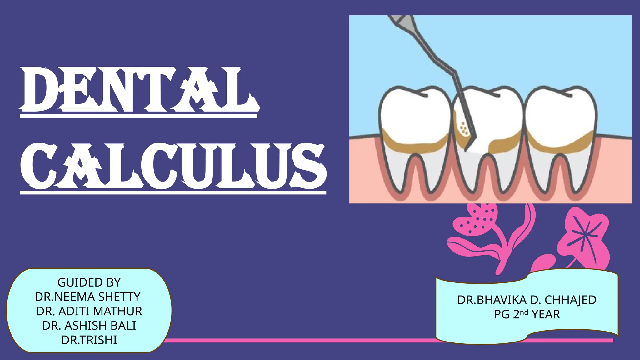
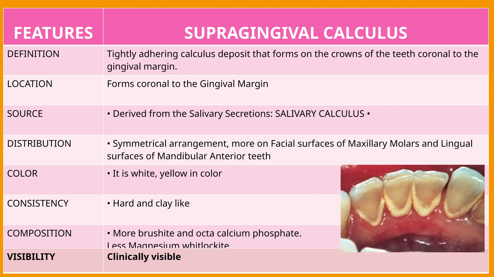
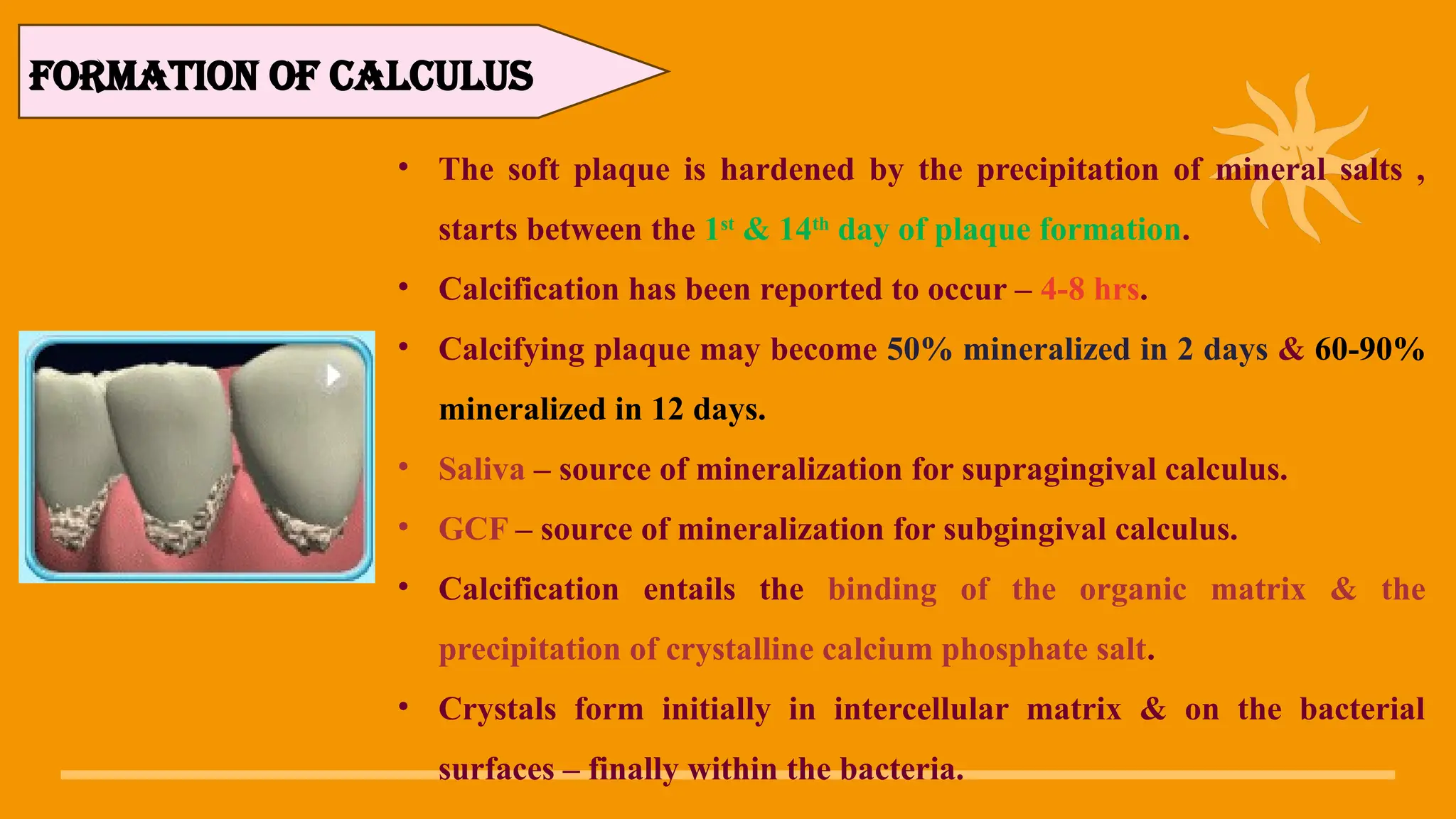
dental
CALCULUS
GUIDED BY
DR.NEEMA SHETTY
DR. ADITI MATHUR
DR. ASHISH BALI
DR.TRISHI
DR.BHAVIKA D. CHHAJED
PG 2nd
YEAR
CONTENT
• INTRODUCTION
• HISTORY
• CLASSIFICATION
• COMPOSITION
• STRUCTURE OF CALCULUS
• FORMATION OF CALCULUS
• THEORIES OF CALCULUS FORMATION
• CACLCULUS ATTACHMENT
• ASSESSMENT AND EVALUATION OF CALCULUS
• ETIOLOGY SIGNIFICANCE OF CALCULUS IN PERIODONTAL DISEASES
• CONCLUSION
• REFERENCES
INTRODUCTION
• The primary cause of gingival inflammation is bacterial plaque.
• Other predisposing factors include calculus,faulty restorations, complications associated
with orthodontic therapy , self inflicted injuries and use of tobacco.
• Calculus is derived from Greek words : Calcis : Limestone
• Tartar : White encrustations inside casks
• Once a tooth erupts, various material gets accumulated on the tooth surface, frequently
called tooth accumulated deposits/materials.
• Soft Deposits : Acquired pellicle, Microbial plaque,Material Alba , Food debris
• Hard Deposits : Calculus
DEFINITION
Calculus consists of mineralized bacterial plaque that forms on the surface of natural
teeth & dental prosthesis.
-CARRANZA 13TH
EDI
Calculus can be defined as a hard concretion that forms on the teeth or dental
prosthesis through the calcification of bacterial plaque.
-PERIOBASICS
SYNONYMS:
Tartar
Disambiguation
Calcis
Odontolithiasis
Fossilized plaue
Introduced the
term ‘TARTAR’
In 1535
PARACELSUS
First to clearly
enunciate the
relationship
between calculus
& periodontal
disease.
In 936-1013AD
ALBUCASIS
First formal
association
between
deposits & oral
disease
In 460-377BC
HIPPOCRATE
S
HISTORY
ACCORDING TO LOCATION:
• Supragingival calculus
• Subgingival calculus
ACCORDING TO SOURCE OF
MINERALIZATION:
• Salivary calculus
• Serumal calculus
CLASSIFICATION
COMPOSITION
INORGANIC CONTENT:
Major inorganic proportions –
76% calcium phosphate
- 3% calcium carbonate
- traces of magnesium phosphate
- other metals
• Principle inorganic components
–
- 39% calcium
- 19% phosphorus
- 2% carbon dioxide
- 1% magnesium
- trace amount of sodium ,
zinc , Strontium ,bromine, copper ,
tungsten , aluminum , silicon ,
iron , fluorine
Crystal forms:
- hydroxyapatite 58%
- magnesium whilockite 21%
- octacalcium phosphate 12%
- brushite 9%
- 97-100% of all supragingival calculus contains
hydroxyapatite & octacalcium phosphate.
- In the mandi. Anterior region – brushite
- In the posterior areas – magnesium whitlockite
ORGANIC CONTENT:
• Mixture of protein-polysaccharide complexes , desquamated epi.
Cells , leukocytes & various types of micro-orgs.
• 1.9% to 9.1% of organic content – carbohydrate consists of galactose ,
glucose , mannose , glucuronic acid , galactosamine.
• Salivary proteins ( 5.9% - 8.2% ) – most of amino acids
• 0.2% lipid – in the form of natural fats , free fatty acids , cholesterol ,
phospholipids.
• Composition of supragingival calculus is similar to that of subgingival
calculus with some difference :
Same hydroxyapatite content , more magnesium whitlockite & less
brushite , octacalcium phosphate.
Ratio of calcium & phosphorus is higher in subgingivally & the sodium
content es with the depth of the periodontal pockets.
FEATURES SUPRAGINGIVAL CALCULUS
DEFINITION Tightly adhering calculus deposit that forms on the crowns of the teeth coronal to the
gingival margin.
LOCATION Forms coronal to the Gingival Margin
SOURCE • Derived from the Salivary Secretions: SALIVARY CALCULUS •
DISTRIBUTION • Symmetrical arrangement, more on Facial surfaces of Maxillary Molars and Lingual
surfaces of Mandibular Anterior teeth
COLOR • It is white, yellow in color
CONSISTENCY • Hard and clay like
COMPOSITION • More brushite and octa calcium phosphate.
Less Magnesium whitlockite
VISIBILITY Clinically visible
FEATURES SUBGINGIVAL CALCULUS
DEFINITION • Calcified deposits that forms on the tooth surface below tre free margin of gingiva
LOCATION Deposits present apical to the crest of the Marginal Gingivaα
SOURCE Derived from the Gingival Exudate: SERUMINAL CALCULUS
DISTRIBUTION • Related to pocket depth, Heavier on Proximal Surfaces
COLOR • Brown/Greenish Black in color
CONSISTENSY • Hard and firm / flint or glass - like
COMPOSITION • Less brushite and octa calcium phosphate.
More magnesium whitlockite
VISIBILTY • Not visible on routine clinical examination
• Supragingival calculus – observed early in life , who do not have access to preventive
dental care.
• First areas of exhibit calculus deposits – facial aspect of maxi. Molars & lingual surfaces
of mandi. Incisors.
• Maximum calculus score – 25-30 years old.
• Calculus accumulation appeared to be symmetric – at 45 years.
• Subgingival calculus appeared 1st
independently or on the interproximal aspects of areas
where supragingival calculus already existed.
PREVALENCE
The following four modes of attachment have been
described
1. Attachment by means of organic pellicle on enamel
2. Mechanical interlocking in cemental resorption
lacunae
3.Penetration of calculus bacteria in cementum.
4. Close adaptation of calculus undersurface depressions
to gently sloping mounds on the unaltered cementum
surface
CALCULUS ATTACHMENT
• The soft plaque is hardened by the precipitation of mineral salts ,
starts between the 1st
& 14th
day of plaque formation.
• Calcification has been reported to occur – 4-8 hrs.
• Calcifying plaque may become 50% mineralized in 2 days & 60-90%
mineralized in 12 days.
• Saliva – source of mineralization for supragingival calculus.
• GCF – source of mineralization for subgingival calculus.
• Calcification entails the binding of the organic matrix & the
precipitation of crystalline calcium phosphate salt.
• Crystals form initially in intercellular matrix & on the bacterial
surfaces – finally within the bacteria.
FORMATION OF CALCULUS
• As calcification progresses, the no. of filamentous bacteria & the foci of calcification
change from basophilic to eosinophilic.
• Calculus – formed in layers – separated by a thin cuticle that becomes embedded in the
calculus as calcification progresses.
• Average daily increment in calculus formers – about 0.10%-0.15% by weight.
• The time required for calculus to reach its maximal level – 10 weeks to 6 moths.
• The decline from maximal accumulation is referred to as “reversal phenomenon”.
RATE OF FORMATION
THEORIES OF CALCULUS FORMATION
BOOSTER MECHANISM
• Bacteria may form phosphatases , which the local conc. Of phosphatases &
thereby lead to calcification.
• It affect the pH of plaque & saliva & destroy protective colloidal action.
• It attach the calculus to the tooth.
• It provides chemicals that induce mineralization.
BACTERIAL THEORY
EPITACTIC THEORY
• According to this theory , seeding agents induce small foci of
calcification that enlarge and coalesce to form a calcified mass referred
to as the epitactic concept or heterogeneous nucleation.
• Amorphous non-crystalline deposits & brushite – transformed to
octacalcium phosphate - then to hydroxyapatite.
• In salivary calculus , brushite – result of the local elevation of Ph , calcium
& phosphate conc. – then maturing process be modified to crystal of
higher calcium to phosphate ratio.
TRANSFORMATION THEORY
• Occurring at specific sites because of the existence of an inhibiting
mechanism at non-calcifying sites.
• During the calcification , inhibitor is parently altered or removed.
• Inhibiting substances – pyrophosphate & enzyme alkaline phosphatase.
INIIBITION THEORY
• According to this theory, calculus formation is the resultant of the action of
phosphatases derived from either oral tissues or oral microorganism on
some salivary phosphate containing complex, most probably phospheric
esters of the hexophosphoric group
ENZYMATIC THEORY
• Oral hygiene index- simplified (John C. Green & Jack R. Vermillion 1994)
• Calculus surface index (Ennever j et al. 1961)
• Calculus surface severity index (Ennever j et al 1961)
• Volpe-manhold method of calculus (A.R.Volpe et al. 1962)
• Calculus index used by national institue of dental research
• Calculus component of periodontal disease index (Ramfjord 1959)
INDICES
CAUSES PERIODONTAL DESTRUCTION IN THE
FOLLOWING MANNER:
• Calculus brings bacterial overlay closer to the supporting
tissues.
• Interfere with local self-cleansing mechanism
• Provide nidus for continuous plaque accumulation.
• Make plaque removal more difficult.
ROLE OF CALCULUS IN PERIODONTAL DISEASE
Conventional calculus detection technique:
1. Direct vision – limited for supragingival calculus only.
2. Compressed air can facilitated the viewing of subgingival calculus.
3. Tactile sense of the operator – by using probe , explorer or a
curette
4. Radiographic technique
DIAGNOSIS
Advancements in calculus detection
technique:
Calculus detection systems
1. Perioscopy (fiberoptic endoscopy based technology)
2. detecTar (spectro-optical technology)
3. Diagnodent (autofluorescence based technology)
Calculus detection & removal system:
1. Perioscan (ultrasound technology)
2. Keylaser (laser based technology)
Agents for softening the mature calculus deposit:
1. ACIDS
Earliest techniques - wooden stick which was moistened with aromatic sulphuric acid
before being introduced into a periodontal pocket to dissolve calculus and to act on
the soft tissues as an astringent (Barker 1872).
Niles (1881) - nitro muriatic acid
Other acids - 20% trichloroacetic acid, bifluoride of mercury and 10% sulphuric acid
DISADVANTAGES - caustic to soft tissues and decalcify tooth structure.
ANTICALCULUS AGENTS:
2. ALKALIS:
Badanes (1929) - argued that it was the action of the mild
alkalis contained in the waters that dissolved the three
principal organic constituents of salivary calculus;
globulin, mucin and calcium oxalate.
• 3. CHELATING AGENTS:
• Chelating agents are used to dissolve crystallized calcium
salts and are capable of combining with calcium to form
stable compounds.
• Sodium hexametaphosphate was found to remove
supragingival calculus from extracted teeth in 10 to 15 days
(Kerr & Field 1944).
• EDTA – (jabro et al. 1992) – application of EDTA gel resulted
in ease of calculus removal.
ANTIMICROBIALS:
1. CHLORHEXIDINE
• Cationic bis-biguanide which acts by being absorbed
onto the bacterial cell wall – leading to damage of
permeability barriors
• At high conc. – precipitation & coagulation of the
cytoplasmic contents occurs (Hennessey 1977).
2. ZINC IONS:
• Reduce plaque acidogenicity (oppermann 1980) &
plaque growth (sexton 1986).
• Inhibits crystal growth by binding to the surface of
solid calcium & phosphate (gilbert 1988).
3. BISPHOSPHONATES:
• Synthetic pyrophosphate analogues – to prevent
calcium deposition by inhibiting crystal growth.
4. TRICLOSAN:
• Non-ionic antibacterial agent with a wide spectrum
of activity against bacteria , fungi , & yeasts.
• When delivered from a dentifrices – bind to oral
mucous membrane & tooth surfaces.
• Used in combination with other anti-calculus
agents.
Anti-calculus agents used in commercial
dentifrices:
1. Triclosan with PVM/MA copolymer
2. Pyrophosphate with PVM/MA copolymer
3. Zinc ions
• Calculus plays an important role in maintaining & accentuating
periodontal disease by keeping plaque in close contact with
the gingival tissue & creating areas where plaque removal is
impossible.
• Therefore, the clinical skill to remove the calculus & other
irritants as a basis for adequate periodontal & prophylactic
therapy.
CONCLUSION:
• Newman and Carranza’s , clinical periodontology , 13th
edition.
• Dr. Nitin Saroch , periobasics , A Textbook of Periodontics & Implantology , 2nd
edition.
• Fairbrother KJ, Heasman PA. Anticalculus agents. J Clin Periodontol. 2000
May;27(5):285-301.
• Shalu bathla , textbook of periodontics , 1st
edition.
• Akcalı A, Lang NP. Dental calculus: the calcified biofilm and its role in disease
development. Periodontol 2000. 2018 Feb;76(1):109-115.
REFERENCES

SEMINAR on the topic Dental calculus.pptx

  • 1.
    dental CALCULUS GUIDED BY DR.NEEMA SHETTY DR.ADITI MATHUR DR. ASHISH BALI DR.TRISHI DR.BHAVIKA D. CHHAJED PG 2nd YEAR
  • 2.
    CONTENT • INTRODUCTION • HISTORY •CLASSIFICATION • COMPOSITION • STRUCTURE OF CALCULUS • FORMATION OF CALCULUS • THEORIES OF CALCULUS FORMATION • CACLCULUS ATTACHMENT • ASSESSMENT AND EVALUATION OF CALCULUS • ETIOLOGY SIGNIFICANCE OF CALCULUS IN PERIODONTAL DISEASES • CONCLUSION • REFERENCES
  • 3.
    INTRODUCTION • The primarycause of gingival inflammation is bacterial plaque. • Other predisposing factors include calculus,faulty restorations, complications associated with orthodontic therapy , self inflicted injuries and use of tobacco. • Calculus is derived from Greek words : Calcis : Limestone • Tartar : White encrustations inside casks • Once a tooth erupts, various material gets accumulated on the tooth surface, frequently called tooth accumulated deposits/materials. • Soft Deposits : Acquired pellicle, Microbial plaque,Material Alba , Food debris • Hard Deposits : Calculus
  • 4.
    DEFINITION Calculus consists ofmineralized bacterial plaque that forms on the surface of natural teeth & dental prosthesis. -CARRANZA 13TH EDI Calculus can be defined as a hard concretion that forms on the teeth or dental prosthesis through the calcification of bacterial plaque. -PERIOBASICS SYNONYMS: Tartar Disambiguation Calcis Odontolithiasis Fossilized plaue
  • 5.
    Introduced the term ‘TARTAR’ In1535 PARACELSUS First to clearly enunciate the relationship between calculus & periodontal disease. In 936-1013AD ALBUCASIS First formal association between deposits & oral disease In 460-377BC HIPPOCRATE S HISTORY
  • 6.
    ACCORDING TO LOCATION: •Supragingival calculus • Subgingival calculus ACCORDING TO SOURCE OF MINERALIZATION: • Salivary calculus • Serumal calculus CLASSIFICATION
  • 7.
    COMPOSITION INORGANIC CONTENT: Major inorganicproportions – 76% calcium phosphate - 3% calcium carbonate - traces of magnesium phosphate - other metals • Principle inorganic components – - 39% calcium - 19% phosphorus - 2% carbon dioxide - 1% magnesium - trace amount of sodium , zinc , Strontium ,bromine, copper , tungsten , aluminum , silicon , iron , fluorine
  • 8.
    Crystal forms: - hydroxyapatite58% - magnesium whilockite 21% - octacalcium phosphate 12% - brushite 9% - 97-100% of all supragingival calculus contains hydroxyapatite & octacalcium phosphate. - In the mandi. Anterior region – brushite - In the posterior areas – magnesium whitlockite
  • 9.
    ORGANIC CONTENT: • Mixtureof protein-polysaccharide complexes , desquamated epi. Cells , leukocytes & various types of micro-orgs. • 1.9% to 9.1% of organic content – carbohydrate consists of galactose , glucose , mannose , glucuronic acid , galactosamine. • Salivary proteins ( 5.9% - 8.2% ) – most of amino acids • 0.2% lipid – in the form of natural fats , free fatty acids , cholesterol , phospholipids.
  • 10.
    • Composition ofsupragingival calculus is similar to that of subgingival calculus with some difference : Same hydroxyapatite content , more magnesium whitlockite & less brushite , octacalcium phosphate. Ratio of calcium & phosphorus is higher in subgingivally & the sodium content es with the depth of the periodontal pockets.
  • 11.
    FEATURES SUPRAGINGIVAL CALCULUS DEFINITIONTightly adhering calculus deposit that forms on the crowns of the teeth coronal to the gingival margin. LOCATION Forms coronal to the Gingival Margin SOURCE • Derived from the Salivary Secretions: SALIVARY CALCULUS • DISTRIBUTION • Symmetrical arrangement, more on Facial surfaces of Maxillary Molars and Lingual surfaces of Mandibular Anterior teeth COLOR • It is white, yellow in color CONSISTENCY • Hard and clay like COMPOSITION • More brushite and octa calcium phosphate. Less Magnesium whitlockite VISIBILITY Clinically visible
  • 12.
    FEATURES SUBGINGIVAL CALCULUS DEFINITION• Calcified deposits that forms on the tooth surface below tre free margin of gingiva LOCATION Deposits present apical to the crest of the Marginal Gingivaα SOURCE Derived from the Gingival Exudate: SERUMINAL CALCULUS DISTRIBUTION • Related to pocket depth, Heavier on Proximal Surfaces COLOR • Brown/Greenish Black in color CONSISTENSY • Hard and firm / flint or glass - like COMPOSITION • Less brushite and octa calcium phosphate. More magnesium whitlockite VISIBILTY • Not visible on routine clinical examination
  • 13.
    • Supragingival calculus– observed early in life , who do not have access to preventive dental care. • First areas of exhibit calculus deposits – facial aspect of maxi. Molars & lingual surfaces of mandi. Incisors. • Maximum calculus score – 25-30 years old. • Calculus accumulation appeared to be symmetric – at 45 years. • Subgingival calculus appeared 1st independently or on the interproximal aspects of areas where supragingival calculus already existed. PREVALENCE
  • 14.
    The following fourmodes of attachment have been described 1. Attachment by means of organic pellicle on enamel 2. Mechanical interlocking in cemental resorption lacunae 3.Penetration of calculus bacteria in cementum. 4. Close adaptation of calculus undersurface depressions to gently sloping mounds on the unaltered cementum surface CALCULUS ATTACHMENT
  • 15.
    • The softplaque is hardened by the precipitation of mineral salts , starts between the 1st & 14th day of plaque formation. • Calcification has been reported to occur – 4-8 hrs. • Calcifying plaque may become 50% mineralized in 2 days & 60-90% mineralized in 12 days. • Saliva – source of mineralization for supragingival calculus. • GCF – source of mineralization for subgingival calculus. • Calcification entails the binding of the organic matrix & the precipitation of crystalline calcium phosphate salt. • Crystals form initially in intercellular matrix & on the bacterial surfaces – finally within the bacteria. FORMATION OF CALCULUS
  • 16.
    • As calcificationprogresses, the no. of filamentous bacteria & the foci of calcification change from basophilic to eosinophilic. • Calculus – formed in layers – separated by a thin cuticle that becomes embedded in the calculus as calcification progresses. • Average daily increment in calculus formers – about 0.10%-0.15% by weight. • The time required for calculus to reach its maximal level – 10 weeks to 6 moths. • The decline from maximal accumulation is referred to as “reversal phenomenon”. RATE OF FORMATION
  • 17.
  • 18.
  • 19.
    • Bacteria mayform phosphatases , which the local conc. Of phosphatases & thereby lead to calcification. • It affect the pH of plaque & saliva & destroy protective colloidal action. • It attach the calculus to the tooth. • It provides chemicals that induce mineralization. BACTERIAL THEORY
  • 20.
    EPITACTIC THEORY • Accordingto this theory , seeding agents induce small foci of calcification that enlarge and coalesce to form a calcified mass referred to as the epitactic concept or heterogeneous nucleation.
  • 21.
    • Amorphous non-crystallinedeposits & brushite – transformed to octacalcium phosphate - then to hydroxyapatite. • In salivary calculus , brushite – result of the local elevation of Ph , calcium & phosphate conc. – then maturing process be modified to crystal of higher calcium to phosphate ratio. TRANSFORMATION THEORY
  • 22.
    • Occurring atspecific sites because of the existence of an inhibiting mechanism at non-calcifying sites. • During the calcification , inhibitor is parently altered or removed. • Inhibiting substances – pyrophosphate & enzyme alkaline phosphatase. INIIBITION THEORY
  • 23.
    • According tothis theory, calculus formation is the resultant of the action of phosphatases derived from either oral tissues or oral microorganism on some salivary phosphate containing complex, most probably phospheric esters of the hexophosphoric group ENZYMATIC THEORY
  • 24.
    • Oral hygieneindex- simplified (John C. Green & Jack R. Vermillion 1994) • Calculus surface index (Ennever j et al. 1961) • Calculus surface severity index (Ennever j et al 1961) • Volpe-manhold method of calculus (A.R.Volpe et al. 1962) • Calculus index used by national institue of dental research • Calculus component of periodontal disease index (Ramfjord 1959) INDICES
  • 25.
    CAUSES PERIODONTAL DESTRUCTIONIN THE FOLLOWING MANNER: • Calculus brings bacterial overlay closer to the supporting tissues. • Interfere with local self-cleansing mechanism • Provide nidus for continuous plaque accumulation. • Make plaque removal more difficult. ROLE OF CALCULUS IN PERIODONTAL DISEASE
  • 26.
    Conventional calculus detectiontechnique: 1. Direct vision – limited for supragingival calculus only. 2. Compressed air can facilitated the viewing of subgingival calculus. 3. Tactile sense of the operator – by using probe , explorer or a curette 4. Radiographic technique DIAGNOSIS
  • 27.
    Advancements in calculusdetection technique: Calculus detection systems 1. Perioscopy (fiberoptic endoscopy based technology) 2. detecTar (spectro-optical technology) 3. Diagnodent (autofluorescence based technology)
  • 28.
    Calculus detection &removal system: 1. Perioscan (ultrasound technology) 2. Keylaser (laser based technology)
  • 29.
    Agents for softeningthe mature calculus deposit: 1. ACIDS Earliest techniques - wooden stick which was moistened with aromatic sulphuric acid before being introduced into a periodontal pocket to dissolve calculus and to act on the soft tissues as an astringent (Barker 1872). Niles (1881) - nitro muriatic acid Other acids - 20% trichloroacetic acid, bifluoride of mercury and 10% sulphuric acid DISADVANTAGES - caustic to soft tissues and decalcify tooth structure. ANTICALCULUS AGENTS:
  • 30.
    2. ALKALIS: Badanes (1929)- argued that it was the action of the mild alkalis contained in the waters that dissolved the three principal organic constituents of salivary calculus; globulin, mucin and calcium oxalate.
  • 31.
    • 3. CHELATINGAGENTS: • Chelating agents are used to dissolve crystallized calcium salts and are capable of combining with calcium to form stable compounds. • Sodium hexametaphosphate was found to remove supragingival calculus from extracted teeth in 10 to 15 days (Kerr & Field 1944). • EDTA – (jabro et al. 1992) – application of EDTA gel resulted in ease of calculus removal.
  • 32.
    ANTIMICROBIALS: 1. CHLORHEXIDINE • Cationicbis-biguanide which acts by being absorbed onto the bacterial cell wall – leading to damage of permeability barriors • At high conc. – precipitation & coagulation of the cytoplasmic contents occurs (Hennessey 1977).
  • 33.
    2. ZINC IONS: •Reduce plaque acidogenicity (oppermann 1980) & plaque growth (sexton 1986). • Inhibits crystal growth by binding to the surface of solid calcium & phosphate (gilbert 1988). 3. BISPHOSPHONATES: • Synthetic pyrophosphate analogues – to prevent calcium deposition by inhibiting crystal growth.
  • 34.
    4. TRICLOSAN: • Non-ionicantibacterial agent with a wide spectrum of activity against bacteria , fungi , & yeasts. • When delivered from a dentifrices – bind to oral mucous membrane & tooth surfaces. • Used in combination with other anti-calculus agents.
  • 35.
    Anti-calculus agents usedin commercial dentifrices: 1. Triclosan with PVM/MA copolymer 2. Pyrophosphate with PVM/MA copolymer 3. Zinc ions
  • 36.
    • Calculus playsan important role in maintaining & accentuating periodontal disease by keeping plaque in close contact with the gingival tissue & creating areas where plaque removal is impossible. • Therefore, the clinical skill to remove the calculus & other irritants as a basis for adequate periodontal & prophylactic therapy. CONCLUSION:
  • 37.
    • Newman andCarranza’s , clinical periodontology , 13th edition. • Dr. Nitin Saroch , periobasics , A Textbook of Periodontics & Implantology , 2nd edition. • Fairbrother KJ, Heasman PA. Anticalculus agents. J Clin Periodontol. 2000 May;27(5):285-301. • Shalu bathla , textbook of periodontics , 1st edition. • Akcalı A, Lang NP. Dental calculus: the calcified biofilm and its role in disease development. Periodontol 2000. 2018 Feb;76(1):109-115. REFERENCES