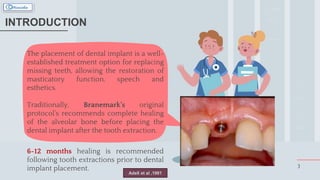
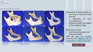
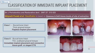
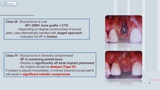
DR. RINISHA SINHA
MDS III POSTGRADUATE TRAINEE
IMMEDIATE
IMPLANT SURGERY
TABLE OF CONTENTS
2
01 04
05
02
03
INTRODUCTION
HISTORY CONCLUSION
POTENTIAL COMPLICATIONS
IMMEDIATE IMPLANTS
The placement of dental implant is a well-
established treatment option for replacing
missing teeth, allowing the restoration of
masticatory function, speech and
esthetics.
Traditionally, Branemark’s original
protocol’s recommends complete healing
of the alveolar bone before placing the
dental implant after the tooth extraction.
6-12 months healing is recommended
following tooth extractions prior to dental
implant placement. 3
INTRODUCTION
Adell et al ,1981
4
Extraction of Teeth results in
• the loss of hard and soft
tissues,
• a reduction of arch
circumference,
• deficient width and height
of the residual ridge
Bone loss occurs
• both buccolingually and
• apicocoronally,
with the first six months
carrying the highest rate of
resorption in either direction.
Atwood, 1983
5
WILSON & WEBER; 1998
MAYFIELD ET AL. 1999
CLASSIFICATION OF IMPLANT PLACEMENT
IMMEDIATE RECENT
MATURE DELAYED
Implant
Placement IMMEDIATE
DELAYED
LATE
HAMMERLE ET AL. 2004
TYPE
I
TYPE
II
TYPE
III
TYPE
IV
ESPOSITO ET AL. 2006
IMMEDIATE
IMMEDIATE
DELAYED
DELAYED
Varying degrees
of resorption and
remodeling
Long waiting period several surgical
interventions
6
Traditional Implant Placement
NOT VERY
ATTRACTIVE
7
German group under the leadership of
Professor WILLY SCHULTE, 1978 who
recommended the so called “TUBINGER
SOFORT IMPLANTANT”.
He proposed the concept of Immediate Implant
Placement using ALUMINUM OXIDE
IMPLANTS
This attempts was unsuccessful not because
of the t/t approach but due to the failure of
biomaterial itself.
Aluminum oxide implants showed high rate of
complications and failures due to implant
fractures. Professor WILLY SCHULTE
8
Schulte and Heimke in 1976
HISTORY
Initial report in the literature
Lazzara in 1989
Illustrated this method of treatment with three
case reports
Gelb in 1993
Validated on a series of 50 consecutive
cases followed over a 3-year period,
providing a survival rate of 98%
Since then, numerous animal studies,
human case reports, and several
randomized controlled studies have
furthered the science of this treatment
modality.
Chen, Wilson et al. 2004
Chen, Beagle et al. 2009
Pre-treatment
alveolar
ridge contours
Chen, Wilson et al. 2004
Outcomes of immediate
placement procedures can
be equally successful as a
delayed approach when
initial primary stability is
achieved.
Barzilay 1993; Schwartz-Arad and Chaushu 1997;
Mayfield 1999; Chen, Wilson et al. 2004; Chen, Beagle et
al. 2009
10
CLASSIFICATION OF IMMEDIATE IMPLANT PLACEMENT
Int J Periodontics and Restorative dent : 2007:27: 313-323
Akiyoshi Funato et al. Classification is based on Osseous & Soft tissue levels of site of extraction.
Class I - Buccal bone Intact
Thick gingival biotype
Flapless Implant placement
Class II - Buccal bone Intact
Thin, scalloped gingival biotype
Immediate implant placement and connective
tissue graft or staged CTG
11
Class III - Buccal bone is Lost
IIP+ GBR+ bone grafts + CTG
- Depending on degree compromises of buccal
plate, case alternatively handled with staged approach.
- Indication for IIP is limited.
Class IV - Buccal bone is Severely compromised
- IIP in remaining palatal bone.
- Results in significantly off Axial Implant placement
- So implant should be delayed (Type IV)
If implant is placed immediately, it inclines towards buccal wall &
will result in significant esthetic compromise.
12
ADVANTAGES
• Preservation of Ridge Contour
• Reduction of Treatment visits, cost and
time
• Maintenance of ideal soft tissue contour
• Improved patient Psychological Outlook
for implant treatment
• Reduction of Surgical Procedure
• Enhanced Healing and Osteogenic
potential
• Simplification of Restorative care
• Optimization of aesthetic and functional
results
Lazzara 1989; Parel and Triplett 1990; Shanaman 1992; Werbitt and
Goldberg 1992; Denissen, Kalk et al. 1993; Schultz 1993; Watzek,
Haider et al. 1995; Missika, Abbou et al. 1997
13
STUDIES IN SUPPORT
Over the past 16 years, numerous studies have confirmed the reliability of implants placed in the post
extraction sockets.
In 1989, Lazzara evaluated the surgical and restorative advantages of implants placed in post
extraction sockets.
Hammerle et al., 2004 – in the consensus report, they stated that Immediate Implant placement has optional
availability for existing bone for implant placement & reduced overall treatment time.
Several prospective studies (Yokuna 1991, Becket et al 1998, 1999, Polizzi, al 2000), retrospective
studies (Held 1993, Gold stein et al 2002 Watzek et al 1995) reported high success rates of implants.
Lindquist et al. 1998, Von Wowern et al. 1990, Dennisen et al. 1993, Werbitt & Goldberg 1992 - Immediate
implant placement in fresh extraction sockets may counteract the alveolar bone resorption that results in
reduction of ridge dimensions.
The overall procedure has proven to have a positive psychological impact on the patient (Gelb 1993,
Cornelini et al. 2000, Kan and Runsaeng 2000).
14
DISADVANTAGES
• Remnants of infection  Contamination
during the initial healing period  Implant
failure
• Extended long axis of implant should be
slightly lingual to incisal edge of the definitive
restorations; also sometimes the morphology of
the socket can compromise initial implant
stability.
• Lack of keratinized mucosa for flap adaptation
 Increased risk of marginal mucosal recession
particularly in thin tissue biotype cases, and
when there is a facial bone dehiscence.
• Technique-sensitive
Lazzara 1989; Parel and Triplett 1990; Shanaman 1992; Werbitt and
Goldberg 1992; Denissen, Kalk et al. 1993; Schultz 1993; Watzek,
Haider et al. 1995; Missika, Abbou et al. 1997
15
STUDIES IN SUPPORT
Studies in human: Araujo et al. 2005, 2006; Botticelli et al. 2006; Studies In animals: Covani et al.
2003, 2004; Botticelli et al. 2004 and Araujo et al. 2005 showed failure of immediate implants to prevent the
resorption of buccal bone.
Mariano sanz et al 2009: the thickness of the buccal bone wall as well as the dimension of the
horizontal gap influenced the hard tissue alterations that occur following immediate implant placement into
extraction sockets.
Daniele botticelli et al. 2010 did an analysis on hard tissue formation adjacent to implants of various size
and configuration immediately placed into extraction sockets and claimed that the installment of root formed
wide implants immediately into extraction sockets will not prevent the resorption of the alveolar crest.
16
INDICATIONS
Traumatically Avulsed Retained Deciduous Teeth Horizontal or Vertical Fracture
Failed Endo-treatment Periodontally Compromised Non-restorable Teeth
17
CONTRAINDICATIONS
Inability to develop
mechanical stability
Proximity to Adjacent teeth Placement outside
alveolar envelope
Presence of Infection
18
PRIMARY STABILITY
Lazzara 1989
The implant needs to engage the bone
• along the lateral walls of the socket without
changing the original socket depth, or
• by engaging bone apical to the original socket
dimensions.
In either of these situations, only one to three
threads of the implant need to be in contact with the
osteotomy site.
19
SALAMA AND SALAMA; 1993
2
0
WHAT IF IT DOESN’T HAPPEN ?? Any implant that can be moved laterally with
finger pressure following placement will have a
poor chance of achieving osseointegration
should be ABORTED
An undersized osteotomy may result in
compression necrosis of the bone, thus
causing implant failure to occur.
Choosing an implant with restorative platform
too large for the planned restoration, only
because the larger implant diameter is able to
achieve stability  concern for esthetics and
horizontal defect dimension
Buser, Martin et al. 2004; Araujo, Wennstrom et al. 2006
21
INFERIOR
ALVEOLAR
NERVE
Buser, von Arx et al. 2000 – Implants should be at least 2 mm superior
to the inferior alveolar nerve during the osteotomy and placement of
the implant
Mandibular second premolar sites frequently have their apex near the
mental foramen and also have a wide socket morphology requiring a
4.8mm implant diameter for stability.
The variability of root morphology for mandibular first and second
molars  avoid the temptation to place the implant into the mesial or
distal root socket to achieve stability  will result in an implant that
results in poor positioning from a restorative perspective.
If an immediate molar implant can be placed, grafting the horizontal
defect dimension with an osseous graft and use of a bioresorbable
membrane will be required, and a healing time to achieve
osseointegration may exceed 16 weeks.
Chen, Wilson et al. 2004
Fugazzotto 2008a, 2008b
22
MAXILLARY
SINUS
the second premolar or first and second molar sites
It is desirable not to place a maxillary implant into the
mesial-buccal, distal-buccal, or palatal root areas to
gain primary stability, only to have an implant
positioned poorly from a restorative perspective.
Certainly, if adequate bone height is available in
maxillary molar sites without penetration into the sinus,
an immediate implant can be inserted, but these
circumstances do not occur frequently.
Fugazzotto and De 2002
Fugazzotto 2008a, 2008b
23
CHECKLIST
• Smile line
• Esthetic Evaluation
• Periodontal biotype
• Probing
• Occlusal Analysis
• Radiographic Evaluation
• Condition of the socket
• Patient psychiatric profile
Shanaman 1992; Werbitt and Goldberg 1992; Denissen, Kalk et al.
1993; Schultz 1993; Watzek, Haider et al. 1995
2
4
CLINICAL REQUIREMENTS TO BE FULFILLED FOR THE SUCCESS
• Absence of any frank active infection,
• Good mechanical anchorage and primary
stability of the implant fixture within the
alveolar socket,
• Atraumatic removal of the unsalvageable
tooth,
• Preservation of the labial plate of bone,
• Use of the appropriate implant design that
corresponds to the socket’s configuration,
• Proper implant position in terms of
angulation & position
Buser, Martin et al. 2004; Araujo, Wennstrom et al. 2006
25
ANATOMIC CONFIGURATIONS AFTER TOOTH EXTRACTION
After a tooth has been extracted, the
resultant defect in the bone may
have several anatomic
configurations that directly
influence implant placement.
Chen, Wilson et al. 2004
26
Loss of all labial bone to the apex of the tooth
• If the bone on the facial or labial aspect of the socket is not present, the
clinician should graft the socket and delay implant placement.
• The predictability of success decreases even when a graft and membrane
are placed, because implant stability at the time of placement is
compromised by the loss of bone.
Loss of a portion (3 to 6mm) of the labial bone.
• Graft is necessary to restore the labial portion of the missing bone.
• If the implant can be placed with its palatal, mesial, and distal surfaces in
contact with the bone and at least 5 mm of apical bone is present to secure
the implant, the lost bone is reconstructed with particulate graft material
27
28
Loss of less than 3 mm of labial bone at the crest
• This is a very common situation when a tooth with extensive caries or a fracture is
extracted.
• After the tooth has been extracted, the crestal resorption is limited to 3 mm from the
planned gingival margin of the final restoration.
• In this situation, the implant is placed at the level of the bone, with attention to implant
collar design. The height of first thread determines the ultimate bone levels adjacent to the
implant.
Lack of bone superior to the apex of the socket, with extreme proximity of adjacent vital
structures, such as the interior alveolar canal, mental foramen, floor of the nose, and floor of
the sinus.
• The mental foramen may be close to the apex of the first or second mandibular premolar
• In the maxilla, preoperative - nasal floor or sinus.
29
Lack of palatal or lingual bone
• This is an uncommon finding,
because palatal or lingual bone is
the last to resorb during
inflammation around a tooth.
• If the palatal or lingual bone is not
present, a graft is necessary before
placement of an implement.
• Another viable option is to delay
grafting for 4 weeks and then place
an onlay graft harvested from the
symphysis.
30
Concavity along the palatal or
labial contour of the
extraction site
• Congenially missing teeth and
retained deciduous teeth in the
anterior maxilla
• The buccal bone may be normal in
shape, with a concavity of the
palatal aspect either along with
entire palatal cortex or 3 to 5mm
from the crest, with an indentation
along with the palatal bone contour
in the axial plane for the implant.
A graft is necessary in this situation,
and in a location with dense palatal
tissue, obtaining an excellent result
is difficult. Onlay grafts are difficult
to place along a palatal concavity.
The continued pressure from
the dense palatal tissue often
causes graft resorpotion
32
Socket that is larger than the proposed diameter of the implant in all dimensions.
• minimum - 5mm of bone is present beyond the apex of the socket
• grafting of the large extraction socket & delayed placement
Socket that is oval in shape, with the long dimension palatal to facial and the short dimension
mesial to distal
• The oval socket typically is found in premolar sites.
• After the implant has been placed, the gap between the implant and labial cortex can be
grafted to prevent epithelial migration
Very thin surrounding bone
• use of periotomes or other extraction devices designed to preserve bone.
• the implant can be placed and the space between the implant and this bone can be grafted
with particulate mineralized bone material..
• If insufficient bone is present to stabilize the implants, a graft is indicated and a delayed
response is planned
• The thinner the bone, the greater the chance of resorption during the post-extraction period
33
Favorable Clinical Conditions
• Thick tissue biotype
• Intact thick facial bone wall
• Sites with no infection
Unfavorable Clinical Conditions
• Thin tissue biotype
• Thin facial bone wall (1mm) or Facial bone
defect
• High lip
• Acute infection – mainly in large apical bone
defects
IMPLANT
MORPHOLOGY
Implant morphological characteristics can influence the treatment outcome of
immediate implant therapy
Root form tapered implants parallel walled implants
• Better Handling And
Stability
• Reduce The Possibility Of
Adjacent Root
• Injury Labial Bone
Perforation
>
Implant : Crown Ratio
2 : 1 or more Favorable
Less than 1 : 1 Unfavorable
36
SURGICAL TECHNIQUE
Sulcular Incisions with Vertical release
Atraumatic Extraction
• Sectioning of Multi-root teeth
• Periotome
Thorough Degranulation
Assessment of Socket Architecture
Widening/Deepening of Osteotomy
Placement of Implants
Placement of Graft/Membrane
Flap Closure
37
ATRAUMATIC EXTRACTION
Local Anesthesia
Care should be taken to minimize the trauma to the gingival 15 scalpel blade: SULCULAR INCISION
Scalpel should be angled to follow the curvature of the tooth closely
Incising interdental papilla should be avoided
Minimised buccolingual luxations with controlled hand motions
A series of thin elevators, periotomes used to separate the bone from the labial, lingual and proximal surface of the
tooth. This allows the removal of tooth without removing the surrounding bone.
3
8
Using the periotome
As gentle pressure is applied
Periotome advances more apically
Periotome – separates the PDL and luxated the
tooth
In molar teeth, if necessary always split tooth with copious
irrigation to section the tooth with the use of airotor hand
piece and remove the individual roots separately
extraction forceps is rotated in a circular movement and the tooth
pulled vertically, without pressure on the labial bone
After tooth removal, a curette or an explorer is used to explore
the location of the buccal plate and confirm its integrity
After the tooth is removed, a spoon shaped curette is used to
remove granulation tissue
Procedural
Delays
If any purulent seen after removing the tooth
No hard or soft tissue grafting procedures should be performed
In some cases, it is not possible to make this determination preoperatively
OSTEOTOMY
PREPARATION
42
2 mm Round drill with copious
irrigation
The drill tip should be positioned along the palatal wall
of the extraction socket,
3 mm to 5 mm coronal to the apical end of the
extraction socket
Directing the drills or osteotomes along the palatal bone in the same long axis
as the tooth socket is recommended to help prevent excess force from being
applied to the labial or buccal wall
Palatal Wall
thicker
denser bone
Force the drill to the labial or
buccal wall
1. Thicker
2. More Stable Bone
3. Increasing Resistance To
Bone Resorption.
Maintain a small gap between the
implant and the labial plate may
facilitate secondary bone fill.
44
IMPROVEMENTS FOR PLACEMENT STABILITY
•The implant
should be seated
in at least two-
thirds in the host
bone
The apex should
be at least 1 mm
to 2 mm longer
than the tooth
being replaced
The implant
diameter at the
cervical area
should be as wide
as possible to
prevent soft tissue
ingrowth
The implant
should be totally
immobilized in
the site without
benefit of graft
material
45
Torque
resistance of
30-40 Ncm
Initial implant
stability
Resonance
Frequency
Analysis
Excessive
torque
Strip the
implant
threads 
Bone necrosis
Primary stability of the
implant should be the
result of mechanical
fixation.
Ideal position Acceptable positions Wrong position
POTENTIAL
COMPLICATIONS
• Cause : Intra operative soft tissue trauma , introduction of the bacterial
organism into the surgical site intraoperatively and postoperatively .
• Mainly caused because –
• in case of periapical pathologies or periodontal infection
• Failure of thorough debridement of the socket
• Failure to remove all the contaminated tissue
• Presence of infection in adjacent tooth
Post operative
infection or edema
• Causes : receptor site – for implant is too large , inadequate host bone , poor
bone density , increased HDD between implant surface and bone
Unstable implant at
the time of
placement
• Presence of infection at the post extraction site Untreated periodontitis
could potentially cross infection to the implant site
• - Local infection is caused by- endodontic lesions
• - root resorption
• - infected root remnants
• - post traumatic lesions
• - Foreign bodies etc
• All these are considered to be transient risk factors
• Sites with acute infection have high risk .
• Chronic infection have medium risk .
• Some studies indicate immediate implant placement into debrided
dentoalveolar sockets.
Local infection at
implant site
• Causes : increased occlusal overload
• Lost graft material
• Peri implant infection
• Measures : -
• Prosthesis Is Lighter In Occlusion
• Passive Closure Of Soft Tissue
• Minimize Flap Opening
Unstable implant
within 3 weeks of
implant placement
• Causes : preoperative evaluation not done properly .
• Preventive measures : ensure there is appropriate soft tissue preoperatively
or consider preoperative or intraoperative or post operative soft tissue
grafting
• poor implant positioning
• membrane exposure during healing
• inadequate bands of keratinized tissue
• after healing
• gingival recession
• implant failure
• unacceptable esthetic outcomes
Inadequate soft
tissue or
inappropriate soft
tissue esthetics
5
0
CONCLUSION
• The immediate implant placement approach has been
studied since the1970s
• Evidence available today indicated that it is a
successful procedure that may offer certain benefit to
patients.
• However, careful planning and case selection are
needed to ensure implant success as well as final
esthetic outcomes.
• Immediate implants and immediate-delayed implants
may offer some advantages over conventional
implants in healed sites in terms of patient satisfaction
and aesthetics possibly by preserving alveolar bone
CONCLUSION FROM A
CLINICAL VIEW-POINT
IIP have a high survival rate between 93.9% to 100%
Implants to be placed 3-5 mm beyond apex for
primary stability
Implants to be placed close to alveolar crest
level (0-3mm)
Consensus regarding HDD filling still not conclusive
Membrane exposure is a question still unanswered
Absolute need for primary closure is still a question ?
Newman, Takei, Klokkevold,
Carranza: Carrazanza’s Clinical
Periodontology, Saunders, 10th
edition.
Color atlas of implant surgery
Author: MICHAEL S. BLOCK
Lindhe, Lang, Karring: Clinical
Periodontology and Implant
Dentistry. Blackwell Munksgaard,
5th edition.
FUNDAMENTALS OF ESTHETIC
IMPLANT DENTISTRY
Abd El Salam El Askary
ITI Treatment Guide volume 3
implant placement in post extraction
sites treatment options
Author : S.CHEN , D.BUSER
Immediate implant placement:
treatment planning and surgical
steps for successful outcome
WILLIAM BECKER & MOSHE
GOLDSTEIN
Dhadse P et al. J Indian Soc
Periodontol, 2014 Jul-Aug; 18(4):
433-440.
Critical review of immediate implant
Loading
Ricardo Gapski; Hom-Lay Wang;
Paulo Mascarenhas; Niklaus P. Lang
Immediate Implant Placement.pptx

Immediate Implant Placement.pptx

  • 1.
    DR. RINISHA SINHA MDSIII POSTGRADUATE TRAINEE IMMEDIATE IMPLANT SURGERY
  • 2.
    TABLE OF CONTENTS 2 0104 05 02 03 INTRODUCTION HISTORY CONCLUSION POTENTIAL COMPLICATIONS IMMEDIATE IMPLANTS
  • 3.
    The placement ofdental implant is a well- established treatment option for replacing missing teeth, allowing the restoration of masticatory function, speech and esthetics. Traditionally, Branemark’s original protocol’s recommends complete healing of the alveolar bone before placing the dental implant after the tooth extraction. 6-12 months healing is recommended following tooth extractions prior to dental implant placement. 3 INTRODUCTION Adell et al ,1981
  • 4.
    4 Extraction of Teethresults in • the loss of hard and soft tissues, • a reduction of arch circumference, • deficient width and height of the residual ridge Bone loss occurs • both buccolingually and • apicocoronally, with the first six months carrying the highest rate of resorption in either direction. Atwood, 1983
  • 5.
    5 WILSON & WEBER;1998 MAYFIELD ET AL. 1999 CLASSIFICATION OF IMPLANT PLACEMENT IMMEDIATE RECENT MATURE DELAYED Implant Placement IMMEDIATE DELAYED LATE HAMMERLE ET AL. 2004 TYPE I TYPE II TYPE III TYPE IV ESPOSITO ET AL. 2006 IMMEDIATE IMMEDIATE DELAYED DELAYED
  • 6.
    Varying degrees of resorptionand remodeling Long waiting period several surgical interventions 6 Traditional Implant Placement NOT VERY ATTRACTIVE
  • 7.
    7 German group underthe leadership of Professor WILLY SCHULTE, 1978 who recommended the so called “TUBINGER SOFORT IMPLANTANT”. He proposed the concept of Immediate Implant Placement using ALUMINUM OXIDE IMPLANTS This attempts was unsuccessful not because of the t/t approach but due to the failure of biomaterial itself. Aluminum oxide implants showed high rate of complications and failures due to implant fractures. Professor WILLY SCHULTE
  • 8.
    8 Schulte and Heimkein 1976 HISTORY Initial report in the literature Lazzara in 1989 Illustrated this method of treatment with three case reports Gelb in 1993 Validated on a series of 50 consecutive cases followed over a 3-year period, providing a survival rate of 98% Since then, numerous animal studies, human case reports, and several randomized controlled studies have furthered the science of this treatment modality. Chen, Wilson et al. 2004 Chen, Beagle et al. 2009
  • 9.
    Pre-treatment alveolar ridge contours Chen, Wilsonet al. 2004 Outcomes of immediate placement procedures can be equally successful as a delayed approach when initial primary stability is achieved. Barzilay 1993; Schwartz-Arad and Chaushu 1997; Mayfield 1999; Chen, Wilson et al. 2004; Chen, Beagle et al. 2009
  • 10.
    10 CLASSIFICATION OF IMMEDIATEIMPLANT PLACEMENT Int J Periodontics and Restorative dent : 2007:27: 313-323 Akiyoshi Funato et al. Classification is based on Osseous & Soft tissue levels of site of extraction. Class I - Buccal bone Intact Thick gingival biotype Flapless Implant placement Class II - Buccal bone Intact Thin, scalloped gingival biotype Immediate implant placement and connective tissue graft or staged CTG
  • 11.
    11 Class III -Buccal bone is Lost IIP+ GBR+ bone grafts + CTG - Depending on degree compromises of buccal plate, case alternatively handled with staged approach. - Indication for IIP is limited. Class IV - Buccal bone is Severely compromised - IIP in remaining palatal bone. - Results in significantly off Axial Implant placement - So implant should be delayed (Type IV) If implant is placed immediately, it inclines towards buccal wall & will result in significant esthetic compromise.
  • 12.
    12 ADVANTAGES • Preservation ofRidge Contour • Reduction of Treatment visits, cost and time • Maintenance of ideal soft tissue contour • Improved patient Psychological Outlook for implant treatment • Reduction of Surgical Procedure • Enhanced Healing and Osteogenic potential • Simplification of Restorative care • Optimization of aesthetic and functional results Lazzara 1989; Parel and Triplett 1990; Shanaman 1992; Werbitt and Goldberg 1992; Denissen, Kalk et al. 1993; Schultz 1993; Watzek, Haider et al. 1995; Missika, Abbou et al. 1997
  • 13.
    13 STUDIES IN SUPPORT Overthe past 16 years, numerous studies have confirmed the reliability of implants placed in the post extraction sockets. In 1989, Lazzara evaluated the surgical and restorative advantages of implants placed in post extraction sockets. Hammerle et al., 2004 – in the consensus report, they stated that Immediate Implant placement has optional availability for existing bone for implant placement & reduced overall treatment time. Several prospective studies (Yokuna 1991, Becket et al 1998, 1999, Polizzi, al 2000), retrospective studies (Held 1993, Gold stein et al 2002 Watzek et al 1995) reported high success rates of implants. Lindquist et al. 1998, Von Wowern et al. 1990, Dennisen et al. 1993, Werbitt & Goldberg 1992 - Immediate implant placement in fresh extraction sockets may counteract the alveolar bone resorption that results in reduction of ridge dimensions. The overall procedure has proven to have a positive psychological impact on the patient (Gelb 1993, Cornelini et al. 2000, Kan and Runsaeng 2000).
  • 14.
    14 DISADVANTAGES • Remnants ofinfection  Contamination during the initial healing period  Implant failure • Extended long axis of implant should be slightly lingual to incisal edge of the definitive restorations; also sometimes the morphology of the socket can compromise initial implant stability. • Lack of keratinized mucosa for flap adaptation  Increased risk of marginal mucosal recession particularly in thin tissue biotype cases, and when there is a facial bone dehiscence. • Technique-sensitive Lazzara 1989; Parel and Triplett 1990; Shanaman 1992; Werbitt and Goldberg 1992; Denissen, Kalk et al. 1993; Schultz 1993; Watzek, Haider et al. 1995; Missika, Abbou et al. 1997
  • 15.
    15 STUDIES IN SUPPORT Studiesin human: Araujo et al. 2005, 2006; Botticelli et al. 2006; Studies In animals: Covani et al. 2003, 2004; Botticelli et al. 2004 and Araujo et al. 2005 showed failure of immediate implants to prevent the resorption of buccal bone. Mariano sanz et al 2009: the thickness of the buccal bone wall as well as the dimension of the horizontal gap influenced the hard tissue alterations that occur following immediate implant placement into extraction sockets. Daniele botticelli et al. 2010 did an analysis on hard tissue formation adjacent to implants of various size and configuration immediately placed into extraction sockets and claimed that the installment of root formed wide implants immediately into extraction sockets will not prevent the resorption of the alveolar crest.
  • 16.
    16 INDICATIONS Traumatically Avulsed RetainedDeciduous Teeth Horizontal or Vertical Fracture Failed Endo-treatment Periodontally Compromised Non-restorable Teeth
  • 17.
    17 CONTRAINDICATIONS Inability to develop mechanicalstability Proximity to Adjacent teeth Placement outside alveolar envelope Presence of Infection
  • 18.
    18 PRIMARY STABILITY Lazzara 1989 Theimplant needs to engage the bone • along the lateral walls of the socket without changing the original socket depth, or • by engaging bone apical to the original socket dimensions. In either of these situations, only one to three threads of the implant need to be in contact with the osteotomy site.
  • 19.
  • 20.
    2 0 WHAT IF ITDOESN’T HAPPEN ?? Any implant that can be moved laterally with finger pressure following placement will have a poor chance of achieving osseointegration should be ABORTED An undersized osteotomy may result in compression necrosis of the bone, thus causing implant failure to occur. Choosing an implant with restorative platform too large for the planned restoration, only because the larger implant diameter is able to achieve stability  concern for esthetics and horizontal defect dimension Buser, Martin et al. 2004; Araujo, Wennstrom et al. 2006
  • 21.
    21 INFERIOR ALVEOLAR NERVE Buser, von Arxet al. 2000 – Implants should be at least 2 mm superior to the inferior alveolar nerve during the osteotomy and placement of the implant Mandibular second premolar sites frequently have their apex near the mental foramen and also have a wide socket morphology requiring a 4.8mm implant diameter for stability. The variability of root morphology for mandibular first and second molars  avoid the temptation to place the implant into the mesial or distal root socket to achieve stability  will result in an implant that results in poor positioning from a restorative perspective. If an immediate molar implant can be placed, grafting the horizontal defect dimension with an osseous graft and use of a bioresorbable membrane will be required, and a healing time to achieve osseointegration may exceed 16 weeks. Chen, Wilson et al. 2004 Fugazzotto 2008a, 2008b
  • 22.
    22 MAXILLARY SINUS the second premolaror first and second molar sites It is desirable not to place a maxillary implant into the mesial-buccal, distal-buccal, or palatal root areas to gain primary stability, only to have an implant positioned poorly from a restorative perspective. Certainly, if adequate bone height is available in maxillary molar sites without penetration into the sinus, an immediate implant can be inserted, but these circumstances do not occur frequently. Fugazzotto and De 2002 Fugazzotto 2008a, 2008b
  • 23.
    23 CHECKLIST • Smile line •Esthetic Evaluation • Periodontal biotype • Probing • Occlusal Analysis • Radiographic Evaluation • Condition of the socket • Patient psychiatric profile Shanaman 1992; Werbitt and Goldberg 1992; Denissen, Kalk et al. 1993; Schultz 1993; Watzek, Haider et al. 1995
  • 24.
    2 4 CLINICAL REQUIREMENTS TOBE FULFILLED FOR THE SUCCESS • Absence of any frank active infection, • Good mechanical anchorage and primary stability of the implant fixture within the alveolar socket, • Atraumatic removal of the unsalvageable tooth, • Preservation of the labial plate of bone, • Use of the appropriate implant design that corresponds to the socket’s configuration, • Proper implant position in terms of angulation & position Buser, Martin et al. 2004; Araujo, Wennstrom et al. 2006
  • 25.
    25 ANATOMIC CONFIGURATIONS AFTERTOOTH EXTRACTION After a tooth has been extracted, the resultant defect in the bone may have several anatomic configurations that directly influence implant placement. Chen, Wilson et al. 2004
  • 26.
    26 Loss of alllabial bone to the apex of the tooth • If the bone on the facial or labial aspect of the socket is not present, the clinician should graft the socket and delay implant placement. • The predictability of success decreases even when a graft and membrane are placed, because implant stability at the time of placement is compromised by the loss of bone. Loss of a portion (3 to 6mm) of the labial bone. • Graft is necessary to restore the labial portion of the missing bone. • If the implant can be placed with its palatal, mesial, and distal surfaces in contact with the bone and at least 5 mm of apical bone is present to secure the implant, the lost bone is reconstructed with particulate graft material
  • 27.
  • 28.
    28 Loss of lessthan 3 mm of labial bone at the crest • This is a very common situation when a tooth with extensive caries or a fracture is extracted. • After the tooth has been extracted, the crestal resorption is limited to 3 mm from the planned gingival margin of the final restoration. • In this situation, the implant is placed at the level of the bone, with attention to implant collar design. The height of first thread determines the ultimate bone levels adjacent to the implant. Lack of bone superior to the apex of the socket, with extreme proximity of adjacent vital structures, such as the interior alveolar canal, mental foramen, floor of the nose, and floor of the sinus. • The mental foramen may be close to the apex of the first or second mandibular premolar • In the maxilla, preoperative - nasal floor or sinus.
  • 29.
    29 Lack of palatalor lingual bone • This is an uncommon finding, because palatal or lingual bone is the last to resorb during inflammation around a tooth. • If the palatal or lingual bone is not present, a graft is necessary before placement of an implement. • Another viable option is to delay grafting for 4 weeks and then place an onlay graft harvested from the symphysis.
  • 30.
    30 Concavity along thepalatal or labial contour of the extraction site • Congenially missing teeth and retained deciduous teeth in the anterior maxilla • The buccal bone may be normal in shape, with a concavity of the palatal aspect either along with entire palatal cortex or 3 to 5mm from the crest, with an indentation along with the palatal bone contour in the axial plane for the implant.
  • 31.
    A graft isnecessary in this situation, and in a location with dense palatal tissue, obtaining an excellent result is difficult. Onlay grafts are difficult to place along a palatal concavity. The continued pressure from the dense palatal tissue often causes graft resorpotion
  • 32.
    32 Socket that islarger than the proposed diameter of the implant in all dimensions. • minimum - 5mm of bone is present beyond the apex of the socket • grafting of the large extraction socket & delayed placement Socket that is oval in shape, with the long dimension palatal to facial and the short dimension mesial to distal • The oval socket typically is found in premolar sites. • After the implant has been placed, the gap between the implant and labial cortex can be grafted to prevent epithelial migration Very thin surrounding bone • use of periotomes or other extraction devices designed to preserve bone. • the implant can be placed and the space between the implant and this bone can be grafted with particulate mineralized bone material.. • If insufficient bone is present to stabilize the implants, a graft is indicated and a delayed response is planned • The thinner the bone, the greater the chance of resorption during the post-extraction period
  • 33.
    33 Favorable Clinical Conditions •Thick tissue biotype • Intact thick facial bone wall • Sites with no infection Unfavorable Clinical Conditions • Thin tissue biotype • Thin facial bone wall (1mm) or Facial bone defect • High lip • Acute infection – mainly in large apical bone defects
  • 34.
  • 35.
    Implant morphological characteristicscan influence the treatment outcome of immediate implant therapy Root form tapered implants parallel walled implants • Better Handling And Stability • Reduce The Possibility Of Adjacent Root • Injury Labial Bone Perforation > Implant : Crown Ratio 2 : 1 or more Favorable Less than 1 : 1 Unfavorable
  • 36.
    36 SURGICAL TECHNIQUE Sulcular Incisionswith Vertical release Atraumatic Extraction • Sectioning of Multi-root teeth • Periotome Thorough Degranulation Assessment of Socket Architecture Widening/Deepening of Osteotomy Placement of Implants Placement of Graft/Membrane Flap Closure
  • 37.
    37 ATRAUMATIC EXTRACTION Local Anesthesia Careshould be taken to minimize the trauma to the gingival 15 scalpel blade: SULCULAR INCISION Scalpel should be angled to follow the curvature of the tooth closely Incising interdental papilla should be avoided Minimised buccolingual luxations with controlled hand motions A series of thin elevators, periotomes used to separate the bone from the labial, lingual and proximal surface of the tooth. This allows the removal of tooth without removing the surrounding bone.
  • 38.
    3 8 Using the periotome Asgentle pressure is applied Periotome advances more apically Periotome – separates the PDL and luxated the tooth In molar teeth, if necessary always split tooth with copious irrigation to section the tooth with the use of airotor hand piece and remove the individual roots separately
  • 39.
    extraction forceps isrotated in a circular movement and the tooth pulled vertically, without pressure on the labial bone After tooth removal, a curette or an explorer is used to explore the location of the buccal plate and confirm its integrity After the tooth is removed, a spoon shaped curette is used to remove granulation tissue Procedural Delays If any purulent seen after removing the tooth No hard or soft tissue grafting procedures should be performed In some cases, it is not possible to make this determination preoperatively
  • 41.
  • 42.
    42 2 mm Rounddrill with copious irrigation The drill tip should be positioned along the palatal wall of the extraction socket, 3 mm to 5 mm coronal to the apical end of the extraction socket
  • 43.
    Directing the drillsor osteotomes along the palatal bone in the same long axis as the tooth socket is recommended to help prevent excess force from being applied to the labial or buccal wall Palatal Wall thicker denser bone Force the drill to the labial or buccal wall 1. Thicker 2. More Stable Bone 3. Increasing Resistance To Bone Resorption. Maintain a small gap between the implant and the labial plate may facilitate secondary bone fill.
  • 44.
    44 IMPROVEMENTS FOR PLACEMENTSTABILITY •The implant should be seated in at least two- thirds in the host bone The apex should be at least 1 mm to 2 mm longer than the tooth being replaced The implant diameter at the cervical area should be as wide as possible to prevent soft tissue ingrowth The implant should be totally immobilized in the site without benefit of graft material
  • 45.
    45 Torque resistance of 30-40 Ncm Initialimplant stability Resonance Frequency Analysis Excessive torque Strip the implant threads  Bone necrosis Primary stability of the implant should be the result of mechanical fixation.
  • 46.
    Ideal position Acceptablepositions Wrong position
  • 47.
  • 48.
    • Cause :Intra operative soft tissue trauma , introduction of the bacterial organism into the surgical site intraoperatively and postoperatively . • Mainly caused because – • in case of periapical pathologies or periodontal infection • Failure of thorough debridement of the socket • Failure to remove all the contaminated tissue • Presence of infection in adjacent tooth Post operative infection or edema • Causes : receptor site – for implant is too large , inadequate host bone , poor bone density , increased HDD between implant surface and bone Unstable implant at the time of placement • Presence of infection at the post extraction site Untreated periodontitis could potentially cross infection to the implant site • - Local infection is caused by- endodontic lesions • - root resorption • - infected root remnants • - post traumatic lesions • - Foreign bodies etc • All these are considered to be transient risk factors • Sites with acute infection have high risk . • Chronic infection have medium risk . • Some studies indicate immediate implant placement into debrided dentoalveolar sockets. Local infection at implant site
  • 49.
    • Causes :increased occlusal overload • Lost graft material • Peri implant infection • Measures : - • Prosthesis Is Lighter In Occlusion • Passive Closure Of Soft Tissue • Minimize Flap Opening Unstable implant within 3 weeks of implant placement • Causes : preoperative evaluation not done properly . • Preventive measures : ensure there is appropriate soft tissue preoperatively or consider preoperative or intraoperative or post operative soft tissue grafting • poor implant positioning • membrane exposure during healing • inadequate bands of keratinized tissue • after healing • gingival recession • implant failure • unacceptable esthetic outcomes Inadequate soft tissue or inappropriate soft tissue esthetics
  • 50.
    5 0 CONCLUSION • The immediateimplant placement approach has been studied since the1970s • Evidence available today indicated that it is a successful procedure that may offer certain benefit to patients. • However, careful planning and case selection are needed to ensure implant success as well as final esthetic outcomes. • Immediate implants and immediate-delayed implants may offer some advantages over conventional implants in healed sites in terms of patient satisfaction and aesthetics possibly by preserving alveolar bone
  • 51.
    CONCLUSION FROM A CLINICALVIEW-POINT IIP have a high survival rate between 93.9% to 100% Implants to be placed 3-5 mm beyond apex for primary stability Implants to be placed close to alveolar crest level (0-3mm) Consensus regarding HDD filling still not conclusive Membrane exposure is a question still unanswered Absolute need for primary closure is still a question ?
  • 52.
    Newman, Takei, Klokkevold, Carranza:Carrazanza’s Clinical Periodontology, Saunders, 10th edition. Color atlas of implant surgery Author: MICHAEL S. BLOCK Lindhe, Lang, Karring: Clinical Periodontology and Implant Dentistry. Blackwell Munksgaard, 5th edition. FUNDAMENTALS OF ESTHETIC IMPLANT DENTISTRY Abd El Salam El Askary ITI Treatment Guide volume 3 implant placement in post extraction sites treatment options Author : S.CHEN , D.BUSER Immediate implant placement: treatment planning and surgical steps for successful outcome WILLIAM BECKER & MOSHE GOLDSTEIN Dhadse P et al. J Indian Soc Periodontol, 2014 Jul-Aug; 18(4): 433-440. Critical review of immediate implant Loading Ricardo Gapski; Hom-Lay Wang; Paulo Mascarenhas; Niklaus P. Lang