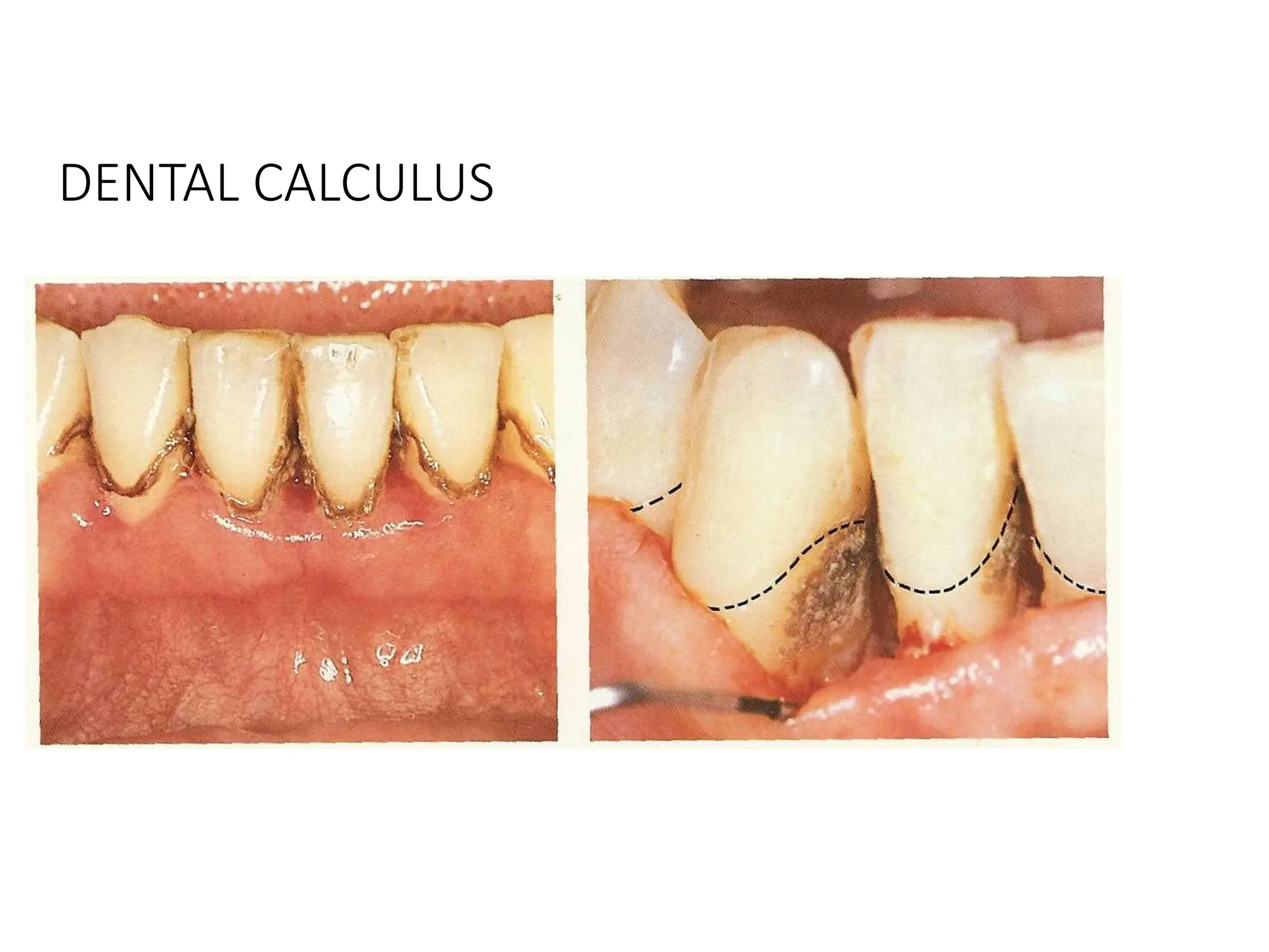
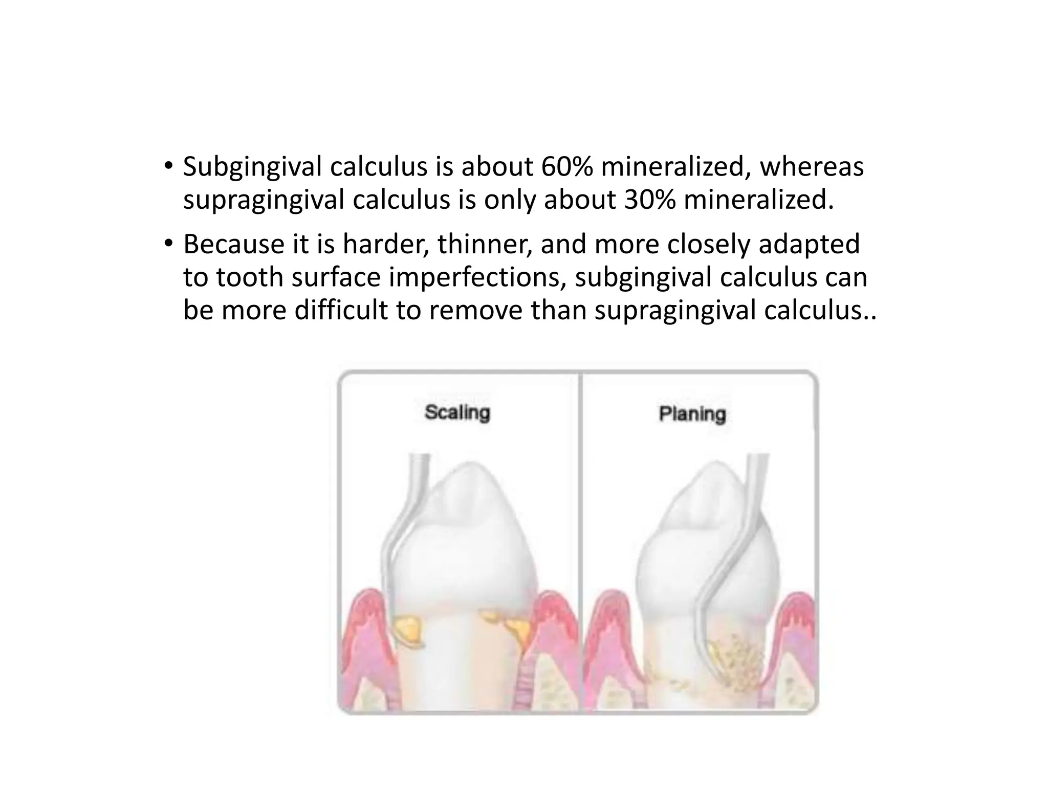
DENTAL CALCULUS
DENTAL CALCULUS
1. Definition
2. Classification
3. Formation
4. Differences
5. Structure and composition..
DENTAL CALCULUS
• Which is mineralized dental biofilm, is a hard, tenacious
mass that forms on the clinical crowns
of natural teeth and on
dentures and other
dental prostheses…
DENTAL CALCULUS
• Is significant in the progression
of inflammatory periodontal
disease
• The rough surface of the
subgingival calculus holds the
disease-producing
bacteria of the
dental biofilm close
to the gingival tissue
and perpetuates the
inflamed state…
DENTAL CALCULUS
• The control of biofilm deposits
by the patient, supplemented
by complete professional
calculus removal, can
reduce or eliminate
gingival inflammation…
DENTAL CALCULUS
• Comprehensive understanding of the characteristics, origin,
development, and methods of prevention of calculus is essential to
patient examination, assessment, treatment, and instruction.
• For successful treatment and prevention, the patient needs to know
the interelationship between biofilm, calculus; and the reasons for
the painstaking manner in which scaling procedures must be carried
out…
DENTAL CALCULUS
Classification
Is classified by its location on a tooth surface as related to the
adjacent free gingival margin, that is,
1. Supragingival
2. Subgingival..
• SUPRAGINGIVAL
located on the clinical crown coronal to the margin
of the gingiva..
• SUBGINGIVAL
Located on the clinical
crown apical to
the margin of the
gingiva.
It extends nearly to the
bottom of the pocket. It
forms on the exposed
root surfaces and on
implants..
DENTAL CALCULUS
FORMATION
1. PELLICLE FORMATION
2. BIOFILM MATURATION
3. MINERALIZATION..
DENTAL CALCULUS
FORMATION……
1. PELLICLE FORMATION
• IS COMPOSED OF MUCOPROTEINS FROM THE SALIVA AND IS AN
ACELLULAR MATERIAL..
DENTAL CALCULUS
FORMATION……
2. BIOFILM MATURATION
• Microorganisms settle in the pellicle layer
• Colonies are formed
• The colonies grow together to form a cohesive biofilm layer..
DENTAL CALCULUS
FORMATION……
3. MINERALIZATION
i. Mineralization Foci (centres) Form
ii. Organic Matrix
iii. Sources of Minerals
iv. Crystal Formation
v. Mechanism of Mineralization..
DENTAL CALCULUS
FORMATION……3. MINERALIZATION
i. Mineralization Foci (centres) Form
• within 24 to 72 hours, more and more mineralization
centres develop close to the underlying tooth surface
• Eventually, the centres grow large enough to touch and
unite..
DENTAL CALCULUS
FORMATION……3. MINERALIZATION
ii. Organic Matrix
• Mineralization first occurs within the intermicrobial matrix
• Filamentous microorganisms provide the matrix for the deposition of
minerals..
DENTAL CALCULUS
FORMATION……3. MINERALIZATION
iii. Sources of minerals
• Supragingival: source of elements is the saliva
• Subgingival: gingival sulcus fluid and inflammatory exudate supply the
minerals..
DENTAL CALCULUS
FORMATION……3. MINERALIZATION
iv. Crystal Formation
• Mineralization consists of crystal formation, namely hydroxyapatite,
octocalcium phosphate, whitlockite and brushite..
. Mechanism of mineralization
Is considered same for supra and subgingival
calculus
Heavy formers have higher salivary levels of
calcium and phosphorus
Light formers have higher levels of parotid
pyrophosphate
Pyrophosphate is an inhibitor of calcification-
used in anticalculus dentrifices..
• Supragingival calculus forms on the tooth above the gingival margin,
and frequently develops opposite the duct orifices of the major
salivary glands.
• It is often found where saliva pools on the inner surfaces of the
lower front teeth, and can form in the fissures of teeth.
• Subgingival calculus forms
from calcium phosphate
and organic materials
derived from serum,
which contribute to
mineralization of plaque
beneath the gumline..
• Subgingival calculus is about 60% mineralized, whereas
supragingival calculus is only about 30% mineralized.
• Because it is harder, thinner, and more closely adapted
to tooth surface imperfections, subgingival calculus can
be more difficult to remove than supragingival calculus..
• The two types of
calculus
may differ in color.
Supragingival
calculus,
which derives its
mineral content
from saliva,
usually appears
as a yellow
to white mass
with a
chalky consistency..
• Subgingival calculus,
which derives its mineral
from the inflammatory
exudate in the sulcus and
gum pocket, appears gray
to black
in color and has a
flint-like consistency.
The dark coloration
may be caused by
bacterial degradation
of components of the
exudate from
bleeding that
accompanies gums
inflammation.
DIFFERENCES BETWEEN SUPRAGINGIVAL AND
SUBGINGIVAL CALCULUS
DIFFERENCES BETWEEN SUPRAGINGIVAL AND
SUBGINGIVAL CALCULUS
DIFFERENCES BETWEEN SUPRAGINGIVAL AND
SUBGINGIVAL CALCULUS
ATTACHMENT OF CALCULUS
1. By means of an acquired pellicle
2. Attachment to minute irregularities in the tooth surface by
mechanical locking into undercuts
3. Attachment by direct contact between calcified intercellular
matrix and the tooth surface..
SIGNIFICANCE OF DENTAL CALCULUS
• Has an important role in the development, promotion and
recurrence of gingival and periodontal infections
• Subgingival biofilm contains pathogenic bacteria that cause
inflammation and destruction in the gingival tissue and lead to loss
of attachment to the tooth surface and development and deepening
of the pocket..
SIGNIFICANCE OF DENTAL CALCULUS……
•With increased pocket depth more biofilm forms.
Greater flow of GSF which contains minerals for
calculus formation
•Subgingival calculus is covered by masses of biofilm
bacteria, which is in contact with the diseased
pocket epithelium and promotes gingivitis and
periodontitis
•With rough surface, permeable structure and
porosity, calculus acts as a reservoir for endotoxins
and tissue breakdown products
• ……….……

LECT 4 DENTAL CALCULUS INCLUDING FORMATION AND CAUSES.pptx

  • 1.
  • 2.
    DENTAL CALCULUS 1. Definition 2.Classification 3. Formation 4. Differences 5. Structure and composition..
  • 3.
    DENTAL CALCULUS • Whichis mineralized dental biofilm, is a hard, tenacious mass that forms on the clinical crowns of natural teeth and on dentures and other dental prostheses…
  • 4.
    DENTAL CALCULUS • Issignificant in the progression of inflammatory periodontal disease • The rough surface of the subgingival calculus holds the disease-producing bacteria of the dental biofilm close to the gingival tissue and perpetuates the inflamed state…
  • 5.
    DENTAL CALCULUS • Thecontrol of biofilm deposits by the patient, supplemented by complete professional calculus removal, can reduce or eliminate gingival inflammation…
  • 6.
    DENTAL CALCULUS • Comprehensiveunderstanding of the characteristics, origin, development, and methods of prevention of calculus is essential to patient examination, assessment, treatment, and instruction.
  • 7.
    • For successfultreatment and prevention, the patient needs to know the interelationship between biofilm, calculus; and the reasons for the painstaking manner in which scaling procedures must be carried out…
  • 8.
    DENTAL CALCULUS Classification Is classifiedby its location on a tooth surface as related to the adjacent free gingival margin, that is, 1. Supragingival 2. Subgingival..
  • 9.
    • SUPRAGINGIVAL located onthe clinical crown coronal to the margin of the gingiva..
  • 10.
    • SUBGINGIVAL Located onthe clinical crown apical to the margin of the gingiva. It extends nearly to the bottom of the pocket. It forms on the exposed root surfaces and on implants..
  • 11.
    DENTAL CALCULUS FORMATION 1. PELLICLEFORMATION 2. BIOFILM MATURATION 3. MINERALIZATION..
  • 12.
    DENTAL CALCULUS FORMATION…… 1. PELLICLEFORMATION • IS COMPOSED OF MUCOPROTEINS FROM THE SALIVA AND IS AN ACELLULAR MATERIAL..
  • 13.
    DENTAL CALCULUS FORMATION…… 2. BIOFILMMATURATION • Microorganisms settle in the pellicle layer • Colonies are formed • The colonies grow together to form a cohesive biofilm layer..
  • 14.
    DENTAL CALCULUS FORMATION…… 3. MINERALIZATION i.Mineralization Foci (centres) Form ii. Organic Matrix iii. Sources of Minerals iv. Crystal Formation v. Mechanism of Mineralization..
  • 15.
    DENTAL CALCULUS FORMATION……3. MINERALIZATION i.Mineralization Foci (centres) Form • within 24 to 72 hours, more and more mineralization centres develop close to the underlying tooth surface • Eventually, the centres grow large enough to touch and unite..
  • 16.
    DENTAL CALCULUS FORMATION……3. MINERALIZATION ii.Organic Matrix • Mineralization first occurs within the intermicrobial matrix • Filamentous microorganisms provide the matrix for the deposition of minerals..
  • 17.
    DENTAL CALCULUS FORMATION……3. MINERALIZATION iii.Sources of minerals • Supragingival: source of elements is the saliva • Subgingival: gingival sulcus fluid and inflammatory exudate supply the minerals..
  • 18.
    DENTAL CALCULUS FORMATION……3. MINERALIZATION iv.Crystal Formation • Mineralization consists of crystal formation, namely hydroxyapatite, octocalcium phosphate, whitlockite and brushite..
  • 19.
    . Mechanism ofmineralization Is considered same for supra and subgingival calculus Heavy formers have higher salivary levels of calcium and phosphorus Light formers have higher levels of parotid pyrophosphate Pyrophosphate is an inhibitor of calcification- used in anticalculus dentrifices..
  • 20.
    • Supragingival calculusforms on the tooth above the gingival margin, and frequently develops opposite the duct orifices of the major salivary glands. • It is often found where saliva pools on the inner surfaces of the lower front teeth, and can form in the fissures of teeth.
  • 21.
    • Subgingival calculusforms from calcium phosphate and organic materials derived from serum, which contribute to mineralization of plaque beneath the gumline..
  • 22.
    • Subgingival calculusis about 60% mineralized, whereas supragingival calculus is only about 30% mineralized. • Because it is harder, thinner, and more closely adapted to tooth surface imperfections, subgingival calculus can be more difficult to remove than supragingival calculus..
  • 23.
    • The twotypes of calculus may differ in color. Supragingival calculus, which derives its mineral content from saliva, usually appears as a yellow to white mass with a chalky consistency..
  • 24.
    • Subgingival calculus, whichderives its mineral from the inflammatory exudate in the sulcus and gum pocket, appears gray to black in color and has a flint-like consistency. The dark coloration may be caused by bacterial degradation of components of the exudate from bleeding that accompanies gums inflammation.
  • 25.
    DIFFERENCES BETWEEN SUPRAGINGIVALAND SUBGINGIVAL CALCULUS
  • 26.
    DIFFERENCES BETWEEN SUPRAGINGIVALAND SUBGINGIVAL CALCULUS
  • 27.
    DIFFERENCES BETWEEN SUPRAGINGIVALAND SUBGINGIVAL CALCULUS
  • 28.
    ATTACHMENT OF CALCULUS 1.By means of an acquired pellicle 2. Attachment to minute irregularities in the tooth surface by mechanical locking into undercuts 3. Attachment by direct contact between calcified intercellular matrix and the tooth surface..
  • 29.
    SIGNIFICANCE OF DENTALCALCULUS • Has an important role in the development, promotion and recurrence of gingival and periodontal infections • Subgingival biofilm contains pathogenic bacteria that cause inflammation and destruction in the gingival tissue and lead to loss of attachment to the tooth surface and development and deepening of the pocket..
  • 30.
    SIGNIFICANCE OF DENTALCALCULUS…… •With increased pocket depth more biofilm forms. Greater flow of GSF which contains minerals for calculus formation •Subgingival calculus is covered by masses of biofilm bacteria, which is in contact with the diseased pocket epithelium and promotes gingivitis and periodontitis •With rough surface, permeable structure and porosity, calculus acts as a reservoir for endotoxins and tissue breakdown products • ……….……