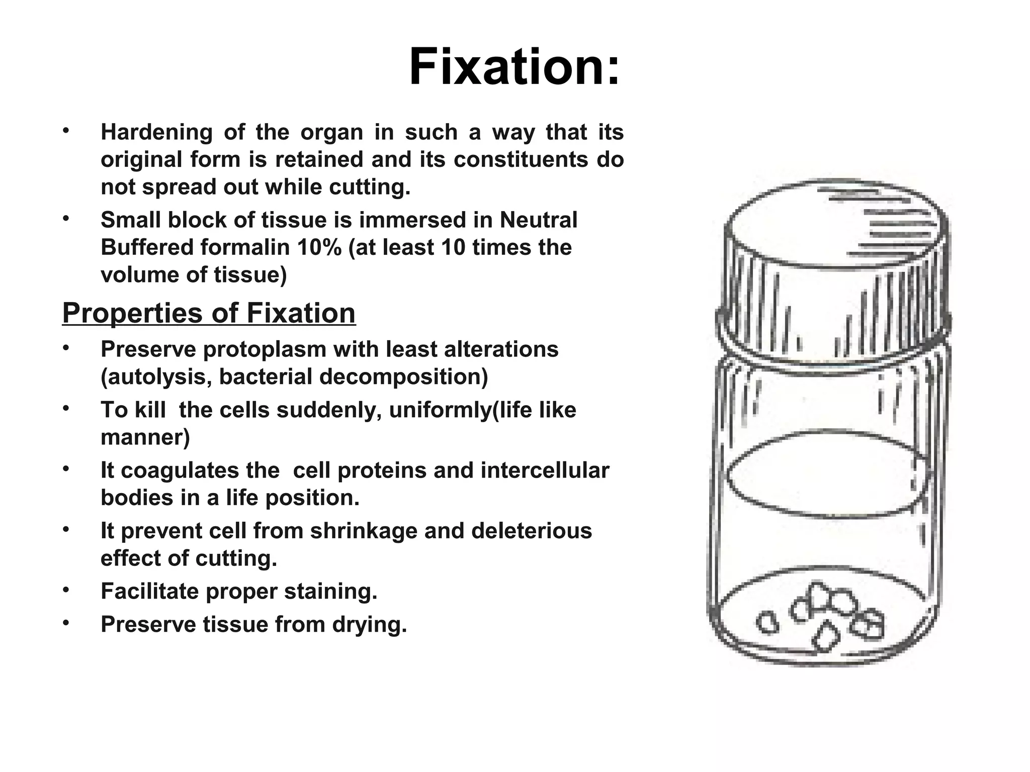
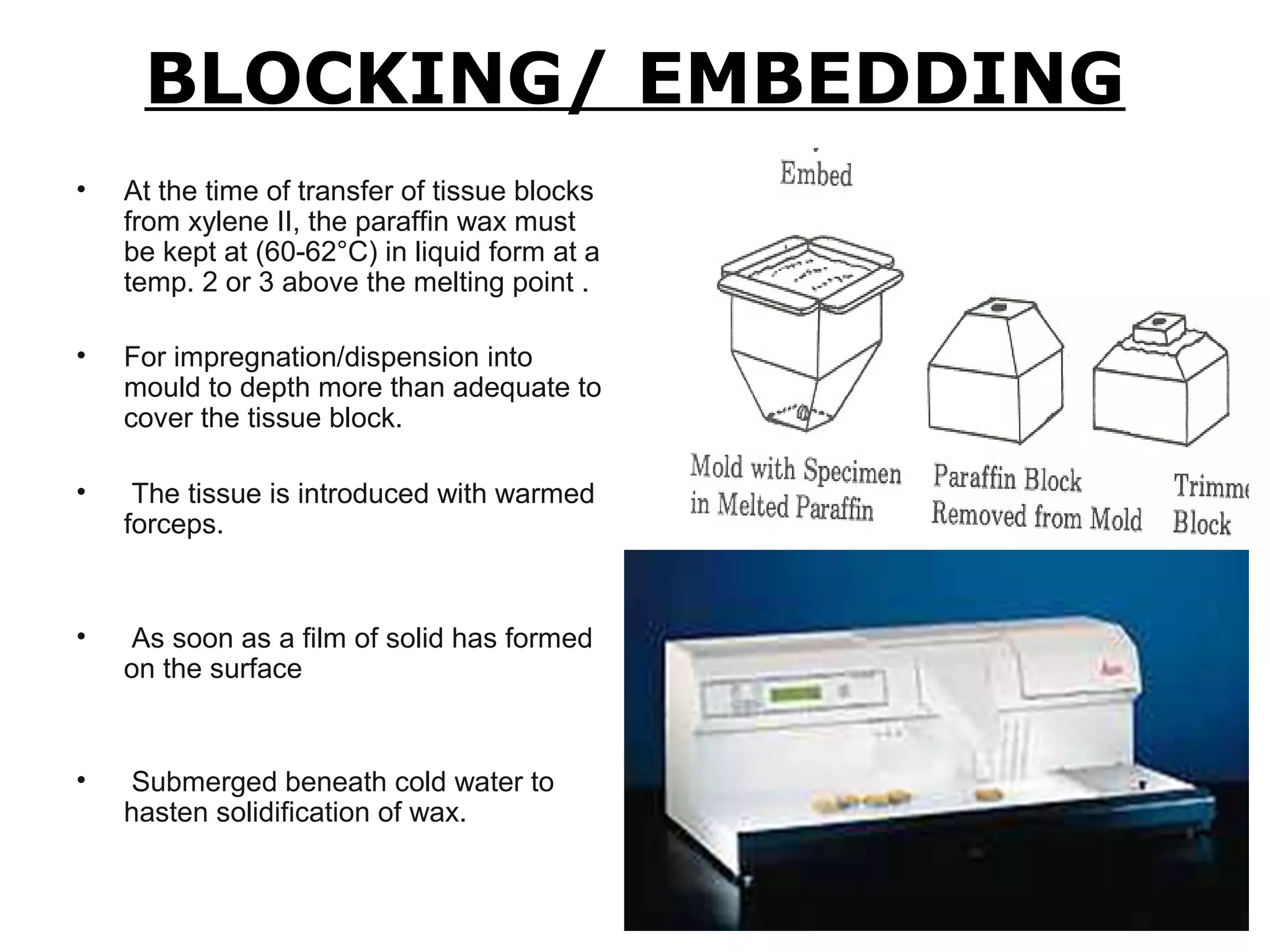
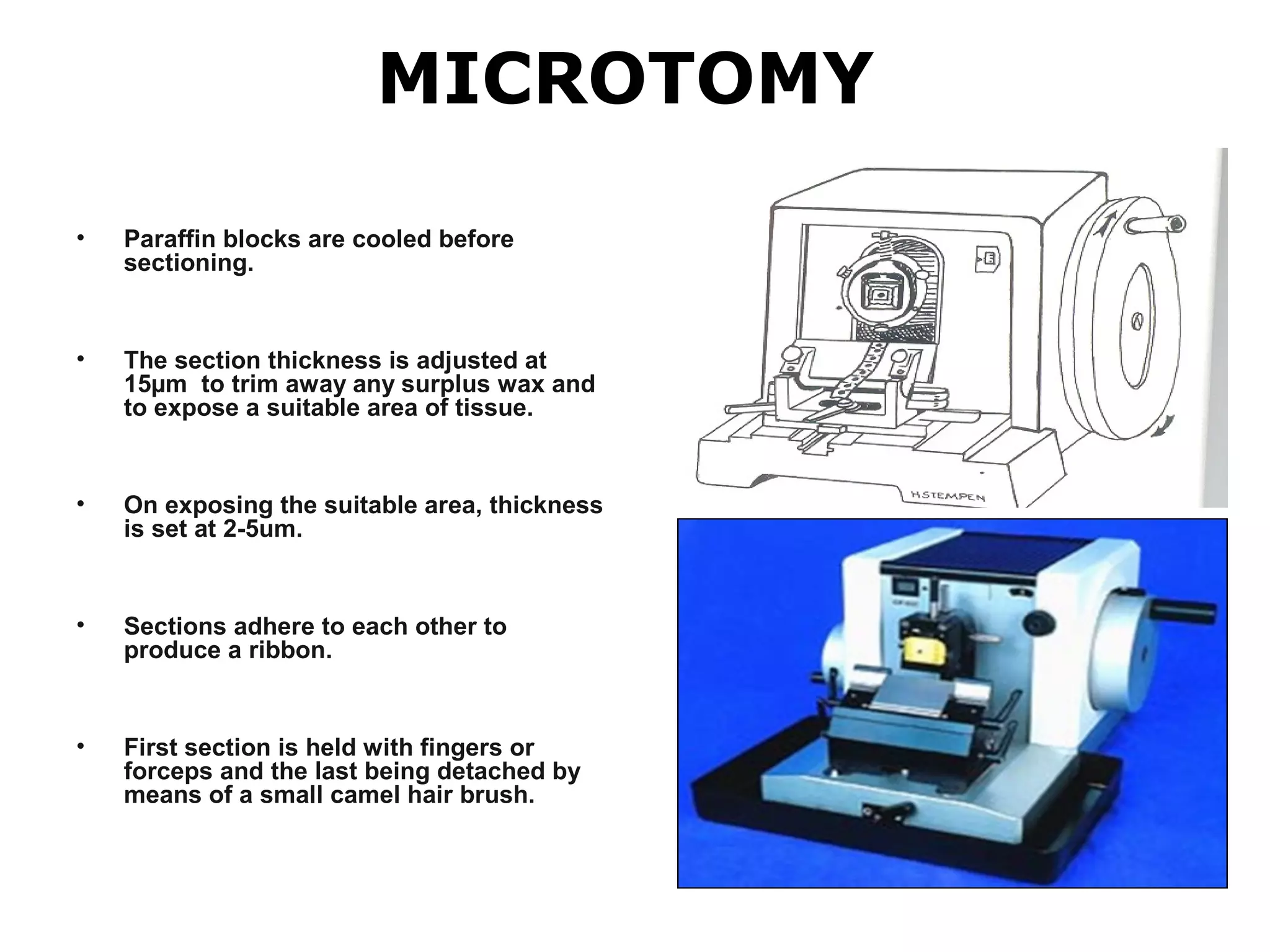
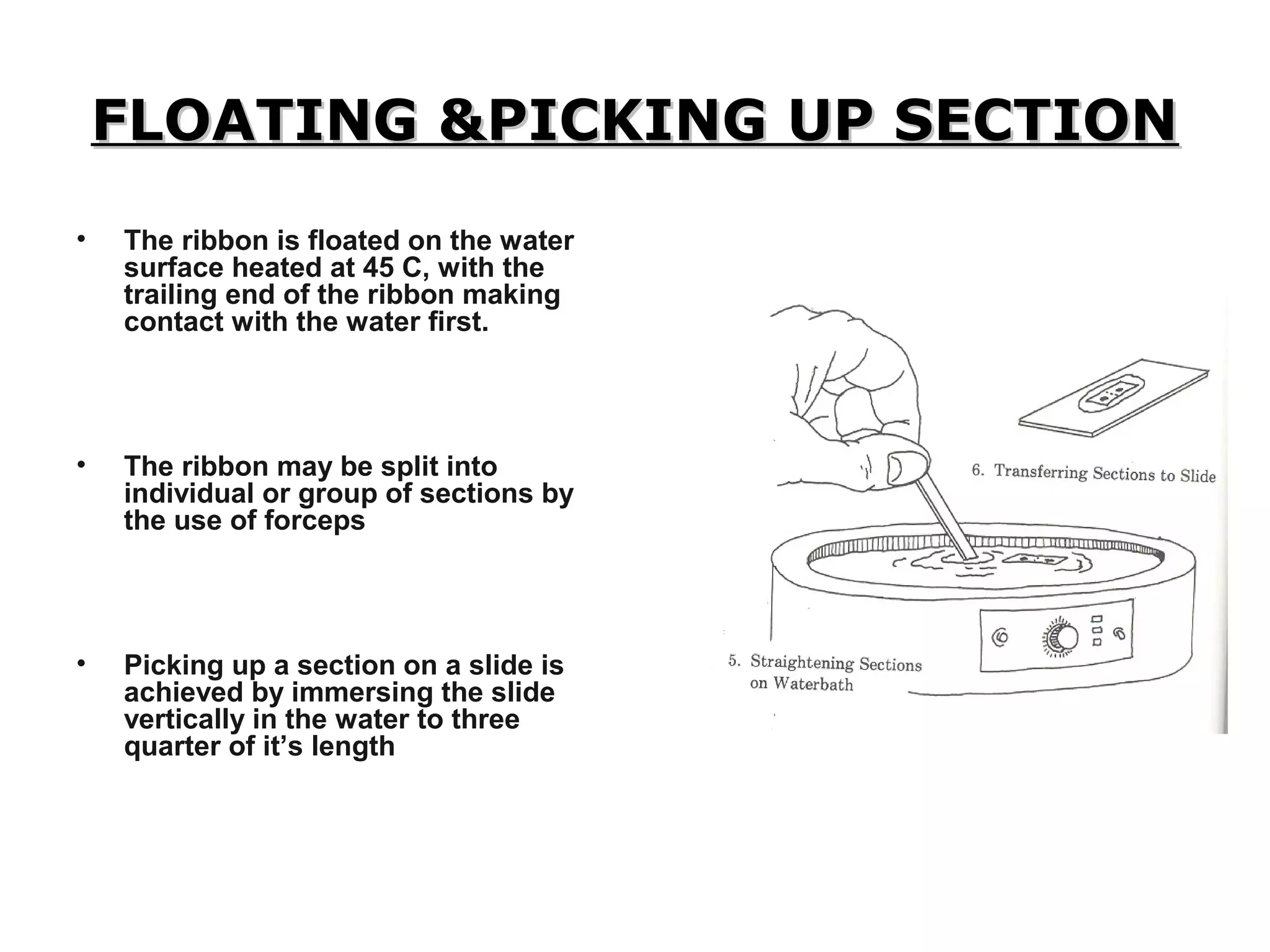
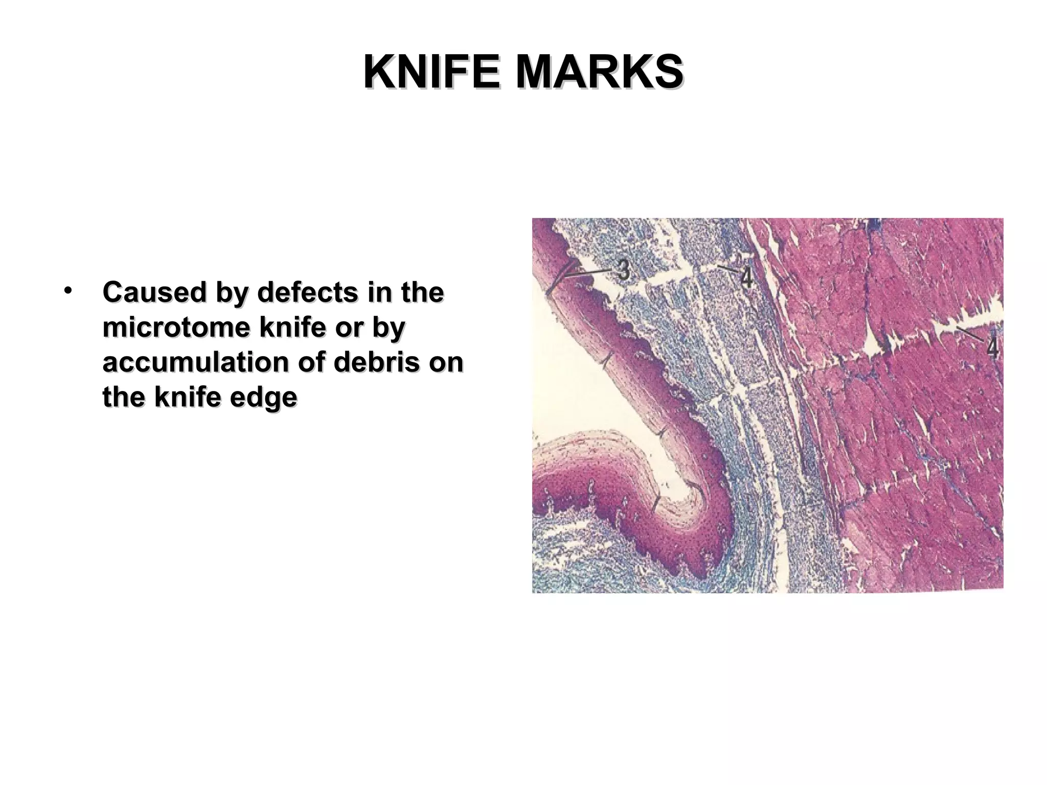
HistopathologicalHistopathological
TechniquesTechniques
HISTOPATHOLOGICAL TECHNIQUES
• Histopathology is the branch of pathology which
concerns with the demonstration of minute structural
alterations in tissues as a result of disease.
• Most of histopathological techniques simulating to those of
applied for study the normal histological structures.
• For the demonstration of minute histological changes, the
tissue must be processed in such a manner that it will provide
maximum information.
• Most convenient way for study of morbid tissue is to use
permanent section.
• A section is prepared by cutting a thin slice from a small piece
of fixed tissue.
• Thin slice is mounted upon a glass slide in a medium of
suitable refractive index and covered with a glass slide.
Scope
• Though the histopathological techniques are labour intensive, cumbersome and
time consuming, particularly when there are automation equipments are not
available; however, their use in diagnosis of diseases is unequivocal.
• Some of the areas where histopathological diagnosis is helpful are described
as follows:
• This is useful in establishing the pathogenesis and pathology of any disease
caused by bacteria, virus, chlamydia, rickettsia, mycoplasma, parasite, toxin,
poisons etc.
• There are certain diseases in which histopathological examination of tissues is
the only alternative to diagnose the disease. e.g. Bovine spongiform
encephalopathy. The agent of this disease takes a very long incubation period and
very difficult to isolate and there is no immune response and inflammation in
animal. Therefore, histopathology remains the only alternative for confirmatory
diagnosis.
• In some cases, the tissues from dead animals are only available material for
laboratory diagnosis. This may occur either due to lack of time or due to
negligence for not collecting the material for serological tests or isolation studies.
Sometimes the transportation of material from remote areas destroys the other
material and the tissues fixed in formalin only remains for making diagnosis. In all
such cases the histopathological examination has its pivotal role.
• The histopathological procedures produce permanent slides, which can be
stored for a longer period and one cannot manipulate the findings; therefore,
it is considered best reliable technique.
• The histopathological techniques are useful in carrying out the
retrospective studies. The unstained slides and blocks can be stored for
indefinite period; which can be examined even after many years for further
studies.
• The presence of causative agents can also be demonstrated in tissue
sections using routine histopathological techniques or special stainings. In
this Gram's staining procedures are used for demonstration of bacteria while
viral inclusions are demonstrated using hematoxylin and eosin or other
staining techniques like Macchiavello's stain or Mann's methylene blue
eosin method. The Negri bodies are demonstrated by Seller's stain in
case of rabies in animals. In such cases, the isolation of causative agent or
their serological examination does not require; since the presence of
causal agent in infected tissues gives a confirmatory diagnosis.
• The detection of chemicals in tissues like enzymes, lipids etc. is
included in histochemical examination; which not only describe the structural
changes but also gives idea about the functional status of the organ.
Collection of Samples:
1. Small piece of tissue (as early as possible)
2. Piece is removed with sharp knife in a particular orientation.
3. Size of piece=1cm to achieve better penetration of fixative.
4. Washing the specimen with normal saline to achieve maximum penetration
of fixative.
5. At the time of tissue collection, it should be kept in mind that the
representative tissue piece should include the part of lesion and a part of
normal tissue,
6. Tissue pieces for histopathological examination should be collected from
all the organs.
7. Tissues should be collected directly in the fixative and not in any other pot
or water.
8. The tissue pieces from hollow organs like intestines, oviduct etc should be
cut transversely and placed on a hard paper.
9. The representative tissue should not be collected from such damaged
portions.
10. The tissues from encapsulated organs should be collected alongwith
capsule or covering.
Labelling:
• Tissue is accompanied by a tag or label, bearing the lab. number given to
specimen at start, through all stages.
• Thin white card with writing in soft pencil withstands all the fluids used in
tissue processing.
Sample collection and preservation
Fixation:
• Hardening of the organ in such a way that its
original form is retained and its constituents do
not spread out while cutting.
• Small block of tissue is immersed in Neutral
Buffered formalin 10% (at least 10 times the
volume of tissue)
Properties of Fixation
• Preserve protoplasm with least alterations
(autolysis, bacterial decomposition)
• To kill the cells suddenly, uniformly(life like
manner)
• It coagulates the cell proteins and intercellular
bodies in a life position.
• It prevent cell from shrinkage and deleterious
effect of cutting.
• Facilitate proper staining.
• Preserve tissue from drying.
Commonly used Fixatives
• 10% Formalin Solution.
• Buffered Neutral Formalin Solution.
• Formalin Alcohol. (glyogen, good cytoplasmic fixation)
• Zenker Fluid. (bone marrow aspirates)
• Bouin’s Fluid. (embryological specimens, Purkinje cells)
• Acetone. (Rabies)
• Ethyl Alcohol. (glycogen, pigments, amyloid, hylaine,
elastic fibers and bacteria)
• Glacial acetic acid. (rapid fixation, swelling of cells)
• Potassium Dichromate. (cytoplasm, not nucleoproteins)
• Heat. 5-10 mm thick piece of tissue, boil in 0.9 percent
Nacl solution for 2-3 min.
10% Formalin Solution
37%-40% Formaldehyde 100 ml
Distilled water 900 ml
Neutral buffered formalin
37%-40% Formaldehyde 100 ml
Distilled water 900 ml
Sodium Phosphate monobasic 4 gm
Sodium Phosphate dibasic (anhydrous) 6.5 gm
Trimming of tissue
• Tissue, enclosed in tissue cassette
and placed in basket of automatic
tissue processor.
Washing of the Tissue
• Washing of tissue under running
tape water overnight (12 hours)
remove excessive Fixative Solution.
PROCESSING
Dehydration
•The purpose of dehydration to remove fixative and
water from the tissue and replace them with
dehydrating fluid.
•There are a variety of compounds many of which are
alcohols. Several are hydrophilic so attract water from
tissue.
Types of dehydrating agents:
•Ethanol
•Methanol
•Acetone
• To minimize tissue distortion from diffusion currents, delicate
specimens are dehydrated in a graded ethanol series from
water through 10%-20%-50%-95%-100% ethanol.
Ethyl alcohol 70% 2 hour
Ethyl alcohol 80% 1 hour
Ethyl alcohol 95% 1 hour
Ethyl alcohol (Absolute 1) .5 hour
Ethyl alcohol (Absolute 2) .5 hour
• To increase the process of dehydration, the tissue blocks
should be agitated either mechanically in an automatic tissue
processor or by shaking the container periodically.
• The volume of alcohol should be at least 50 times more than
the tissue placed for dehydration.
Clearing:
• It involves removal of dehydrating agent and replacement by
some fluid which is miscible with dehydrating agent and
embedding medium.
 Choice of a clearing agent depends upon the following:Choice of a clearing agent depends upon the following:
• The type of tissues to be processed, and the type
of processing to be undertaken.
• The processor system to be used.
• Intended processing conditions such as temperature,
vacuum and pressure.
• Safety factors.
• Cost and convenience.
• Speedy removal of dehydrating agent .
• Ease of removal by molten paraffin wax .
• Minimal tissue damage .
• Like ethanol, xylene should also be kept in tightly
stoppered bottle to prevent the evaporation.
Equal parts of Absolute alcohol +Xylene .5 hour
Xylene I 1 hour
Xylene II 1 hour
• If xylene is not available then benzene may be used for 3
hr as its action of clearing is slower than xylene.
• On complete clearing, the tissue becomes transparent,
then they should be transferred in paraffin wax for
impregnation.
Wax Impregnation/Infiltration
•It is the process by which tissues are
surrounded by a medium such as agar,
gelatin, or wax which when solidified will
provide sufficient external support during
sectioning.
•Tissue is impregnated with melted wax.
•Xylene +Paraffin 1 hour
•Paraffin wax I 2 hours
•Paraffin wax II 3 hours
BLOCKING/ EMBEDDING
• At the time of transfer of tissue blocks
from xylene II, the paraffin wax must
be kept at (60-62°C) in liquid form at a
temp. 2 or 3 above the melting point .
• For impregnation/dispension into
mould to depth more than adequate to
cover the tissue block.
• The tissue is introduced with warmed
forceps.
• As soon as a film of solid has formed
on the surface
• Submerged beneath cold water to
hasten solidification of wax.
MICROTOMY
• Paraffin blocks are cooled before
sectioning.
• The section thickness is adjusted at
15µm to trim away any surplus wax and
to expose a suitable area of tissue.
• On exposing the suitable area, thickness
is set at 2-5um.
• Sections adhere to each other to
produce a ribbon.
• First section is held with fingers or
forceps and the last being detached by
means of a small camel hair brush.
FLOATING &PICKING UP SECTIONFLOATING &PICKING UP SECTION
• The ribbon is floated on the water
surface heated at 45 C, with the
trailing end of the ribbon making
contact with the water first.
• The ribbon may be split into
individual or group of sections by
the use of forceps
• Picking up a section on a slide is
achieved by immersing the slide
vertically in the water to three
quarter of it’s length
DRYING SECTIONSDRYING SECTIONS
• The slides are placed on the hot
plate adjusted at melting point of
wax for ten minutes.
• Drying in oven at the same
temperature should be done for 45
minutes.
PRE STAINING PREPARATIONPRE STAINING PREPARATION
Deparaffinization with XyleneDeparaffinization with Xylene
• Xylene IXylene I 3 minutes3 minutes
• Xylene IIXylene II 3 minutes3 minutes
RehydrationRehydration
• Absolute ethyl alcoholAbsolute ethyl alcohol II 3 minutes3 minutes
• Absolute ethyl alcoholAbsolute ethyl alcohol IIII 3 minutes3 minutes
• Ethyl alcohol 90%Ethyl alcohol 90% 2 minutes2 minutes
• Ethyl alcohol 70%Ethyl alcohol 70% 2 minutes2 minutes
• Distilled waterDistilled water 3 minutes3 minutes
• Wash in tap water and rinse in Distilled waterWash in tap water and rinse in Distilled water
STAININGSTAINING
• Harris haematoxylinHarris haematoxylin 15 min15 min
• Distilled waterDistilled water 5 min5 min
• Acid alcohol 70%Acid alcohol 70% 4-10 dips4-10 dips
• Ammonia AlcoholAmmonia Alcohol 5 min5 min
• Wash in running tap waterWash in running tap water 5 min or less5 min or less
• EosinEosin 2 min2 min
• Wash in running tap waterWash in running tap water 1-5 minutes1-5 minutes
POST STAINING PROCEDURESPOST STAINING PROCEDURES
• Ethyl alcohol 95%Ethyl alcohol 95% 2 min2 min
• Absolute Ethyl alcoholAbsolute Ethyl alcohol 2 min2 min
• Absolute Ethyl alcoholAbsolute Ethyl alcohol 3 min3 min
• Xylene IXylene I 3 min3 min
• Xylene IIXylene II 3 min3 min
Harris HaematoxylinHarris Haematoxylin
Haematoxylin crystalsHaematoxylin crystals 1 gm1 gm
Alcohol 95%Alcohol 95% 10 ml10 ml
Potassium alumPotassium alum
(Aluminum potassium sulphate)(Aluminum potassium sulphate) 20 gm20 gm
Distilled waterDistilled water 200 ml200 ml
Mercuric oxideMercuric oxide 0.5 gm0.5 gm
Glacial acetic acidGlacial acetic acid 8.0 ml8.0 ml
Eosin solutionEosin solution
• EosinEosin 1 gm1 gm
• Distilled waterDistilled water 100 ml100 ml
• Acetic acidAcetic acid 0.05 ml0.05 ml
Acid alcohol:Acid alcohol:
• Ethyl alcohol 70%Ethyl alcohol 70% 990 ml990 ml
• HCl (concentrated)HCl (concentrated) 10 ml10 ml
Dilute ammonia solutionDilute ammonia solution
• Distilled waterDistilled water 250 ml250 ml
• Ammonia (concentrated)Ammonia (concentrated) 2 drops2 drops
• Acid dyeAcid dye: When the staining property is in: When the staining property is in
the acid radical of the neutral salt, thethe acid radical of the neutral salt, the
stain is called an acid dye e.g.,stain is called an acid dye e.g., eosineosin,,
picric acid, azo dyes, tryan red, orange G.picric acid, azo dyes, tryan red, orange G.
• Basic dyeBasic dye: The coloring property is in the: The coloring property is in the
basic radical of the neutral salt, the stain isbasic radical of the neutral salt, the stain is
called a basic dye e.g.,called a basic dye e.g., hematoxylinhematoxylin,,
toluidine blue, methylene blue.toluidine blue, methylene blue.
PRECAUTIONSPRECAUTIONS
• Rapid fixation of fresh specimen is essential as post-
mortem degeneration markedly alters the appearance of the
various cell types in tissue making interpretation
unrewarding.
• Quality of nuclear staining begins to deteriorate and
precipitate is formed in stored stain after a few months. At
this stage, stain should be filtered and staining time should
be increased.
• It is better to prepare a fresh batch of stain every month
Preparation of Haematoxylin:
• Haematoxylin is dissolved in the absolute alcohol
• Alum is dissolved in the warm distilled water
• Haematoxylin solution is added to the alum solution and
rapidly brought to boil
• Mercuric oxide is then added
• Stain is rapidly cooled by plunging the flask into cold water
and acetic acid is added when cold
MOUNTINGMOUNTING
• Mounting with canada balsam,Mounting with canada balsam,
apply coverslip and examine underapply coverslip and examine under
the microscope.the microscope.
HELPFUL HINTSHELPFUL HINTS
• Lenses of your microscope should be cleanLenses of your microscope should be clean
• Make sure that fine adjustment knob is near the middle of its range ofMake sure that fine adjustment knob is near the middle of its range of
rotationrotation
• To start your observation using the lowest power objectiveTo start your observation using the lowest power objective
• By knowing the app. dia. Of RBC in the section, you can estimate theBy knowing the app. dia. Of RBC in the section, you can estimate the
size of other tissue components.size of other tissue components.
• Goat: 2.4 umGoat: 2.4 um
• Dog: 4.9umDog: 4.9um
• Chichen: 9.4umChichen: 9.4um
• Average value based on total of 20-30 cell measured from differentAverage value based on total of 20-30 cell measured from different
slide preparationsslide preparations
ARTIFACTSARTIFACTS
FoldFold
Knife marksKnife marks
SpacesSpaces
Stain precipitatesStain precipitates
ShrinkageShrinkage
Air bubblesAir bubbles
examples of commonly occurring imperfections seen inexamples of commonly occurring imperfections seen in
slide preparationsslide preparations
STAIN PRECIPITATESSTAIN PRECIPITATES
• Occasionally , solutionOccasionally , solution
accumulate precipitate thataccumulate precipitate that
may stick to the surfacemay stick to the surface
tissue section duringtissue section during
staining procedurestaining procedure
SEPARATION (SPACES)SEPARATION (SPACES)
• Tissue may be subjected toTissue may be subjected to
excessive pressure, tensionexcessive pressure, tension
or shrinkage duringor shrinkage during
processing resulting inprocessing resulting in
separations of tissueseparations of tissue
KNIFE MARKSKNIFE MARKS
• Caused by defects in theCaused by defects in the
microtome knife or bymicrotome knife or by
accumulation of debris onaccumulation of debris on
the knife edgethe knife edge
FOLDSFOLDS
• Folds occurs when theFolds occurs when the
tissue sections fail to spreadtissue sections fail to spread
properlyproperly
Histopathological technique

Histopathological technique

  • 1.
  • 2.
    HISTOPATHOLOGICAL TECHNIQUES • Histopathologyis the branch of pathology which concerns with the demonstration of minute structural alterations in tissues as a result of disease. • Most of histopathological techniques simulating to those of applied for study the normal histological structures. • For the demonstration of minute histological changes, the tissue must be processed in such a manner that it will provide maximum information. • Most convenient way for study of morbid tissue is to use permanent section. • A section is prepared by cutting a thin slice from a small piece of fixed tissue. • Thin slice is mounted upon a glass slide in a medium of suitable refractive index and covered with a glass slide.
  • 3.
    Scope • Though thehistopathological techniques are labour intensive, cumbersome and time consuming, particularly when there are automation equipments are not available; however, their use in diagnosis of diseases is unequivocal. • Some of the areas where histopathological diagnosis is helpful are described as follows: • This is useful in establishing the pathogenesis and pathology of any disease caused by bacteria, virus, chlamydia, rickettsia, mycoplasma, parasite, toxin, poisons etc. • There are certain diseases in which histopathological examination of tissues is the only alternative to diagnose the disease. e.g. Bovine spongiform encephalopathy. The agent of this disease takes a very long incubation period and very difficult to isolate and there is no immune response and inflammation in animal. Therefore, histopathology remains the only alternative for confirmatory diagnosis. • In some cases, the tissues from dead animals are only available material for laboratory diagnosis. This may occur either due to lack of time or due to negligence for not collecting the material for serological tests or isolation studies. Sometimes the transportation of material from remote areas destroys the other material and the tissues fixed in formalin only remains for making diagnosis. In all such cases the histopathological examination has its pivotal role.
  • 4.
    • The histopathologicalprocedures produce permanent slides, which can be stored for a longer period and one cannot manipulate the findings; therefore, it is considered best reliable technique. • The histopathological techniques are useful in carrying out the retrospective studies. The unstained slides and blocks can be stored for indefinite period; which can be examined even after many years for further studies. • The presence of causative agents can also be demonstrated in tissue sections using routine histopathological techniques or special stainings. In this Gram's staining procedures are used for demonstration of bacteria while viral inclusions are demonstrated using hematoxylin and eosin or other staining techniques like Macchiavello's stain or Mann's methylene blue eosin method. The Negri bodies are demonstrated by Seller's stain in case of rabies in animals. In such cases, the isolation of causative agent or their serological examination does not require; since the presence of causal agent in infected tissues gives a confirmatory diagnosis. • The detection of chemicals in tissues like enzymes, lipids etc. is included in histochemical examination; which not only describe the structural changes but also gives idea about the functional status of the organ.
  • 5.
    Collection of Samples: 1.Small piece of tissue (as early as possible) 2. Piece is removed with sharp knife in a particular orientation. 3. Size of piece=1cm to achieve better penetration of fixative. 4. Washing the specimen with normal saline to achieve maximum penetration of fixative. 5. At the time of tissue collection, it should be kept in mind that the representative tissue piece should include the part of lesion and a part of normal tissue, 6. Tissue pieces for histopathological examination should be collected from all the organs. 7. Tissues should be collected directly in the fixative and not in any other pot or water. 8. The tissue pieces from hollow organs like intestines, oviduct etc should be cut transversely and placed on a hard paper. 9. The representative tissue should not be collected from such damaged portions. 10. The tissues from encapsulated organs should be collected alongwith capsule or covering. Labelling: • Tissue is accompanied by a tag or label, bearing the lab. number given to specimen at start, through all stages. • Thin white card with writing in soft pencil withstands all the fluids used in tissue processing. Sample collection and preservation
  • 6.
    Fixation: • Hardening ofthe organ in such a way that its original form is retained and its constituents do not spread out while cutting. • Small block of tissue is immersed in Neutral Buffered formalin 10% (at least 10 times the volume of tissue) Properties of Fixation • Preserve protoplasm with least alterations (autolysis, bacterial decomposition) • To kill the cells suddenly, uniformly(life like manner) • It coagulates the cell proteins and intercellular bodies in a life position. • It prevent cell from shrinkage and deleterious effect of cutting. • Facilitate proper staining. • Preserve tissue from drying.
  • 7.
    Commonly used Fixatives •10% Formalin Solution. • Buffered Neutral Formalin Solution. • Formalin Alcohol. (glyogen, good cytoplasmic fixation) • Zenker Fluid. (bone marrow aspirates) • Bouin’s Fluid. (embryological specimens, Purkinje cells) • Acetone. (Rabies) • Ethyl Alcohol. (glycogen, pigments, amyloid, hylaine, elastic fibers and bacteria) • Glacial acetic acid. (rapid fixation, swelling of cells) • Potassium Dichromate. (cytoplasm, not nucleoproteins) • Heat. 5-10 mm thick piece of tissue, boil in 0.9 percent Nacl solution for 2-3 min.
  • 8.
    10% Formalin Solution 37%-40%Formaldehyde 100 ml Distilled water 900 ml Neutral buffered formalin 37%-40% Formaldehyde 100 ml Distilled water 900 ml Sodium Phosphate monobasic 4 gm Sodium Phosphate dibasic (anhydrous) 6.5 gm
  • 9.
    Trimming of tissue •Tissue, enclosed in tissue cassette and placed in basket of automatic tissue processor. Washing of the Tissue • Washing of tissue under running tape water overnight (12 hours) remove excessive Fixative Solution.
  • 10.
    PROCESSING Dehydration •The purpose ofdehydration to remove fixative and water from the tissue and replace them with dehydrating fluid. •There are a variety of compounds many of which are alcohols. Several are hydrophilic so attract water from tissue. Types of dehydrating agents: •Ethanol •Methanol •Acetone
  • 11.
    • To minimizetissue distortion from diffusion currents, delicate specimens are dehydrated in a graded ethanol series from water through 10%-20%-50%-95%-100% ethanol. Ethyl alcohol 70% 2 hour Ethyl alcohol 80% 1 hour Ethyl alcohol 95% 1 hour Ethyl alcohol (Absolute 1) .5 hour Ethyl alcohol (Absolute 2) .5 hour • To increase the process of dehydration, the tissue blocks should be agitated either mechanically in an automatic tissue processor or by shaking the container periodically. • The volume of alcohol should be at least 50 times more than the tissue placed for dehydration.
  • 12.
    Clearing: • It involvesremoval of dehydrating agent and replacement by some fluid which is miscible with dehydrating agent and embedding medium.  Choice of a clearing agent depends upon the following:Choice of a clearing agent depends upon the following: • The type of tissues to be processed, and the type of processing to be undertaken. • The processor system to be used. • Intended processing conditions such as temperature, vacuum and pressure. • Safety factors. • Cost and convenience. • Speedy removal of dehydrating agent . • Ease of removal by molten paraffin wax . • Minimal tissue damage .
  • 13.
    • Like ethanol,xylene should also be kept in tightly stoppered bottle to prevent the evaporation. Equal parts of Absolute alcohol +Xylene .5 hour Xylene I 1 hour Xylene II 1 hour • If xylene is not available then benzene may be used for 3 hr as its action of clearing is slower than xylene. • On complete clearing, the tissue becomes transparent, then they should be transferred in paraffin wax for impregnation.
  • 14.
    Wax Impregnation/Infiltration •It isthe process by which tissues are surrounded by a medium such as agar, gelatin, or wax which when solidified will provide sufficient external support during sectioning. •Tissue is impregnated with melted wax. •Xylene +Paraffin 1 hour •Paraffin wax I 2 hours •Paraffin wax II 3 hours
  • 15.
    BLOCKING/ EMBEDDING • Atthe time of transfer of tissue blocks from xylene II, the paraffin wax must be kept at (60-62°C) in liquid form at a temp. 2 or 3 above the melting point . • For impregnation/dispension into mould to depth more than adequate to cover the tissue block. • The tissue is introduced with warmed forceps. • As soon as a film of solid has formed on the surface • Submerged beneath cold water to hasten solidification of wax.
  • 16.
    MICROTOMY • Paraffin blocksare cooled before sectioning. • The section thickness is adjusted at 15µm to trim away any surplus wax and to expose a suitable area of tissue. • On exposing the suitable area, thickness is set at 2-5um. • Sections adhere to each other to produce a ribbon. • First section is held with fingers or forceps and the last being detached by means of a small camel hair brush.
  • 17.
    FLOATING &PICKING UPSECTIONFLOATING &PICKING UP SECTION • The ribbon is floated on the water surface heated at 45 C, with the trailing end of the ribbon making contact with the water first. • The ribbon may be split into individual or group of sections by the use of forceps • Picking up a section on a slide is achieved by immersing the slide vertically in the water to three quarter of it’s length
  • 18.
    DRYING SECTIONSDRYING SECTIONS •The slides are placed on the hot plate adjusted at melting point of wax for ten minutes. • Drying in oven at the same temperature should be done for 45 minutes.
  • 19.
    PRE STAINING PREPARATIONPRESTAINING PREPARATION Deparaffinization with XyleneDeparaffinization with Xylene • Xylene IXylene I 3 minutes3 minutes • Xylene IIXylene II 3 minutes3 minutes RehydrationRehydration • Absolute ethyl alcoholAbsolute ethyl alcohol II 3 minutes3 minutes • Absolute ethyl alcoholAbsolute ethyl alcohol IIII 3 minutes3 minutes • Ethyl alcohol 90%Ethyl alcohol 90% 2 minutes2 minutes • Ethyl alcohol 70%Ethyl alcohol 70% 2 minutes2 minutes • Distilled waterDistilled water 3 minutes3 minutes • Wash in tap water and rinse in Distilled waterWash in tap water and rinse in Distilled water
  • 20.
    STAININGSTAINING • Harris haematoxylinHarrishaematoxylin 15 min15 min • Distilled waterDistilled water 5 min5 min • Acid alcohol 70%Acid alcohol 70% 4-10 dips4-10 dips • Ammonia AlcoholAmmonia Alcohol 5 min5 min • Wash in running tap waterWash in running tap water 5 min or less5 min or less • EosinEosin 2 min2 min • Wash in running tap waterWash in running tap water 1-5 minutes1-5 minutes
  • 21.
    POST STAINING PROCEDURESPOSTSTAINING PROCEDURES • Ethyl alcohol 95%Ethyl alcohol 95% 2 min2 min • Ethyl alcohol 95%Ethyl alcohol 95% 2 min2 min • Absolute Ethyl alcoholAbsolute Ethyl alcohol 2 min2 min • Absolute Ethyl alcoholAbsolute Ethyl alcohol 3 min3 min • Xylene IXylene I 3 min3 min • Xylene IIXylene II 3 min3 min
  • 22.
    Harris HaematoxylinHarris Haematoxylin HaematoxylincrystalsHaematoxylin crystals 1 gm1 gm Alcohol 95%Alcohol 95% 10 ml10 ml Potassium alumPotassium alum (Aluminum potassium sulphate)(Aluminum potassium sulphate) 20 gm20 gm Distilled waterDistilled water 200 ml200 ml Mercuric oxideMercuric oxide 0.5 gm0.5 gm Glacial acetic acidGlacial acetic acid 8.0 ml8.0 ml
  • 23.
    Eosin solutionEosin solution •EosinEosin 1 gm1 gm • Distilled waterDistilled water 100 ml100 ml • Acetic acidAcetic acid 0.05 ml0.05 ml Acid alcohol:Acid alcohol: • Ethyl alcohol 70%Ethyl alcohol 70% 990 ml990 ml • HCl (concentrated)HCl (concentrated) 10 ml10 ml Dilute ammonia solutionDilute ammonia solution • Distilled waterDistilled water 250 ml250 ml • Ammonia (concentrated)Ammonia (concentrated) 2 drops2 drops
  • 24.
    • Acid dyeAciddye: When the staining property is in: When the staining property is in the acid radical of the neutral salt, thethe acid radical of the neutral salt, the stain is called an acid dye e.g.,stain is called an acid dye e.g., eosineosin,, picric acid, azo dyes, tryan red, orange G.picric acid, azo dyes, tryan red, orange G. • Basic dyeBasic dye: The coloring property is in the: The coloring property is in the basic radical of the neutral salt, the stain isbasic radical of the neutral salt, the stain is called a basic dye e.g.,called a basic dye e.g., hematoxylinhematoxylin,, toluidine blue, methylene blue.toluidine blue, methylene blue.
  • 25.
    PRECAUTIONSPRECAUTIONS • Rapid fixationof fresh specimen is essential as post- mortem degeneration markedly alters the appearance of the various cell types in tissue making interpretation unrewarding. • Quality of nuclear staining begins to deteriorate and precipitate is formed in stored stain after a few months. At this stage, stain should be filtered and staining time should be increased. • It is better to prepare a fresh batch of stain every month Preparation of Haematoxylin: • Haematoxylin is dissolved in the absolute alcohol • Alum is dissolved in the warm distilled water • Haematoxylin solution is added to the alum solution and rapidly brought to boil • Mercuric oxide is then added • Stain is rapidly cooled by plunging the flask into cold water and acetic acid is added when cold
  • 26.
    MOUNTINGMOUNTING • Mounting withcanada balsam,Mounting with canada balsam, apply coverslip and examine underapply coverslip and examine under the microscope.the microscope.
  • 27.
    HELPFUL HINTSHELPFUL HINTS •Lenses of your microscope should be cleanLenses of your microscope should be clean • Make sure that fine adjustment knob is near the middle of its range ofMake sure that fine adjustment knob is near the middle of its range of rotationrotation • To start your observation using the lowest power objectiveTo start your observation using the lowest power objective • By knowing the app. dia. Of RBC in the section, you can estimate theBy knowing the app. dia. Of RBC in the section, you can estimate the size of other tissue components.size of other tissue components. • Goat: 2.4 umGoat: 2.4 um • Dog: 4.9umDog: 4.9um • Chichen: 9.4umChichen: 9.4um • Average value based on total of 20-30 cell measured from differentAverage value based on total of 20-30 cell measured from different slide preparationsslide preparations
  • 28.
    ARTIFACTSARTIFACTS FoldFold Knife marksKnife marks SpacesSpaces StainprecipitatesStain precipitates ShrinkageShrinkage Air bubblesAir bubbles examples of commonly occurring imperfections seen inexamples of commonly occurring imperfections seen in slide preparationsslide preparations
  • 29.
    STAIN PRECIPITATESSTAIN PRECIPITATES •Occasionally , solutionOccasionally , solution accumulate precipitate thataccumulate precipitate that may stick to the surfacemay stick to the surface tissue section duringtissue section during staining procedurestaining procedure
  • 30.
    SEPARATION (SPACES)SEPARATION (SPACES) •Tissue may be subjected toTissue may be subjected to excessive pressure, tensionexcessive pressure, tension or shrinkage duringor shrinkage during processing resulting inprocessing resulting in separations of tissueseparations of tissue
  • 31.
    KNIFE MARKSKNIFE MARKS •Caused by defects in theCaused by defects in the microtome knife or bymicrotome knife or by accumulation of debris onaccumulation of debris on the knife edgethe knife edge
  • 32.
    FOLDSFOLDS • Folds occurswhen theFolds occurs when the tissue sections fail to spreadtissue sections fail to spread properlyproperly