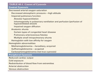
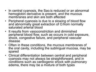
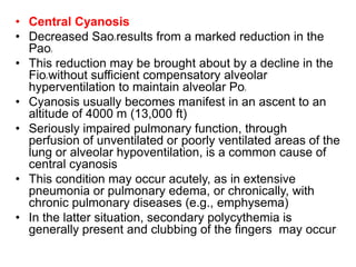
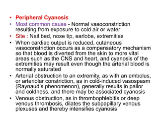
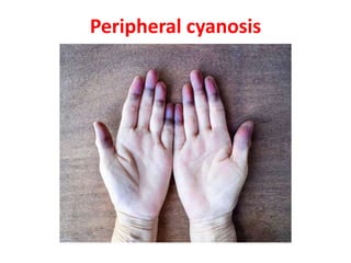
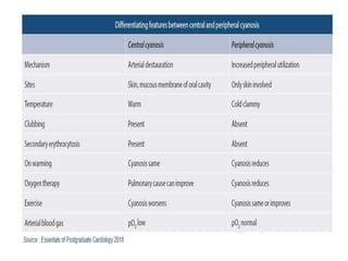
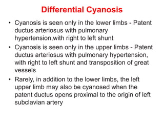
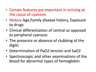
CYANOSIS
Dr.G.VENKATA RAMANA
MBBS DNB FAMILY MEDICINE
• Bluish color of the skin and mucous membranes
resulting from an increased quantity of reduced
hemoglobin (i.e., deoxygenated hemoglobin) or
of hemoglobin derivatives (e.g., methemoglobin
or sulfhemoglobin) in the small blood vessels of
those tissues
• It is usually most marked in the lips, nail beds,
ears, and malar eminences
• Types
• Central
• Peripheral
• Differential
• The increase in the quantity of reduced hemoglobin in
the mucocutaneous vessels that produces cyanosis may
be brought about either by an increase in the quantity of
venous blood as a result of dilation of the venules
(including precapillary venules) or by a reduction in the
Sao2 in the capillary blood
• In general, cyanosis becomes apparent when the
concentration of reduced hemoglobin in capillary blood
exceeds 40 g/L (4 g/dL)
• It is the absolute, rather than the relative, quantity of
reduced hemoglobin that is important in producing
cyanosis
• Thus, in a patient with severe anemia, the relative
quantity of reduced hemoglobin in the venous blood may
be very large when considered in relation to the total
quantity of hemoglobin in the blood
• However, since the concentration of the latter is
markedly reduced, the absolute quantity of reduced
hemoglobin may still be low, and, therefore, patients with
severe anemia and even marked arterial desaturation
may not display cyanosis
• Conversely, the higher the total hemoglobin content, the
greater is the tendency toward cyanosis; thus, patients
with marked polycythemia tend to be cyanotic at higher
levels of Sao2 than patients with normal hematocrit
values
• Likewise, local passive congestion, which causes an
increase in the total quantity of reduced hemoglobin in
the vessels in a given area, may cause cyanosis
• The degree of cyanosis is modified by the color of
the cutaneous pigment and the thickness of the
skin, as well as by the state of the cutaneous
capillaries
• In some instances, central cyanosis can be
detected reliably when the Sao2 has fallen to 85%;
in others, particularly in dark-skinned persons,it
may not be detected until it has declined to 75%
• In the latter case, examination of the mucous
membranes in the oral cavity and the
conjunctivae rather than examination of the skin
is more helpful in the detection of cyanosis
• In central cyanosis, the Sao2 is reduced or an abnormal
hemoglobin derivative is present, and the mucous
membranes and skin are both affected
• Peripheral cyanosis is due to a slowing of blood flow
and abnormally great extraction of O2 from normally
saturated arterial blood
• It results from vasoconstriction and diminished
peripheral blood flow, such as occurs in cold exposure,
shock, congestive failure, and peripheral vascular
disease
• Often in these conditions, the mucous membranes of
the oral cavity, including the sublingual mucosa, may be
spared
• Clinical differentiation between central and peripheral
cyanosis may not always be straightforward, and in
conditions such as cardiogenic shock with pulmonary
edema, there may be a mixture of both types
• Central Cyanosis
• Decreased Sao2 results from a marked reduction in the
Pao2
• This reduction may be brought about by a decline in the
Fio2 without sufficient compensatory alveolar
hyperventilation to maintain alveolar Po2
• Cyanosis usually becomes manifest in an ascent to an
altitude of 4000 m (13,000 ft)
• Seriously impaired pulmonary function, through
perfusion of unventilated or poorly ventilated areas of the
lung or alveolar hypoventilation, is a common cause of
central cyanosis
• This condition may occur acutely, as in extensive
pneumonia or pulmonary edema, or chronically, with
chronic pulmonary diseases (e.g., emphysema)
• In the latter situation, secondary polycythemia is
generally present and clubbing of the fingers may occur
• Another cause of reduced Sao2 is shunting of systemic
venous blood into the arterial circuit
• Certain forms of congenital heart disease are associated
with cyanosis on this basis
• Pulmonary arteriovenous fistulae may be congenital or
acquired, solitary or multiple, and microscopic or massive
• The severity of cyanosis produced by these fistulae
depends on their size and number
• They occur with some frequency in hereditary hemorrhagic
telangiectasia
• Sao2 reduction and cyanosis may also occur in some
patients with cirrhosis, presumably as a consequence of
pulmonary arteriovenous fistulae or portal vein–pulmonary
vein anastomoses
• In patients with cardiac or pulmonary right-to-left shunts, the
presence and severity of cyanosis depend on the size of the
shunt relative to the systemic flow and on the Hb-O2 saturation
of the venous blood.
• With increased extraction of O2 from the blood by the
exercising muscles, the venous blood returning to the right
side of the heart is more unsaturated than at rest, and
shunting of this blood intensifies the cyanosis
• Secondary polycythemia occurs frequently in patients in this
setting and contributes to the cyanosis
• Cyanosis can be caused by small quantities of circulating
methemoglobin (Hb Fe3+) and by even smaller quantities of
sulfhemoglobin
• Both of these hemoglobin derivatives impair oxygen delivery
to the tissues
• Although they are uncommon causes of cyanosis, these
abnormal hemoglobin species should be sought by
spectroscopy when cyanosis is not readily explained by
malfunction of the circulatory or respiratory systems
• Generally, digital clubbing does not occur with them
• Peripheral Cyanosis
• Most common cause - Normal vasoconstriction
resulting from exposure to cold air or water
• Site : Nail bed, nose tip, earlobe, extremities
• When cardiac output is reduced, cutaneous
vasoconstriction occurs as a compensatory mechanism
so that blood is diverted from the skin to more vital
areas such as the CNS and heart, and cyanosis of the
extremities may result even though the arterial blood is
normally saturated
• Arterial obstruction to an extremity, as with an embolus,
or arteriolar constriction, as in cold-induced vasospasm
(Raynaud’s phenomenon), generally results in pallor
and coldness, and there may be associated cyanosis
• Venous obstruction, as in thrombophlebitis or deep
venous thrombosis, dilates the subpapillary venous
plexuses and thereby intensifies cyanosis
Peripheral cyanosis
Differential Cyanosis
• Cyanosis is seen only in the lower limbs - Patent
ductus arteriosus with pulmonary
hypertension,with right to left shunt
• Cyanosis is seen only in the upper limbs - Patent
ductus arteriosus with pulmonary hypertension,
with right to left shunt and transposition of great
vessels
• Rarely, in addition to the lower limbs, the left
upper limb may also be cyanosed when the
patent ductus opens proximal to the origin of left
subclavian artery
Methemoglobin Sulfhemoglobin
Common causes Ingested medication (dapsone,
antimalarial agents)
Local anesthetic
Nitrates or nitrites
Ingested medication
(dapsone, phenacetin,
sulfonamides)
Nitrites
Typical
presentation
Cyanosis
Neurologic symptoms from
headache to coma
Respiratory depression
Cyanosis
Color of the blood Chocolate brown Green
Pulse oximetry Inaccurate Inaccurate
Arterial blood gas PaO2 normal
Methemoglobin is detected on
most machines
PaO2 normal
Confirmatory
testing
Blood gas (arterial or venous) Spectrophotometry or
Gas chromatography
Management Methylene blue (MB) or
Ascorbic acid
Supportive (no antidote)
Exchange transfusion in
severe cases
Lack of response to MB
Carboxyhemoglobin
Common causes Inhaled CO gas from fires, gas-powered
generators, kerosene heaters
Smoking
Typical presentation Neurologic symptoms from headache to coma
Chest pain from myocardial ischemia
Headache, malaise
Color of the blood Cherry red
Pulse oximetry Inaccurate
Arterial blood gas PaO2 normal
Carboxyhemoglobin is detected on most
machines
Confirmatory testing Co-oximetry using arterial blood (in stable
patients, venous samples are accurate)
Management High-flow or hyperbaric oxygen
• There are two types of cyanosis – Iron-replete and iron-
deplete
• Iron-replete compensated erythrocytosis establish equilibrium
with haematocrits and rarely result in symptoms of hyper-
viscosity because iron-replete cells are deformable
• Iron-deplete cells are less deformable and the
decompensated erythrocytosis fails to establish equilibrium
with unstable, rising haematocrits, resulting in recurrent
hyper-viscosity symptoms
• Phelebotomy for symptoms of hyper-viscosity which is not
due to dehydration or iron deficiency is a simple outpatient
procedure involving removal of 500 ml of blood over 45
minutes with isovolumetric replacement of isotonic saline
• Iron repletion in iron-deplete cyanosis must be done gradually
• Certain features are important in arriving at
the cause of cyanosis
• History Age,Family disease history, Exposure
to drugs
• Clinical differentiation of central as opposed
to peripheral cyanosis
• The presence or absence of clubbing of the
digits
• Determination of PaO2 tension and SaO2
• Spectroscopic and other examinations of the
blood for abnormal types of hemoglobin
LAB TESTS
• CBC
• CHEST XRAY
• ECG
• 2DECHO
• ABG
• OXIMETRIC STUDIES
• SPECTROSCOPE
• HB ELECTROPHORESIS
cyanosis.pptx
cyanosis.pptx

cyanosis.pptx

  • 1.
  • 2.
    • Bluish colorof the skin and mucous membranes resulting from an increased quantity of reduced hemoglobin (i.e., deoxygenated hemoglobin) or of hemoglobin derivatives (e.g., methemoglobin or sulfhemoglobin) in the small blood vessels of those tissues • It is usually most marked in the lips, nail beds, ears, and malar eminences • Types • Central • Peripheral • Differential
  • 3.
    • The increasein the quantity of reduced hemoglobin in the mucocutaneous vessels that produces cyanosis may be brought about either by an increase in the quantity of venous blood as a result of dilation of the venules (including precapillary venules) or by a reduction in the Sao2 in the capillary blood • In general, cyanosis becomes apparent when the concentration of reduced hemoglobin in capillary blood exceeds 40 g/L (4 g/dL) • It is the absolute, rather than the relative, quantity of reduced hemoglobin that is important in producing cyanosis • Thus, in a patient with severe anemia, the relative quantity of reduced hemoglobin in the venous blood may be very large when considered in relation to the total quantity of hemoglobin in the blood
  • 4.
    • However, sincethe concentration of the latter is markedly reduced, the absolute quantity of reduced hemoglobin may still be low, and, therefore, patients with severe anemia and even marked arterial desaturation may not display cyanosis • Conversely, the higher the total hemoglobin content, the greater is the tendency toward cyanosis; thus, patients with marked polycythemia tend to be cyanotic at higher levels of Sao2 than patients with normal hematocrit values • Likewise, local passive congestion, which causes an increase in the total quantity of reduced hemoglobin in the vessels in a given area, may cause cyanosis
  • 5.
    • The degreeof cyanosis is modified by the color of the cutaneous pigment and the thickness of the skin, as well as by the state of the cutaneous capillaries • In some instances, central cyanosis can be detected reliably when the Sao2 has fallen to 85%; in others, particularly in dark-skinned persons,it may not be detected until it has declined to 75% • In the latter case, examination of the mucous membranes in the oral cavity and the conjunctivae rather than examination of the skin is more helpful in the detection of cyanosis
  • 7.
    • In centralcyanosis, the Sao2 is reduced or an abnormal hemoglobin derivative is present, and the mucous membranes and skin are both affected • Peripheral cyanosis is due to a slowing of blood flow and abnormally great extraction of O2 from normally saturated arterial blood • It results from vasoconstriction and diminished peripheral blood flow, such as occurs in cold exposure, shock, congestive failure, and peripheral vascular disease • Often in these conditions, the mucous membranes of the oral cavity, including the sublingual mucosa, may be spared • Clinical differentiation between central and peripheral cyanosis may not always be straightforward, and in conditions such as cardiogenic shock with pulmonary edema, there may be a mixture of both types
  • 8.
    • Central Cyanosis •Decreased Sao2 results from a marked reduction in the Pao2 • This reduction may be brought about by a decline in the Fio2 without sufficient compensatory alveolar hyperventilation to maintain alveolar Po2 • Cyanosis usually becomes manifest in an ascent to an altitude of 4000 m (13,000 ft) • Seriously impaired pulmonary function, through perfusion of unventilated or poorly ventilated areas of the lung or alveolar hypoventilation, is a common cause of central cyanosis • This condition may occur acutely, as in extensive pneumonia or pulmonary edema, or chronically, with chronic pulmonary diseases (e.g., emphysema) • In the latter situation, secondary polycythemia is generally present and clubbing of the fingers may occur
  • 9.
    • Another causeof reduced Sao2 is shunting of systemic venous blood into the arterial circuit • Certain forms of congenital heart disease are associated with cyanosis on this basis • Pulmonary arteriovenous fistulae may be congenital or acquired, solitary or multiple, and microscopic or massive • The severity of cyanosis produced by these fistulae depends on their size and number • They occur with some frequency in hereditary hemorrhagic telangiectasia • Sao2 reduction and cyanosis may also occur in some patients with cirrhosis, presumably as a consequence of pulmonary arteriovenous fistulae or portal vein–pulmonary vein anastomoses
  • 10.
    • In patientswith cardiac or pulmonary right-to-left shunts, the presence and severity of cyanosis depend on the size of the shunt relative to the systemic flow and on the Hb-O2 saturation of the venous blood. • With increased extraction of O2 from the blood by the exercising muscles, the venous blood returning to the right side of the heart is more unsaturated than at rest, and shunting of this blood intensifies the cyanosis • Secondary polycythemia occurs frequently in patients in this setting and contributes to the cyanosis • Cyanosis can be caused by small quantities of circulating methemoglobin (Hb Fe3+) and by even smaller quantities of sulfhemoglobin • Both of these hemoglobin derivatives impair oxygen delivery to the tissues • Although they are uncommon causes of cyanosis, these abnormal hemoglobin species should be sought by spectroscopy when cyanosis is not readily explained by malfunction of the circulatory or respiratory systems • Generally, digital clubbing does not occur with them
  • 12.
    • Peripheral Cyanosis •Most common cause - Normal vasoconstriction resulting from exposure to cold air or water • Site : Nail bed, nose tip, earlobe, extremities • When cardiac output is reduced, cutaneous vasoconstriction occurs as a compensatory mechanism so that blood is diverted from the skin to more vital areas such as the CNS and heart, and cyanosis of the extremities may result even though the arterial blood is normally saturated • Arterial obstruction to an extremity, as with an embolus, or arteriolar constriction, as in cold-induced vasospasm (Raynaud’s phenomenon), generally results in pallor and coldness, and there may be associated cyanosis • Venous obstruction, as in thrombophlebitis or deep venous thrombosis, dilates the subpapillary venous plexuses and thereby intensifies cyanosis
  • 13.
  • 16.
    Differential Cyanosis • Cyanosisis seen only in the lower limbs - Patent ductus arteriosus with pulmonary hypertension,with right to left shunt • Cyanosis is seen only in the upper limbs - Patent ductus arteriosus with pulmonary hypertension, with right to left shunt and transposition of great vessels • Rarely, in addition to the lower limbs, the left upper limb may also be cyanosed when the patent ductus opens proximal to the origin of left subclavian artery
  • 17.
    Methemoglobin Sulfhemoglobin Common causesIngested medication (dapsone, antimalarial agents) Local anesthetic Nitrates or nitrites Ingested medication (dapsone, phenacetin, sulfonamides) Nitrites Typical presentation Cyanosis Neurologic symptoms from headache to coma Respiratory depression Cyanosis Color of the blood Chocolate brown Green Pulse oximetry Inaccurate Inaccurate Arterial blood gas PaO2 normal Methemoglobin is detected on most machines PaO2 normal Confirmatory testing Blood gas (arterial or venous) Spectrophotometry or Gas chromatography Management Methylene blue (MB) or Ascorbic acid Supportive (no antidote) Exchange transfusion in severe cases Lack of response to MB
  • 18.
    Carboxyhemoglobin Common causes InhaledCO gas from fires, gas-powered generators, kerosene heaters Smoking Typical presentation Neurologic symptoms from headache to coma Chest pain from myocardial ischemia Headache, malaise Color of the blood Cherry red Pulse oximetry Inaccurate Arterial blood gas PaO2 normal Carboxyhemoglobin is detected on most machines Confirmatory testing Co-oximetry using arterial blood (in stable patients, venous samples are accurate) Management High-flow or hyperbaric oxygen
  • 19.
    • There aretwo types of cyanosis – Iron-replete and iron- deplete • Iron-replete compensated erythrocytosis establish equilibrium with haematocrits and rarely result in symptoms of hyper- viscosity because iron-replete cells are deformable • Iron-deplete cells are less deformable and the decompensated erythrocytosis fails to establish equilibrium with unstable, rising haematocrits, resulting in recurrent hyper-viscosity symptoms • Phelebotomy for symptoms of hyper-viscosity which is not due to dehydration or iron deficiency is a simple outpatient procedure involving removal of 500 ml of blood over 45 minutes with isovolumetric replacement of isotonic saline • Iron repletion in iron-deplete cyanosis must be done gradually
  • 20.
    • Certain featuresare important in arriving at the cause of cyanosis • History Age,Family disease history, Exposure to drugs • Clinical differentiation of central as opposed to peripheral cyanosis • The presence or absence of clubbing of the digits • Determination of PaO2 tension and SaO2 • Spectroscopic and other examinations of the blood for abnormal types of hemoglobin
  • 22.
    LAB TESTS • CBC •CHEST XRAY • ECG • 2DECHO • ABG • OXIMETRIC STUDIES • SPECTROSCOPE • HB ELECTROPHORESIS