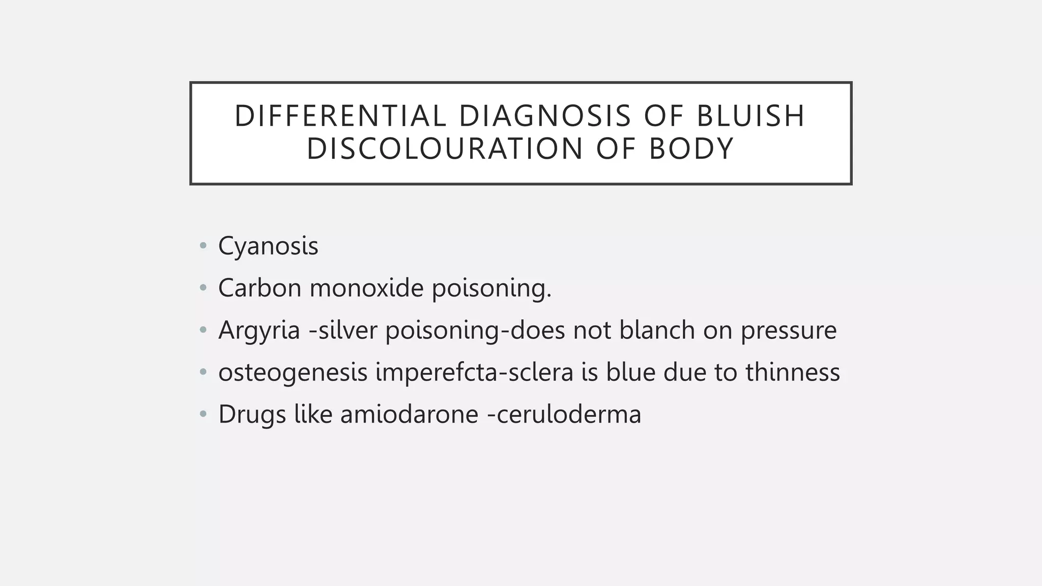
APPROACH TO CYANOSIS
Sarath tk
116 batch
DEFINITION
• Cyanosis is derived from the word ‘CYAN’,which comes from ‘Kyanous’,the greek
word for blue.
• Cyanosis is the bluish discolouration of the skin and mucous membrane due
to the presence of increased amount of reduced
haemoglobin/deoxyhaemoglobin (>5g/dl)or of haemoglobin derivatives
(sulphhaemoglobin and methaemoglobin) in the capillary blood.
• Cyanosis is normally detected when the oxygen saturation is (SaO2) is <85%
TYPES
1.Peripheral cyanosis
2.Central cyanosis
PERIPHERAL CYANOSIS
• Arterial blood is normally saturated but there is oxygen unsaturation
at the venous end of capillary. may be due to sluggish peripheral
circulation
• Excessive extraction of oxygen by the peripheral tissues from the
arterial blood .
• SITES- Tip of nose
Ear lobules
Outer aspect of lips,cheeks and chin
• Tips of fingers and toes.
• Nail bed of fingers and toes.
• Palms and soles.
• Tongue remains unaffected
CAUSES
• Exposure to cold air or cold water (vasoconstriction)
• Reduced cardiac output(Congestive cardiac failure)
• Shock or peripheral circulatory failure
• Hyperviscosity syndromes(Polycythemia, Multiple myeloma)
• Mitral stenosis( Lips, tip of nose and cheeks cyanosed in mitral
facies)
• Venous obstruction (SVC syndrome)-local cyanosis
• Arterial sclerosis ( Atherosclerosis)
CENTRAL CYANOSIS
• Pathological condition caused by decreased arterial oxygen
saturation either due to defective oxygenation in lungs or
admixture of venous and arterial blood.
• Usually detected when the oxygen saturation of arterial
blood goes below 80 - 85%.
• it involves highly vascularized tissues through which blood
flow is at brisk.
• Cardiac output is normal and patients have warm extremities.
SITES
• Tongue(margins and
undersurface)
• Inner aspect of lips
• Mucous membrane of gums ,soft
palate, cheeks.
• Lower palpebral conjunctiva.
• Also the sites mentioned in
peripheral cyanosis ( both can
occur together)
CAUSES
• Cyanotic congenital heart diseases eg: Tetralogy of fallot
,transposition of great aorta,Total anomalous pulmonary venous
return,Truncus arteriosus,Tricuspid atresia.
• Eisenmenger’s syndrome(ASD,VSD,PDA with reversal of shunt)
• Single ventricle.
• Acute pulmonary edema ( due to left sided heart failure)-most
common cause.
• Lobar pneumonia
• Asthma
• COPD ,cor pulmonale
• Congenital diaphragmatic hernia.
• Birth asphyxias.
• Seizures.
• ICH
OTHERS-Polycythemia,methaemoglobinemia,metabolic
disease,infections(sepsis)
DIFFERENTIAL CYANOSIS
• Hands red (less blue)and feet blue seen in PDA with
reversal of shunt
• I t requires pulmonary vascular resistance elevated to
a systemic level and a patent ductus arteriosus.
• Desaturated blood from the ductus enters the aorta
distal to subclavian artery,sparing the brachiocephalic
circulation.
MIXED CYANOSIS
• Central and peripheral cyanosis present in a single
patient.
• Seen in -Acute MI with acute LVF
CCF due to left sided heart failure
Rarely in polycythemia
ENTEROGENOUS OR PIGMENT CYANOSIS
• Cyanosis due to excessive
sulphaemoglogin(>0.5g/dl)or methaemoglobin
(>1.5g/dl)in blood.
• Causes are hereditary haemoglobin M
disease,poisoning by aniline dyes ,drugs like nitrates
and nitrites(nitroglycerin,sodium nitroprusside
etc),carboxyhaemoglobinaemia (a cherry red flush in
smokers)
DIFFERENTIAL DIAGNOSIS OF BLUISH
DISCOLOURATION OF BODY
• Cyanosis
• Carbon monoxide poisoning.
• Argyria -silver poisoning-does not blanch on pressure
• osteogenesis imperefcta-sclera is blue due to thinness
• Drugs like amiodarone -ceruloderma
MANAGEMENT
• AIM:
To differentiate physiological and pathological
cyanosis
To differentiate cardiac from non cardiac causes.
Find causes which needs urgent referral or treatment.
• Do: Complete maternal and newborn history
Perform a full physical examination.
Investigations
INVESTIGATION
• PULSE OXIMETRY
• Standard methord pf monitoring infants with rds cyanosis
• Normal >=95%
• Mesurements usually not performed when child is crying or moving as it
reduces the quality of signal
ARTERIAL BLOOD GAS
• Arterial PO2: to confirm central cyanosis :SaO2 not as good
an indicator dueto Increase fetal Hb affinity for O2 (left-shift)
• Increase PaCO2: may indicate pulmonary or CNS disorders,
heart failure
• Decrease pH: sepsis, circulatory shock, severe hypoxemia
• Methemoglobinemia: Decrease SaO2, normal PaO2,
chocolate-brown blood
HYPEROXIA TEST
• Hyperoxia test is the initial method to distinguish CCHD from pulmonary
disease.
• The test consists in measuring an arterial blood gas at room air and 100%
inspired oxygen after 10 minutes.
• Neonates with congenital heart disease are usually not able to increase PaO2
above 100 mm Hg during 100% oxygen administration
• . In patients with pulmonary disease, PaO2 generally increased greater than or
equal to 100 mm Hg with 100% oxygen as ventilation-perfusion discrepancies
are overcome.
• A positive result indicates the cardiac origin and further cardiac workup is
indicated to rule out CCHD
BLOOD INVESTIGATIONS
• INCREASE OR DECREASE WBS INDICATES SEPSIS
• HAEMATOCRIT>65%:POLYCYTHEMIA
• FURTHER X RAY AND ECG HELPS CONFIRM CARDIAC OR PULMONARY
CAUSE
TREATMENT
• Warming of the affected area - in peripheral cyanosis
• Oxygenation and adequate ventilation.
(Partial press of oxygen normalises completely during artificial ventilation
in infant with CNS disorder)
.IV fluids- children with feeding difficulty
. If sepsis is suspected or another specific cause is not identified ,start on
broad spectrum antibiotics and then obtain a full septic screening.
.Drugs- Prostaglandin E1
For ductal dependant CHD- iv infusion of PGE1 to maintain patency 0.1mcg/kg/min.
. Surgery
THANK YOU

APPROACH TO CYANOSIS .pptx

  • 1.
  • 2.
    DEFINITION • Cyanosis isderived from the word ‘CYAN’,which comes from ‘Kyanous’,the greek word for blue. • Cyanosis is the bluish discolouration of the skin and mucous membrane due to the presence of increased amount of reduced haemoglobin/deoxyhaemoglobin (>5g/dl)or of haemoglobin derivatives (sulphhaemoglobin and methaemoglobin) in the capillary blood. • Cyanosis is normally detected when the oxygen saturation is (SaO2) is <85%
  • 3.
  • 4.
    PERIPHERAL CYANOSIS • Arterialblood is normally saturated but there is oxygen unsaturation at the venous end of capillary. may be due to sluggish peripheral circulation • Excessive extraction of oxygen by the peripheral tissues from the arterial blood .
  • 5.
    • SITES- Tipof nose Ear lobules Outer aspect of lips,cheeks and chin • Tips of fingers and toes. • Nail bed of fingers and toes. • Palms and soles. • Tongue remains unaffected
  • 6.
    CAUSES • Exposure tocold air or cold water (vasoconstriction) • Reduced cardiac output(Congestive cardiac failure) • Shock or peripheral circulatory failure • Hyperviscosity syndromes(Polycythemia, Multiple myeloma) • Mitral stenosis( Lips, tip of nose and cheeks cyanosed in mitral facies) • Venous obstruction (SVC syndrome)-local cyanosis • Arterial sclerosis ( Atherosclerosis)
  • 7.
    CENTRAL CYANOSIS • Pathologicalcondition caused by decreased arterial oxygen saturation either due to defective oxygenation in lungs or admixture of venous and arterial blood. • Usually detected when the oxygen saturation of arterial blood goes below 80 - 85%. • it involves highly vascularized tissues through which blood flow is at brisk. • Cardiac output is normal and patients have warm extremities.
  • 8.
    SITES • Tongue(margins and undersurface) •Inner aspect of lips • Mucous membrane of gums ,soft palate, cheeks. • Lower palpebral conjunctiva. • Also the sites mentioned in peripheral cyanosis ( both can occur together)
  • 9.
    CAUSES • Cyanotic congenitalheart diseases eg: Tetralogy of fallot ,transposition of great aorta,Total anomalous pulmonary venous return,Truncus arteriosus,Tricuspid atresia. • Eisenmenger’s syndrome(ASD,VSD,PDA with reversal of shunt) • Single ventricle. • Acute pulmonary edema ( due to left sided heart failure)-most common cause. • Lobar pneumonia • Asthma
  • 10.
    • COPD ,corpulmonale • Congenital diaphragmatic hernia. • Birth asphyxias. • Seizures. • ICH OTHERS-Polycythemia,methaemoglobinemia,metabolic disease,infections(sepsis)
  • 12.
    DIFFERENTIAL CYANOSIS • Handsred (less blue)and feet blue seen in PDA with reversal of shunt • I t requires pulmonary vascular resistance elevated to a systemic level and a patent ductus arteriosus. • Desaturated blood from the ductus enters the aorta distal to subclavian artery,sparing the brachiocephalic circulation.
  • 13.
    MIXED CYANOSIS • Centraland peripheral cyanosis present in a single patient. • Seen in -Acute MI with acute LVF CCF due to left sided heart failure Rarely in polycythemia
  • 14.
    ENTEROGENOUS OR PIGMENTCYANOSIS • Cyanosis due to excessive sulphaemoglogin(>0.5g/dl)or methaemoglobin (>1.5g/dl)in blood. • Causes are hereditary haemoglobin M disease,poisoning by aniline dyes ,drugs like nitrates and nitrites(nitroglycerin,sodium nitroprusside etc),carboxyhaemoglobinaemia (a cherry red flush in smokers)
  • 15.
    DIFFERENTIAL DIAGNOSIS OFBLUISH DISCOLOURATION OF BODY • Cyanosis • Carbon monoxide poisoning. • Argyria -silver poisoning-does not blanch on pressure • osteogenesis imperefcta-sclera is blue due to thinness • Drugs like amiodarone -ceruloderma
  • 16.
    MANAGEMENT • AIM: To differentiatephysiological and pathological cyanosis To differentiate cardiac from non cardiac causes. Find causes which needs urgent referral or treatment.
  • 17.
    • Do: Completematernal and newborn history Perform a full physical examination. Investigations
  • 27.
    INVESTIGATION • PULSE OXIMETRY •Standard methord pf monitoring infants with rds cyanosis • Normal >=95% • Mesurements usually not performed when child is crying or moving as it reduces the quality of signal
  • 28.
    ARTERIAL BLOOD GAS •Arterial PO2: to confirm central cyanosis :SaO2 not as good an indicator dueto Increase fetal Hb affinity for O2 (left-shift) • Increase PaCO2: may indicate pulmonary or CNS disorders, heart failure • Decrease pH: sepsis, circulatory shock, severe hypoxemia • Methemoglobinemia: Decrease SaO2, normal PaO2, chocolate-brown blood
  • 29.
    HYPEROXIA TEST • Hyperoxiatest is the initial method to distinguish CCHD from pulmonary disease. • The test consists in measuring an arterial blood gas at room air and 100% inspired oxygen after 10 minutes. • Neonates with congenital heart disease are usually not able to increase PaO2 above 100 mm Hg during 100% oxygen administration • . In patients with pulmonary disease, PaO2 generally increased greater than or equal to 100 mm Hg with 100% oxygen as ventilation-perfusion discrepancies are overcome. • A positive result indicates the cardiac origin and further cardiac workup is indicated to rule out CCHD
  • 30.
    BLOOD INVESTIGATIONS • INCREASEOR DECREASE WBS INDICATES SEPSIS • HAEMATOCRIT>65%:POLYCYTHEMIA
  • 31.
    • FURTHER XRAY AND ECG HELPS CONFIRM CARDIAC OR PULMONARY CAUSE
  • 32.
    TREATMENT • Warming ofthe affected area - in peripheral cyanosis • Oxygenation and adequate ventilation. (Partial press of oxygen normalises completely during artificial ventilation in infant with CNS disorder) .IV fluids- children with feeding difficulty . If sepsis is suspected or another specific cause is not identified ,start on broad spectrum antibiotics and then obtain a full septic screening.
  • 33.
    .Drugs- Prostaglandin E1 Forductal dependant CHD- iv infusion of PGE1 to maintain patency 0.1mcg/kg/min. . Surgery
  • 34.