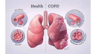
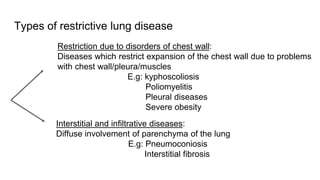
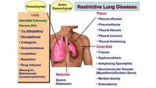
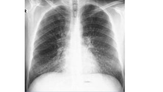
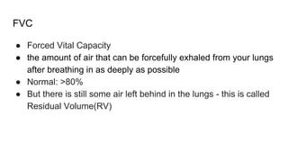
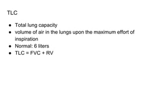
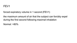
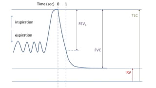
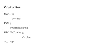
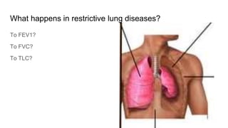
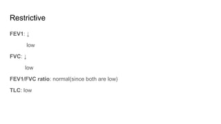
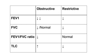
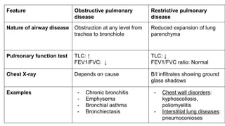
Obstructive vs Restrictive
Pulmonary Disease
Dr. Salman Ansari(MBBS)
Tutor, Dept. of Pathology
Kanachur Institute of Medical Sciences
● Obstructive lung diseases
● Restrictive lung diseases
● Obstructive vs restrictive lung diseases
Obstructive lung diseases
Increase in resistance to airflow
Due to partial or complete obstruction at any level from the trachea to the
bronchioles
Examples of obstructive airway diseases:
- Emphysema
- Chronic Bronchitis
- Bronchial asthma
- Bronchiectasis
Restrictive pulmonary diseases
Reduced expansion of lung parenchyma and decreased total lung
capacity
X-ray shows ground-glass shadows
Types of restrictive lung disease
Restriction due to disorders of chest wall:
Diseases which restrict expansion of the chest wall due to problems
with chest wall/pleura/muscles
E.g: kyphoscoliosis
Poliomyelitis
Pleural diseases
Severe obesity
Interstitial and infiltrative diseases:
Diffuse involvement of parenchyma of the lung
E.g: Pneumoconiosis
Interstitial fibrosis
Spirometry: basic definitions
1. FVC
2. TLC
3. FEV1
FVC
● Forced Vital Capacity
● the amount of air that can be forcefully exhaled from your lungs
after breathing in as deeply as possible
● Normal: >80%
● But there is still some air left behind in the lungs - this is called
Residual Volume(RV)
TLC
● Total lung capacity
● volume of air in the lungs upon the maximum effort of
inspiration
● Normal: 6 liters
● TLC = FVC + RV
FEV1
forced expiratory volume in 1 second (FEV1)
the maximum amount of air that the subject can forcibly expel
during the first second following maximal inhalation
Normal: >80%
What happens in obstructive lung diseases?
To FEV1?
To FVC?
To TLC?
Obstructive
FEV1: ↓↓
Very low
FVC: ↓
low/almost normal
FEV1/FVC ratio: ↓↓
Very low
TLC: high
What happens in restrictive lung diseases?
To FEV1?
To FVC?
To TLC?
Restrictive
FEV1: ↓
low
FVC: ↓
low
FEV1/FVC ratio: normal(since both are low)
TLC: low
Obstructive Restrictive
FEV1 ↓ ↓ ↓
FVC ↓ /Normal ↓
FEV1/FVC ratio ↓ Normal
TLC ↑ ↓
Feature Obstructive pulmonary
disease
Restrictive pulmonary
disease
Nature of airway disease Obstruction at any level from
trachea to bronchiole
Reduced expansion of lung
parenchyma
Pulmonary function test TLC: ↑
FEV1/FVC: ↓
TLC: ↓
FEV1/FVC ratio: Normal
Chest X-ray Depends on cause B/l infiltrates showing ground
glass shadows
Examples - Chronic bronchitis
- Emphysema
- Bronchial asthma
- Bronchiectasis
- Chest wall disorders:
kyphoscoliosis,
poliomyelitis
- Interstitial lung diseases:
pneumoconioses
For notes,
scan:
References:
● Ramadas Nayak - Textbook of Pathology for Allied
Health Sciences
Questions:
salman.s.ansari92@gmail.com
For PPT, scan:

Obstructive vs Restrictive Pulmonary Disease - Pathology - ATOT

  • 1.
    Obstructive vs Restrictive PulmonaryDisease Dr. Salman Ansari(MBBS) Tutor, Dept. of Pathology Kanachur Institute of Medical Sciences
  • 2.
    ● Obstructive lungdiseases ● Restrictive lung diseases ● Obstructive vs restrictive lung diseases
  • 3.
    Obstructive lung diseases Increasein resistance to airflow Due to partial or complete obstruction at any level from the trachea to the bronchioles Examples of obstructive airway diseases: - Emphysema - Chronic Bronchitis - Bronchial asthma - Bronchiectasis
  • 6.
    Restrictive pulmonary diseases Reducedexpansion of lung parenchyma and decreased total lung capacity X-ray shows ground-glass shadows
  • 7.
    Types of restrictivelung disease Restriction due to disorders of chest wall: Diseases which restrict expansion of the chest wall due to problems with chest wall/pleura/muscles E.g: kyphoscoliosis Poliomyelitis Pleural diseases Severe obesity Interstitial and infiltrative diseases: Diffuse involvement of parenchyma of the lung E.g: Pneumoconiosis Interstitial fibrosis
  • 10.
  • 11.
    FVC ● Forced VitalCapacity ● the amount of air that can be forcefully exhaled from your lungs after breathing in as deeply as possible ● Normal: >80% ● But there is still some air left behind in the lungs - this is called Residual Volume(RV)
  • 12.
    TLC ● Total lungcapacity ● volume of air in the lungs upon the maximum effort of inspiration ● Normal: 6 liters ● TLC = FVC + RV
  • 13.
    FEV1 forced expiratory volumein 1 second (FEV1) the maximum amount of air that the subject can forcibly expel during the first second following maximal inhalation Normal: >80%
  • 15.
    What happens inobstructive lung diseases? To FEV1? To FVC? To TLC?
  • 16.
    Obstructive FEV1: ↓↓ Very low FVC:↓ low/almost normal FEV1/FVC ratio: ↓↓ Very low TLC: high
  • 17.
    What happens inrestrictive lung diseases? To FEV1? To FVC? To TLC?
  • 18.
    Restrictive FEV1: ↓ low FVC: ↓ low FEV1/FVCratio: normal(since both are low) TLC: low
  • 19.
    Obstructive Restrictive FEV1 ↓↓ ↓ FVC ↓ /Normal ↓ FEV1/FVC ratio ↓ Normal TLC ↑ ↓
  • 20.
    Feature Obstructive pulmonary disease Restrictivepulmonary disease Nature of airway disease Obstruction at any level from trachea to bronchiole Reduced expansion of lung parenchyma Pulmonary function test TLC: ↑ FEV1/FVC: ↓ TLC: ↓ FEV1/FVC ratio: Normal Chest X-ray Depends on cause B/l infiltrates showing ground glass shadows Examples - Chronic bronchitis - Emphysema - Bronchial asthma - Bronchiectasis - Chest wall disorders: kyphoscoliosis, poliomyelitis - Interstitial lung diseases: pneumoconioses
  • 21.
    For notes, scan: References: ● RamadasNayak - Textbook of Pathology for Allied Health Sciences Questions: salman.s.ansari92@gmail.com For PPT, scan: