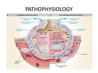
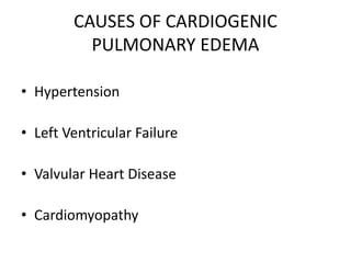
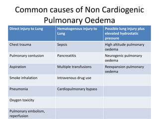
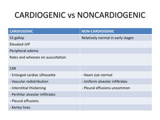
PULMONARY OEDEMA
Prajjwal Malla
MDGP Resident 3rd year
Definition
• A condition characterized by fluid accumulation in the
lungs caused by extravasation of fluid from pulmonary
vasculature into the interstitium and alveoli of the
lungs.
Etiopathogenesis
• Imbalance of the following forces
– Increased pulmonary capillary pressure
– Decreased plasma oncotic pressure
– Increased negative interstitial pressure
• Damage to the alveolar-capillary barrier
• Lymphatic obstruction
• Idiopathic mechanism
CLASSIFICATION
• CARDIOGENIC
• NON CARDIOGENIC
PATHOPHYSIOLOGY
CAUSES OF CARDIOGENIC
PULMONARY EDEMA
• Hypertension
• Left Ventricular Failure
• Valvular Heart Disease
• Cardiomyopathy
Common causes of Non Cardiogenic
Pulmonary Oedema
Direct injury to Lung Hematogenous injury to
Lung
Possible lung injury plus
elevated hydrostatic
pressure
Chest trauma Sepsis High altitude pulmonary
oedema
Pulmonary contusion Pancreatitis Neurogenic pulmonary
oedema
Aspiration Multiple transfusions Reexpansion pulmonary
oedema
Smoke inhalation Intravenous drug use
Pneumonia Cardiopulmonary bypass
Oxygen toxicity
Pulmonary embolism,
reperfusion
CARDIOGENIC vs NONCARDIOGENIC
CARDIOGENIC NON-CARDIOGENIC
S3 gallop Relatively normal in early stages
Elevated JVP
Peripheral edema
Rales and wheezes on auscultation
CXR
- Enlarged cardiac silhouette - Heart size normal
- Vascular redistribution - Uniform alveolar infiltrates
- Interstitial thickening - Pleural effusions uncommon
- Perihilar alveolar infiltrates
- Pleural effusions
- Kerley lines
Approach to a Patient with Pulmonary
Oedema
• Exertional dyspnoea √ Palpitation
• Orthopnoea √ Excessive sweating
• Aspiration of food or √ Skin colour change – pale skin
foreign body
• Direct chest injuries √ Chest pain (if cardiogenic)
• Walking high altitude √ Rapid weight gain (cardiogenic)
• Chest pain (right or left) √ Fatigue
• Leg pain or swelling (Pulmonary embolism) √ Loss of appetite
• Cough with blood tinged sputum √ Smoking history
LABORATORY INVESTIGATIONS
• Routine CBC
• Renal Function Test
• Liver Function Test
• Arterial blood gas analysis
• Serum cardiac biomarkers
• Ultrasound
• Echocardiograpy
Investigation…..
• Pulmonary artery catheterization is indicated
when
– Cause remain uncertain
– Pulmonary edema refractory to therapy
– Pulmonary edema accompanied by hypotension
Complications
• Pulmonary edema, especially acute, can lead to fatal
respiratory distress or cardiac arrest due to hypoxia.
• Assisted ventilation is provided if signs of respiratory
fatigue - lethargy, fatigue, diaphoresis, worsening anxiety
• Sudden cardiac death secondary to cardiac arrhythmia-
continuous monitoring of the heart rhythm
• Non-cardiogenic PE – resolves much less quickly, and
most require mechanical ventilation
TREATMENT APPROACH
• EMERGENCY MANAGEMENT
– Upright sitting posture
– Support of oxygenation and ventilation
– Oxygen therapy
– Positive pressure ventilation
• REDUCTION OF PRELOAD AND INOTROPIC DRUGS
– Loop diuretics
– Nitrates
– Morphine
– Dopamine
TREATMENT APPROACH…..
• Condition that complicate PE must be
corrected
– Infection
– Renal failure
– Anemia
OXYGEN THERAPY
• Is essential to ensure adequate O2 delivery to peripheral
tissues, including heart.
• When hypoxemia (PO2<60 mmHg) without hypercapnia
– O2 given either by nasal prongs or Venturi mask with
reservoir.
• If PO2 cannot be maintained at or near 60 mmHg despite
inhalation of 100% O2 at 20L/min, or if progressive
hypercapnia, mechanical ventillation is needed
NON INVASIVE VENTILLATION
• Continuous Positive Airway Pressure (CPAP)
• Non-invasive Intermittent Positive-pressure Ventillation (NIPPV)
• CPAP maintains the same positive-pressure support throughout
the respiratory cycle
• NIPPV increases airway pressure more during inspiration than
during expiration
• compared to CPAP, NIPPV produces greater improvements in
oxygenation and carbon dioxide clearance and a greater reduction
in the work of breathing in patients with pulmonary oedema.
• Intubation and mechanical ventilation
REDUCTION OF PRELOAD
• Diuretics : 0.5-1.0 mg/kg
• The loop diuretics - furosemide, bumetanide, and
torsemide are effective in most forms of
pulmonary edema, even in the presence of
hypoalbuminemia, hyponatremia, or
hypochloremia.
• Nitrates :
– Nitroglycerin and isosorbide dinitrate - venodilators but have
coronary vasodilating properties as well.
• Sublingual nitroglycerin (0.4 mg × 3 every 5 min)
– first-line therapy for acute cardiogenic pulmonary edema.
• Morphine :
– Given in 2- to 4-mg IV boluses, morphine is a transient
venodilator that reduces preload while relieving dyspnea and
anxiety.
• ACE inhibitors :
– reduce both afterload and preload and are
recommended for hypertensive patients.
• Physical Methods:
– In nonhypotensive patients, venous return can be
reduced by use of the sitting position with the legs
dangling along the side of the bed
• Inotropics :
– agents are indicated in patients with cardiogenic
pulmonary edema and severe LV dysfunction
• Digitalis Glycosides:
– useful for control of ventricular rate in patients
with rapid atrial fibrillation or flutter and LV
dysfunction
• Sinus tachycardia or atrial fibrillation can result
from elevated left atrial pressure and sympathetic
stimulation.
• may require cardioversion
• Reexpansion pulmonary edema can develop after
removal of longstanding pleural space air or fluid.
• These patients may develop hypotension or oliguria
resulting from rapid fluid shifts into the lung.
• Diuretics and preload reduction are contraindicated
• Intravascular volume repletion often is needed while
supporting oxygenation and gas exchange.
• High-altitude pulmonary edema
– can be prevented by use of dexamethasone, calcium
channel–blocking drugs, or long-acting inhaled β2-
adrenergic agonists.
• Treatment
– descent from altitude
– bed rest
– oxygen
– inhaled nitric oxide.
– Nifedipine may also be effective.
Prognosis
• In-hospital death rates - cardiogenic PE- as
high as 15-20%
• Myocardial infarction, associated hypotension,
and a history of frequent hospitalizations for
CPE generally increase the mortality risk
Take Home Message
• Clinical suspicion should be made when a patient with
acute onset shortness of breath presents in ER.
• Try to find the cause for the condition.
• Prompt positioning and proper treatment would help
relief the patient and could be life saving.
• Prognosis depends upon the severity of the condition.
References
- Harrison’s Principles of Internal Medicine 18th
edition
- Uptodate
- Medscape
- Online journals

Pulmonary oedema

  • 1.
  • 2.
    Definition • A conditioncharacterized by fluid accumulation in the lungs caused by extravasation of fluid from pulmonary vasculature into the interstitium and alveoli of the lungs.
  • 3.
    Etiopathogenesis • Imbalance ofthe following forces – Increased pulmonary capillary pressure – Decreased plasma oncotic pressure – Increased negative interstitial pressure • Damage to the alveolar-capillary barrier • Lymphatic obstruction • Idiopathic mechanism
  • 4.
  • 5.
  • 6.
    CAUSES OF CARDIOGENIC PULMONARYEDEMA • Hypertension • Left Ventricular Failure • Valvular Heart Disease • Cardiomyopathy
  • 7.
    Common causes ofNon Cardiogenic Pulmonary Oedema Direct injury to Lung Hematogenous injury to Lung Possible lung injury plus elevated hydrostatic pressure Chest trauma Sepsis High altitude pulmonary oedema Pulmonary contusion Pancreatitis Neurogenic pulmonary oedema Aspiration Multiple transfusions Reexpansion pulmonary oedema Smoke inhalation Intravenous drug use Pneumonia Cardiopulmonary bypass Oxygen toxicity Pulmonary embolism, reperfusion
  • 8.
    CARDIOGENIC vs NONCARDIOGENIC CARDIOGENICNON-CARDIOGENIC S3 gallop Relatively normal in early stages Elevated JVP Peripheral edema Rales and wheezes on auscultation CXR - Enlarged cardiac silhouette - Heart size normal - Vascular redistribution - Uniform alveolar infiltrates - Interstitial thickening - Pleural effusions uncommon - Perihilar alveolar infiltrates - Pleural effusions - Kerley lines
  • 11.
    Approach to aPatient with Pulmonary Oedema • Exertional dyspnoea √ Palpitation • Orthopnoea √ Excessive sweating • Aspiration of food or √ Skin colour change – pale skin foreign body • Direct chest injuries √ Chest pain (if cardiogenic) • Walking high altitude √ Rapid weight gain (cardiogenic) • Chest pain (right or left) √ Fatigue • Leg pain or swelling (Pulmonary embolism) √ Loss of appetite • Cough with blood tinged sputum √ Smoking history
  • 12.
    LABORATORY INVESTIGATIONS • RoutineCBC • Renal Function Test • Liver Function Test • Arterial blood gas analysis • Serum cardiac biomarkers • Ultrasound • Echocardiograpy
  • 13.
    Investigation….. • Pulmonary arterycatheterization is indicated when – Cause remain uncertain – Pulmonary edema refractory to therapy – Pulmonary edema accompanied by hypotension
  • 14.
    Complications • Pulmonary edema,especially acute, can lead to fatal respiratory distress or cardiac arrest due to hypoxia. • Assisted ventilation is provided if signs of respiratory fatigue - lethargy, fatigue, diaphoresis, worsening anxiety • Sudden cardiac death secondary to cardiac arrhythmia- continuous monitoring of the heart rhythm • Non-cardiogenic PE – resolves much less quickly, and most require mechanical ventilation
  • 15.
    TREATMENT APPROACH • EMERGENCYMANAGEMENT – Upright sitting posture – Support of oxygenation and ventilation – Oxygen therapy – Positive pressure ventilation • REDUCTION OF PRELOAD AND INOTROPIC DRUGS – Loop diuretics – Nitrates – Morphine – Dopamine
  • 17.
    TREATMENT APPROACH….. • Conditionthat complicate PE must be corrected – Infection – Renal failure – Anemia
  • 18.
    OXYGEN THERAPY • Isessential to ensure adequate O2 delivery to peripheral tissues, including heart. • When hypoxemia (PO2<60 mmHg) without hypercapnia – O2 given either by nasal prongs or Venturi mask with reservoir. • If PO2 cannot be maintained at or near 60 mmHg despite inhalation of 100% O2 at 20L/min, or if progressive hypercapnia, mechanical ventillation is needed
  • 19.
    NON INVASIVE VENTILLATION •Continuous Positive Airway Pressure (CPAP) • Non-invasive Intermittent Positive-pressure Ventillation (NIPPV) • CPAP maintains the same positive-pressure support throughout the respiratory cycle • NIPPV increases airway pressure more during inspiration than during expiration • compared to CPAP, NIPPV produces greater improvements in oxygenation and carbon dioxide clearance and a greater reduction in the work of breathing in patients with pulmonary oedema. • Intubation and mechanical ventilation
  • 20.
    REDUCTION OF PRELOAD •Diuretics : 0.5-1.0 mg/kg • The loop diuretics - furosemide, bumetanide, and torsemide are effective in most forms of pulmonary edema, even in the presence of hypoalbuminemia, hyponatremia, or hypochloremia.
  • 21.
    • Nitrates : –Nitroglycerin and isosorbide dinitrate - venodilators but have coronary vasodilating properties as well. • Sublingual nitroglycerin (0.4 mg × 3 every 5 min) – first-line therapy for acute cardiogenic pulmonary edema. • Morphine : – Given in 2- to 4-mg IV boluses, morphine is a transient venodilator that reduces preload while relieving dyspnea and anxiety.
  • 22.
    • ACE inhibitors: – reduce both afterload and preload and are recommended for hypertensive patients. • Physical Methods: – In nonhypotensive patients, venous return can be reduced by use of the sitting position with the legs dangling along the side of the bed
  • 23.
    • Inotropics : –agents are indicated in patients with cardiogenic pulmonary edema and severe LV dysfunction • Digitalis Glycosides: – useful for control of ventricular rate in patients with rapid atrial fibrillation or flutter and LV dysfunction
  • 24.
    • Sinus tachycardiaor atrial fibrillation can result from elevated left atrial pressure and sympathetic stimulation. • may require cardioversion
  • 25.
    • Reexpansion pulmonaryedema can develop after removal of longstanding pleural space air or fluid. • These patients may develop hypotension or oliguria resulting from rapid fluid shifts into the lung. • Diuretics and preload reduction are contraindicated • Intravascular volume repletion often is needed while supporting oxygenation and gas exchange.
  • 26.
    • High-altitude pulmonaryedema – can be prevented by use of dexamethasone, calcium channel–blocking drugs, or long-acting inhaled β2- adrenergic agonists. • Treatment – descent from altitude – bed rest – oxygen – inhaled nitric oxide. – Nifedipine may also be effective.
  • 27.
    Prognosis • In-hospital deathrates - cardiogenic PE- as high as 15-20% • Myocardial infarction, associated hypotension, and a history of frequent hospitalizations for CPE generally increase the mortality risk
  • 28.
    Take Home Message •Clinical suspicion should be made when a patient with acute onset shortness of breath presents in ER. • Try to find the cause for the condition. • Prompt positioning and proper treatment would help relief the patient and could be life saving. • Prognosis depends upon the severity of the condition.
  • 29.
    References - Harrison’s Principlesof Internal Medicine 18th edition - Uptodate - Medscape - Online journals

Editor's Notes

  • #5 Pulmonary capillary wedge pressure >18 mmHg Acute cardiac event
  • #13 Findings of B-lines on ultrasonography In the further differentiation of CPE from ARDS, moderately or severely decreased left ventricular function, left-sided pleural effusion (> 20 mm), and a large inferior vena cava minimal diameter (> 23 mm) were predictive of CPE.