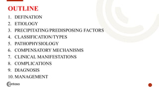
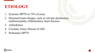
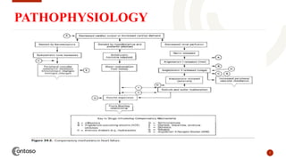
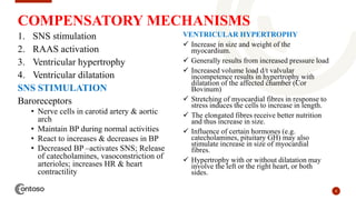
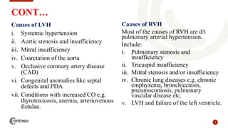
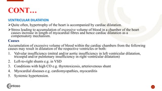
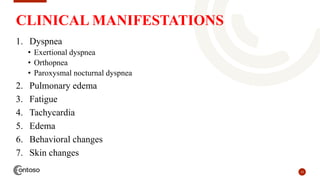
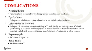
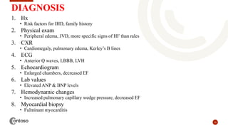
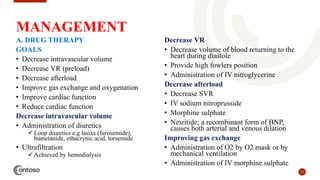
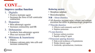
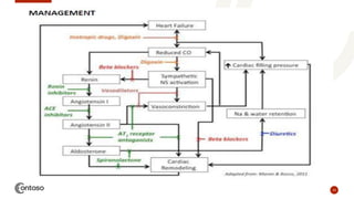
CVS PATHOLOGY –
HEART FAILURE
SAMOEI – EGERTON UNIVERSITY, MBCHB
2
OUTLINE
1. DEFINATION
2. ETIOLOGY
3. PRECIPITATING/PREDISPOSING FACTORS
4. CLASSIFICATION/TYPES
5. PATHOPHYSIOLOGY
6. COMPENSATORY MECHANISMS
7. CLINICAL MANIFESTATIONS
8. COMPLICATIONS
9. DIAGNOSIS
10. MANAGEMENT
DEFINATION;
• Is a pathophysiologic state whereby any
structural or functional cardiac d/o impairs the
ability of the ventricle to fill with or eject blood
to meet the metabolic demands of the body.
• This means less oxygen is reaching the organs
and muscles which can make one feel tired and
short of breath.
ļ‚§ The term CHF is used for the chronic form of
heart failure in which the patient has evidence of
congestion in peripheral circulation and of lungs.
ļ‚§ CHF is the end-result of various forms of serious
heart diseases.
3
HEART FAILURE
4
ETIOLOGY
1. Systemic HPTN in 75% of cases.
2. Structural heart changes, such as valvular dysfunction,
cardiomyopathy, inflammatory, heart diseases.
3. Arrhythmias
4. Coronary Artery Disease (CAD)
5. Pulmonary HPTN
5
PRECIPITATING FACTORS
1. Congenital heart defects
2. Severe lung disease
3. Diabetes
4. Severe anemia
5. Overactive thyroid (hyperthyroidism)
6. Hypervolemia
6
CLASSIFICATION OF HEART FAILURE
1. According to the course of disease
1. Acute HF
2. Chronic HF
2. According to the severity
1. Mild HF or complete compensation
2. Moderate HF or incomplete
compensation
3. Severe HF or decompensation
3. According to the location of HF
1. Left-side heart failure(LHF)
2. Right-side heart failure (RHF)
3. Biventricular failure (whole heart
failure)
4. According to the function impaired
1. Systolic failure
2. Diastolic failure
5. According to the CO
1. Low-output HF
2. High-output HF
6. According to area affected
1. Backward HF
2. Forward HF
7
PATHOPHYSIOLOGY
8
COMPENSATORY MECHANISMS
1. SNS stimulation
2. RAAS activation
3. Ventricular hypertrophy
4. Ventricular dilatation
SNS STIMULATION
Baroreceptors
• Nerve cells in carotid artery & aortic
arch
• Maintain BP during normal activities
• React to increases & decreases in BP
• Decreased BP –activates SNS; Release
of catecholamines, vasoconstriction of
arterioles; increases HR & heart
contractility
VENTRICULAR HYPERTROPHY
 Increase in size and weight of the
myocardium.
 Generally results from increased pressure load
 Increased volume load d/t valvular
incompetence results in hypertrophy with
dilatation of the affected chamber (Cor
Bovinum)
 Stretching of myocardial fibres in response to
stress induces the cells to increase in length.
 The elongated fibres receive better nutrition
and thus increase in size.
 Influence of certain hormones (e.g.
catecholamines, pituitary GH) may also
stimulate increase in size of myocardial
fibres.
 Hypertrophy with or without dilatation may
involve the left or the right heart, or both
sides.
9
CONT…
Causes of LVH
i. Systemic hypertension
ii. Aortic stenosis and insufficiency
iii. Mitral insufficiency
iv. Coarctation of the aorta
v. Occlusive coronary artery disease
(CAD)
vi. Congenital anomalies like septal
defects and PDA
vii. Conditions with increased CO e.g.
thyrotoxicosis, anemia, arteriovenous
fistulae.
Causes of RVH
Most of the causes of RVH are d/t
pulmonary arterial hypertension.
Include:
i. Pulmonary stenosis and
insufficiency
ii. Tricuspid insufficiency
iii. Mitral stenosis and/or insufficiency
iv. Chronic lung diseases e.g. chronic
emphysema, bronchiectasis,
pneumoconiosis, pulmonary
vascular disease etc.
v. LVH and failure of the left ventricle.
10
CONT…
VENTRICULAR DILATATION
Quite often, hypertrophy of the heart is accompanied by cardiac dilatation.
Stress leading to accumulation of excessive volume of blood in a chamber of the heart
causes increase in length of myocardial fibres and hence cardiac dilatation as a
compensatory mechanism.
Causes
Accumulation of excessive volume of blood within the cardiac chambers from the following
causes may result in dilatation of the respective ventricles or both:
1. Valvular insufficiency (mitral and/or aortic insufficiency in left ventricular dilatation,
tricuspid and/or pulmonary insufficiency in right ventricular dilatation)
2. Left-to-right shunts e.g. in VSD
3. Conditions with high CO e.g. thyrotoxicosis, arteriovenous shunt
4. Myocardial diseases e.g. cardiomyopathies, myocarditis
5. Systemic hypertension.
11
CONT…
12
COUNTERREGULATORY MECHANISMS
Body will try to maintain a balance in compensatory mechanism
• Heart muscles produce Natriuretic peptides
Atrial Natriuretic peptide(ANP)
b-type natriuretic peptide(BNP)
• These are endothelin and aldosterone antagonists which will cause
diuresis and vasodilation, preventing hypertrophy.
• Nitric oxide released by vascular endothelium causes vasodilation
13
CLINICAL MANIFESTATIONS
1. Dyspnea
• Exertional dyspnea
• Orthopnea
• Paroxysmal nocturnal dyspnea
2. Pulmonary edema
3. Fatigue
4. Tachycardia
5. Edema
6. Behavioral changes
7. Skin changes
14
CONT…
RIGHT-SIDED HEART FAILURE
1. Jugular venous distention (JVD)
2. Hepatomegaly
3. Anasarca
4. Edema (Pitting type)
5. Ascitis
6. Weight gain
7. Tachycardia
8. Murmurs
LEFT-SIDED HEART FAILURE
1. Pulmonary edema (crackles)
2. Pleural effusion
3. S3 & S4 heart sounds
4. Pulses ulternans
5. Changes in mental status
6. Displaced PMI, LV heaves
7. Dyspnea & orthopnea
15
COMLICATIONS
1. Pleural effusion
• Resulting from increased hydrostatic pressure in pulmonary capillaries
2. Dysrhythmias
• Enlargement of chambers cause alteration in normal electrical pathway
3. Left ventricular thrombus
• Enlarged LV decreases ventricular filling, CO and finally SV causing stasis of blood
(particularly in the atrial appendage) that increases chance of thrombus formation which
can shed emboli and cause strokes and manifestations of infarction in other organs.
4. Hepatomegaly
• d/t venous congestion
5. Renal failure
• d/t diminished CO
16
DIAGNOSIS
1. Hx
• Risk factors for IHD, family history
2. Physical exam
• Peripheral edema, JVD, more specific signs of HF than rales
3. CXR
• Cardiomegaly, pulmonary edema, Kerley’s B lines
4. ECG
• Anterior Q waves, LBBB, LVH
5. Echocardiogram
• Enlarged chambers, decreased EF
6. Lab values
• Elevated ANP & BNP levels
7. Hemodynamic changes
• Increased pulmonary capillary wedge pressure, decreased EF
8. Myocardial biopsy
• Fulminant myocarditis
17
MANAGEMENT
A. DRUG THERAPY
GOALS
• Decrease intravascular volume
• Decrease VR (preload)
• Decrease afterload
• Improve gas exchange and oxygenation
• Improve cardiac function
• Reduce cardiac function
Decrease intravascular volume
• Administration of diuretics
 Loop diuretics e.g lasixs (furosemide),
bumetanide, ethacrynic acid, torsemide
• Ultrafiltration
 Achieved by hemodialysis
Decrease VR
• Decrease volume of blood returning to the
heart during diastole
• Provide high fowlers position
• Administration of IV nitroglycerine
Decrease afterload
• Decrease SVR
• IV sodium nitroprusside
• Morphine sulphate
• Nesritide; a recombinant form of BNP,
causes both arterial and venous dilation
Improving gas exchange
• Administration of O2 by O2 mask or by
mechanical ventilation
• Administration of IV morphine sulphate
18
CONT…
Improve cardiac function
1. Digitalis
• Positive inotropic agent
• Increases the force of left ventricular
contraction
2. Dopamine
• Beta adrenergic agonist
• Increases contractility and HR
3. Dobutamine
• Synthetic beta adrenergic agonist
• Does not increase SVR
4. Milrinone & Inamrinone
• PDEI’s
• Enhance calcium entry into cells and
increase contractility
Reducing anxiety
• Morphine sulphate
• Benzodiazepines
N/B – About diuretics
All diuretics increase urine volume and sodium
excretion but differ in pharmacologic properties
Thiazide diuretics
ļ‚§ Effective and commonly used in HF
ļ‚§ Depletes potassium
Loop diuretics
ļ‚§ Increase sodium excretion
ļ‚§ Rapidly acting IV agent
Potassium sparing diuretics
ļ‚§ Help avoid incidences of hypokalemia
ļ‚§ Weaker diuretic agent
ļ‚§ Amiloride, spironolactone, triamtrene
19
20
CONT…
B. NUTRITIONAL THERAPY
• Diet education and weight
management is critical
• Dietary restriction of sodium
Mild HF- 2.5g salt
Severe HF- 500-1000g of salt
• Instruct to follow DASH diet
• Fluid restriction
• Weigh regularly same time, weight
gain of 1.4kg over the past 2 days
should be informed to health care
provider
C. NON-PHARMACOLOGICAL
MNGMT
1. Intraaortic balloon pump
(IABP)
2. Pacemaker therapy/cardiac
resynchronization therapy
(CRT)
3. Left ventricular assist device
4. Artificial heart
5. Heart transplantation
21
LEFT SIDED HEART FAILURE
• It is initiated by stress to the left heart.
Causes
• Ischemic heart disease (IHD)
• Systemic hypertension
• Mitral or aortic valve disease
• Myocardial diseases e.g. cardiomyopathies, myocarditis
• Restrictive pericarditis
22
CONT…
MORPHOLOGIC FEATURES
HEART
Depends on the underlying disease process
• Myocardial infarction or valvular deformities may be present.
• Massive hypertrophy and cardiac dilatation, With the exception of failure due to
mitral valve stenosis or restrictive cardiomyopathies
• Left ventricular dilation can result in mitral insufficiency and left atrial enlargement,
which is associated with an increased incidence of atrial fibrillation.
LUNGS
• Pulmonary congestion and edema
• Pleural effusion d/t increased hydrostatic pressure
• The lungs are heavy and boggy
• Microscopically show perivascular and interstitial transudates, alveolar septal edema,
and accumulation of edema fluid in the alveolar spaces.
23
CONT…
CLINICAL FEATURES
1. Dyspnea (shortness of breath) on exertion and orthopnea (dyspnea when
recumbent)
2. Diminished CO
3. Pulmonary edema.
4. Cough, as a consequence of fluid transudation into air spaces.
5. Paroxysmal nocturnal dyspnea; patients wake up from sleep with extreme dyspnea
and feelings of suffocation.
6. Enlarged heart (cardiomegaly) & Tachycardia
7. S3 & S4
8. Mitral regurgitation and a systolic murmur d/t progressive ventricular dilation
9. Afib & increased incidence of thrombus formation
10. Diminished cerebral perfusion; hypoxic encephalopathy marked by irritability,
diminished cognition, and restlessness that can progress to stupor and coma.
24
CONT…
Tx
DRUG THERAPY
1. Diuretics
ļ‚§ Thiazide diuretics; 1st line drugs
ļ‚§ Loop diuretics
ļ‚§ K+ sparing diuretics used in combination
2. Positive inotropes
ļ‚§ Digitalis (be careful with side effects)
3. Vasodilators
ļ‚§ Nitroglycerine, sodium nitroprusside
4. ACEIs
ļ‚§ 1st line therapy in Tx of CHF
ļ‚§ Enalapril, captopril
5. Angiotensin receptor blockers (ARB)
ļ‚§ In patients who do not tolerate ACEIs
ļ‚§ Losartan, telmisartan
6. Human BNP
ļ‚§ Nesritide
7. Beta blockers
ļ‚§ Atenolol, Metoprolol
NUTRITIONAL THERAPY
1. Diet education and weight mngmt
2. Dietary restriction of sodium
3. Fluid restriction
NON-PHARMACOLOGICAL THERAPY
1. Cardiac resynchronization therapy (CRT)-
(exogenous pacing of both the right and left
ventricles)
2. Cardiac contractility modulation (exogenous
stimulation of cardiac muscle)
25
RIGHT-SIDED HEART FAILURE
• Occurs more often as a consequence of left-
sided HF.
• However, some conditions affect the right
ventricle primarily, producing right-sided
heart failure.
CAUSES
1. As a consequence of left ventricular failure.
2. Cor pulmonale in which right heart failure
occurs due to intrinsic lung diseases.
3. Pulmonary or tricuspid valvular disease.
4. Pulmonary hypertension secondary to
pulmonary thromboembolism.
5. Myocardial disease affecting right heart.
6. Congenital heart disease with left-to-right
shunt.
ļ‚§ Whatever be the underlying cause,
the clinical manifestations of right-
sided HF are upstream of the right
heart such as systemic (d/t caval
blood) and portal venous
congestion, and reduced CO.
ļ‚§ Accordingly, the pathologic
changes are as under:
a) Systemic venous congestion in
different tissues and organs e.g.
subcutaneous edema on dependent
parts, passive congestion of the liver,
spleen, and kidneys, ascites,
hydrothorax, congestion of leg veins
and neck veins.
b) Reduced CO resulting in circulatory
stagnation causing anoxia, cyanosis
and coldness of extremities.
26
ACUTE HEART FAILURE
 Sudden and rapid development of heart failure
There is sudden reduction in CO resulting in systemic hypotension.
Edema does not occur.
Instead, a state of cardiogenic shock and cerebral hypoxia develops.
Occurs in the following conditions:
1. Larger MI
2. Valve rupture
3. Cardiac tamponade
4. Massive pulmonary embolism
5. Acute viral myocarditis
6. Acute bacterial toxemia.
27
CHRONIC HEART FAILURE
Heart failure develops slowly.
Compensatory mechanisms like tachycardia, cardiac dilatation and
cardiac hypertrophy try to make adjustments so as to maintain adequate
CO.
Often results in well-maintained arterial pressure
There is accumulation of edema.
Observed in the following states:
1. Myocardial ischemia from atherosclerotic CAD
2. Multivalvular heart disease
3. Systemic arterial hypertension
4. Chronic lung diseases resulting in hypoxia and pulmonary arterial hypertension
5. Progression of acute into chronic failure.
BACKWARD AND FORWARD HEART FAILURE
According to this concept, either
of the ventricles fails to eject
blood normally, resulting in rise
of EDV in the ventricle and
increase in volume and pressure
in the atrium which is
transmitted backward producing
elevated pressure in the veins.
28
BACKWARD HEART FAILURE According to this hypothesis,
clinical manifestations result
directly from failure of the heart
to pump blood causing
diminished flow of blood to the
tissues, especially diminished
renal
BACKWARD HEART FAILURE
29
SYSTOLIC & DIASTOLIC HEART FAILURE
SYSTOLIC HEART FAILURE
• Is the heart failure d/t decreased
ability of heart to contract.
• May involve right heart or left heart
or both.
• It is caused either by muscular
weakness or valvular defect.
• Ventricles may be filled with blood
but cannot pump it out with sufficient
force.
• Ejection fraction decreases to about
20%.
• So the amount of blood pumped to the
body and to the lungs is decreased.
• As a result, more amount of blood remains
in ventricle.
• Later the blood starts accumulating in
lungs or systemic veins or both.
• Usually the ventricle enlarges in systolic
heart failure.
DIASTOLIC HEART FAILURE
• Is the heart failure that occurs when the
ventricles cannot relax properly due to the
stiffening of cardiac muscle.
• So, there is reduction in ventricular filling
and cardiac output.
30
COMPENSATED & DECOMPENSATED HF
COMPENSATED HEART FAILURE
• Is the heart failure with adequate cardiac
output.
• Heart tries to maintain cardiac output by
normal compensatory mechanisms such
as increase in heart rate, increase in force
of ventricular contraction and ventricular
hypertrophy.
• In compensated heart failure, the
symptoms are stable and features of fluid
retention and pulmonary edema are
absent.
• Eventually, in most of the patients the
heart can no longer meet the demand
even by compensatory mechanisms and
this condition leads to decompensated
heart failure.
DECOMPENSATED HEART
FAILURE
• Is the heart failure with
inadequate cardiac output.
• It is characterized by
deterioration and sudden and
drastic worsening of cardiac
function, resulting in death.
31
VENTRICULAR ASSIST DEVICE
32
CRT PACEMAKER
33
INTRA-AORTIC BALLOON PUMP
34
THANKYOU !
NESHKDOC@GMAIL.COM
HTTP://WWW.EGERTONFSH.COM/

HEART FAILURE

  • 1.
    CVS PATHOLOGY – HEARTFAILURE SAMOEI – EGERTON UNIVERSITY, MBCHB
  • 2.
    2 OUTLINE 1. DEFINATION 2. ETIOLOGY 3.PRECIPITATING/PREDISPOSING FACTORS 4. CLASSIFICATION/TYPES 5. PATHOPHYSIOLOGY 6. COMPENSATORY MECHANISMS 7. CLINICAL MANIFESTATIONS 8. COMPLICATIONS 9. DIAGNOSIS 10. MANAGEMENT
  • 3.
    DEFINATION; • Is apathophysiologic state whereby any structural or functional cardiac d/o impairs the ability of the ventricle to fill with or eject blood to meet the metabolic demands of the body. • This means less oxygen is reaching the organs and muscles which can make one feel tired and short of breath. ļ‚§ The term CHF is used for the chronic form of heart failure in which the patient has evidence of congestion in peripheral circulation and of lungs. ļ‚§ CHF is the end-result of various forms of serious heart diseases. 3 HEART FAILURE
  • 4.
    4 ETIOLOGY 1. Systemic HPTNin 75% of cases. 2. Structural heart changes, such as valvular dysfunction, cardiomyopathy, inflammatory, heart diseases. 3. Arrhythmias 4. Coronary Artery Disease (CAD) 5. Pulmonary HPTN
  • 5.
    5 PRECIPITATING FACTORS 1. Congenitalheart defects 2. Severe lung disease 3. Diabetes 4. Severe anemia 5. Overactive thyroid (hyperthyroidism) 6. Hypervolemia
  • 6.
    6 CLASSIFICATION OF HEARTFAILURE 1. According to the course of disease 1. Acute HF 2. Chronic HF 2. According to the severity 1. Mild HF or complete compensation 2. Moderate HF or incomplete compensation 3. Severe HF or decompensation 3. According to the location of HF 1. Left-side heart failure(LHF) 2. Right-side heart failure (RHF) 3. Biventricular failure (whole heart failure) 4. According to the function impaired 1. Systolic failure 2. Diastolic failure 5. According to the CO 1. Low-output HF 2. High-output HF 6. According to area affected 1. Backward HF 2. Forward HF
  • 7.
  • 8.
    8 COMPENSATORY MECHANISMS 1. SNSstimulation 2. RAAS activation 3. Ventricular hypertrophy 4. Ventricular dilatation SNS STIMULATION Baroreceptors • Nerve cells in carotid artery & aortic arch • Maintain BP during normal activities • React to increases & decreases in BP • Decreased BP –activates SNS; Release of catecholamines, vasoconstriction of arterioles; increases HR & heart contractility VENTRICULAR HYPERTROPHY  Increase in size and weight of the myocardium.  Generally results from increased pressure load  Increased volume load d/t valvular incompetence results in hypertrophy with dilatation of the affected chamber (Cor Bovinum)  Stretching of myocardial fibres in response to stress induces the cells to increase in length.  The elongated fibres receive better nutrition and thus increase in size.  Influence of certain hormones (e.g. catecholamines, pituitary GH) may also stimulate increase in size of myocardial fibres.  Hypertrophy with or without dilatation may involve the left or the right heart, or both sides.
  • 9.
    9 CONT… Causes of LVH i.Systemic hypertension ii. Aortic stenosis and insufficiency iii. Mitral insufficiency iv. Coarctation of the aorta v. Occlusive coronary artery disease (CAD) vi. Congenital anomalies like septal defects and PDA vii. Conditions with increased CO e.g. thyrotoxicosis, anemia, arteriovenous fistulae. Causes of RVH Most of the causes of RVH are d/t pulmonary arterial hypertension. Include: i. Pulmonary stenosis and insufficiency ii. Tricuspid insufficiency iii. Mitral stenosis and/or insufficiency iv. Chronic lung diseases e.g. chronic emphysema, bronchiectasis, pneumoconiosis, pulmonary vascular disease etc. v. LVH and failure of the left ventricle.
  • 10.
    10 CONT… VENTRICULAR DILATATION Quite often,hypertrophy of the heart is accompanied by cardiac dilatation. Stress leading to accumulation of excessive volume of blood in a chamber of the heart causes increase in length of myocardial fibres and hence cardiac dilatation as a compensatory mechanism. Causes Accumulation of excessive volume of blood within the cardiac chambers from the following causes may result in dilatation of the respective ventricles or both: 1. Valvular insufficiency (mitral and/or aortic insufficiency in left ventricular dilatation, tricuspid and/or pulmonary insufficiency in right ventricular dilatation) 2. Left-to-right shunts e.g. in VSD 3. Conditions with high CO e.g. thyrotoxicosis, arteriovenous shunt 4. Myocardial diseases e.g. cardiomyopathies, myocarditis 5. Systemic hypertension.
  • 11.
  • 12.
    12 COUNTERREGULATORY MECHANISMS Body willtry to maintain a balance in compensatory mechanism • Heart muscles produce Natriuretic peptides Atrial Natriuretic peptide(ANP) b-type natriuretic peptide(BNP) • These are endothelin and aldosterone antagonists which will cause diuresis and vasodilation, preventing hypertrophy. • Nitric oxide released by vascular endothelium causes vasodilation
  • 13.
    13 CLINICAL MANIFESTATIONS 1. Dyspnea •Exertional dyspnea • Orthopnea • Paroxysmal nocturnal dyspnea 2. Pulmonary edema 3. Fatigue 4. Tachycardia 5. Edema 6. Behavioral changes 7. Skin changes
  • 14.
    14 CONT… RIGHT-SIDED HEART FAILURE 1.Jugular venous distention (JVD) 2. Hepatomegaly 3. Anasarca 4. Edema (Pitting type) 5. Ascitis 6. Weight gain 7. Tachycardia 8. Murmurs LEFT-SIDED HEART FAILURE 1. Pulmonary edema (crackles) 2. Pleural effusion 3. S3 & S4 heart sounds 4. Pulses ulternans 5. Changes in mental status 6. Displaced PMI, LV heaves 7. Dyspnea & orthopnea
  • 15.
    15 COMLICATIONS 1. Pleural effusion •Resulting from increased hydrostatic pressure in pulmonary capillaries 2. Dysrhythmias • Enlargement of chambers cause alteration in normal electrical pathway 3. Left ventricular thrombus • Enlarged LV decreases ventricular filling, CO and finally SV causing stasis of blood (particularly in the atrial appendage) that increases chance of thrombus formation which can shed emboli and cause strokes and manifestations of infarction in other organs. 4. Hepatomegaly • d/t venous congestion 5. Renal failure • d/t diminished CO
  • 16.
    16 DIAGNOSIS 1. Hx • Riskfactors for IHD, family history 2. Physical exam • Peripheral edema, JVD, more specific signs of HF than rales 3. CXR • Cardiomegaly, pulmonary edema, Kerley’s B lines 4. ECG • Anterior Q waves, LBBB, LVH 5. Echocardiogram • Enlarged chambers, decreased EF 6. Lab values • Elevated ANP & BNP levels 7. Hemodynamic changes • Increased pulmonary capillary wedge pressure, decreased EF 8. Myocardial biopsy • Fulminant myocarditis
  • 17.
    17 MANAGEMENT A. DRUG THERAPY GOALS •Decrease intravascular volume • Decrease VR (preload) • Decrease afterload • Improve gas exchange and oxygenation • Improve cardiac function • Reduce cardiac function Decrease intravascular volume • Administration of diuretics  Loop diuretics e.g lasixs (furosemide), bumetanide, ethacrynic acid, torsemide • Ultrafiltration  Achieved by hemodialysis Decrease VR • Decrease volume of blood returning to the heart during diastole • Provide high fowlers position • Administration of IV nitroglycerine Decrease afterload • Decrease SVR • IV sodium nitroprusside • Morphine sulphate • Nesritide; a recombinant form of BNP, causes both arterial and venous dilation Improving gas exchange • Administration of O2 by O2 mask or by mechanical ventilation • Administration of IV morphine sulphate
  • 18.
    18 CONT… Improve cardiac function 1.Digitalis • Positive inotropic agent • Increases the force of left ventricular contraction 2. Dopamine • Beta adrenergic agonist • Increases contractility and HR 3. Dobutamine • Synthetic beta adrenergic agonist • Does not increase SVR 4. Milrinone & Inamrinone • PDEI’s • Enhance calcium entry into cells and increase contractility Reducing anxiety • Morphine sulphate • Benzodiazepines N/B – About diuretics All diuretics increase urine volume and sodium excretion but differ in pharmacologic properties Thiazide diuretics ļ‚§ Effective and commonly used in HF ļ‚§ Depletes potassium Loop diuretics ļ‚§ Increase sodium excretion ļ‚§ Rapidly acting IV agent Potassium sparing diuretics ļ‚§ Help avoid incidences of hypokalemia ļ‚§ Weaker diuretic agent ļ‚§ Amiloride, spironolactone, triamtrene
  • 19.
  • 20.
    20 CONT… B. NUTRITIONAL THERAPY •Diet education and weight management is critical • Dietary restriction of sodium Mild HF- 2.5g salt Severe HF- 500-1000g of salt • Instruct to follow DASH diet • Fluid restriction • Weigh regularly same time, weight gain of 1.4kg over the past 2 days should be informed to health care provider C. NON-PHARMACOLOGICAL MNGMT 1. Intraaortic balloon pump (IABP) 2. Pacemaker therapy/cardiac resynchronization therapy (CRT) 3. Left ventricular assist device 4. Artificial heart 5. Heart transplantation
  • 21.
    21 LEFT SIDED HEARTFAILURE • It is initiated by stress to the left heart. Causes • Ischemic heart disease (IHD) • Systemic hypertension • Mitral or aortic valve disease • Myocardial diseases e.g. cardiomyopathies, myocarditis • Restrictive pericarditis
  • 22.
    22 CONT… MORPHOLOGIC FEATURES HEART Depends onthe underlying disease process • Myocardial infarction or valvular deformities may be present. • Massive hypertrophy and cardiac dilatation, With the exception of failure due to mitral valve stenosis or restrictive cardiomyopathies • Left ventricular dilation can result in mitral insufficiency and left atrial enlargement, which is associated with an increased incidence of atrial fibrillation. LUNGS • Pulmonary congestion and edema • Pleural effusion d/t increased hydrostatic pressure • The lungs are heavy and boggy • Microscopically show perivascular and interstitial transudates, alveolar septal edema, and accumulation of edema fluid in the alveolar spaces.
  • 23.
    23 CONT… CLINICAL FEATURES 1. Dyspnea(shortness of breath) on exertion and orthopnea (dyspnea when recumbent) 2. Diminished CO 3. Pulmonary edema. 4. Cough, as a consequence of fluid transudation into air spaces. 5. Paroxysmal nocturnal dyspnea; patients wake up from sleep with extreme dyspnea and feelings of suffocation. 6. Enlarged heart (cardiomegaly) & Tachycardia 7. S3 & S4 8. Mitral regurgitation and a systolic murmur d/t progressive ventricular dilation 9. Afib & increased incidence of thrombus formation 10. Diminished cerebral perfusion; hypoxic encephalopathy marked by irritability, diminished cognition, and restlessness that can progress to stupor and coma.
  • 24.
    24 CONT… Tx DRUG THERAPY 1. Diuretics ļ‚§Thiazide diuretics; 1st line drugs ļ‚§ Loop diuretics ļ‚§ K+ sparing diuretics used in combination 2. Positive inotropes ļ‚§ Digitalis (be careful with side effects) 3. Vasodilators ļ‚§ Nitroglycerine, sodium nitroprusside 4. ACEIs ļ‚§ 1st line therapy in Tx of CHF ļ‚§ Enalapril, captopril 5. Angiotensin receptor blockers (ARB) ļ‚§ In patients who do not tolerate ACEIs ļ‚§ Losartan, telmisartan 6. Human BNP ļ‚§ Nesritide 7. Beta blockers ļ‚§ Atenolol, Metoprolol NUTRITIONAL THERAPY 1. Diet education and weight mngmt 2. Dietary restriction of sodium 3. Fluid restriction NON-PHARMACOLOGICAL THERAPY 1. Cardiac resynchronization therapy (CRT)- (exogenous pacing of both the right and left ventricles) 2. Cardiac contractility modulation (exogenous stimulation of cardiac muscle)
  • 25.
    25 RIGHT-SIDED HEART FAILURE •Occurs more often as a consequence of left- sided HF. • However, some conditions affect the right ventricle primarily, producing right-sided heart failure. CAUSES 1. As a consequence of left ventricular failure. 2. Cor pulmonale in which right heart failure occurs due to intrinsic lung diseases. 3. Pulmonary or tricuspid valvular disease. 4. Pulmonary hypertension secondary to pulmonary thromboembolism. 5. Myocardial disease affecting right heart. 6. Congenital heart disease with left-to-right shunt. ļ‚§ Whatever be the underlying cause, the clinical manifestations of right- sided HF are upstream of the right heart such as systemic (d/t caval blood) and portal venous congestion, and reduced CO. ļ‚§ Accordingly, the pathologic changes are as under: a) Systemic venous congestion in different tissues and organs e.g. subcutaneous edema on dependent parts, passive congestion of the liver, spleen, and kidneys, ascites, hydrothorax, congestion of leg veins and neck veins. b) Reduced CO resulting in circulatory stagnation causing anoxia, cyanosis and coldness of extremities.
  • 26.
    26 ACUTE HEART FAILURE Sudden and rapid development of heart failure There is sudden reduction in CO resulting in systemic hypotension. Edema does not occur. Instead, a state of cardiogenic shock and cerebral hypoxia develops. Occurs in the following conditions: 1. Larger MI 2. Valve rupture 3. Cardiac tamponade 4. Massive pulmonary embolism 5. Acute viral myocarditis 6. Acute bacterial toxemia.
  • 27.
    27 CHRONIC HEART FAILURE Heartfailure develops slowly. Compensatory mechanisms like tachycardia, cardiac dilatation and cardiac hypertrophy try to make adjustments so as to maintain adequate CO. Often results in well-maintained arterial pressure There is accumulation of edema. Observed in the following states: 1. Myocardial ischemia from atherosclerotic CAD 2. Multivalvular heart disease 3. Systemic arterial hypertension 4. Chronic lung diseases resulting in hypoxia and pulmonary arterial hypertension 5. Progression of acute into chronic failure.
  • 28.
    BACKWARD AND FORWARDHEART FAILURE According to this concept, either of the ventricles fails to eject blood normally, resulting in rise of EDV in the ventricle and increase in volume and pressure in the atrium which is transmitted backward producing elevated pressure in the veins. 28 BACKWARD HEART FAILURE According to this hypothesis, clinical manifestations result directly from failure of the heart to pump blood causing diminished flow of blood to the tissues, especially diminished renal BACKWARD HEART FAILURE
  • 29.
    29 SYSTOLIC & DIASTOLICHEART FAILURE SYSTOLIC HEART FAILURE • Is the heart failure d/t decreased ability of heart to contract. • May involve right heart or left heart or both. • It is caused either by muscular weakness or valvular defect. • Ventricles may be filled with blood but cannot pump it out with sufficient force. • Ejection fraction decreases to about 20%. • So the amount of blood pumped to the body and to the lungs is decreased. • As a result, more amount of blood remains in ventricle. • Later the blood starts accumulating in lungs or systemic veins or both. • Usually the ventricle enlarges in systolic heart failure. DIASTOLIC HEART FAILURE • Is the heart failure that occurs when the ventricles cannot relax properly due to the stiffening of cardiac muscle. • So, there is reduction in ventricular filling and cardiac output.
  • 30.
    30 COMPENSATED & DECOMPENSATEDHF COMPENSATED HEART FAILURE • Is the heart failure with adequate cardiac output. • Heart tries to maintain cardiac output by normal compensatory mechanisms such as increase in heart rate, increase in force of ventricular contraction and ventricular hypertrophy. • In compensated heart failure, the symptoms are stable and features of fluid retention and pulmonary edema are absent. • Eventually, in most of the patients the heart can no longer meet the demand even by compensatory mechanisms and this condition leads to decompensated heart failure. DECOMPENSATED HEART FAILURE • Is the heart failure with inadequate cardiac output. • It is characterized by deterioration and sudden and drastic worsening of cardiac function, resulting in death.
  • 31.
  • 32.
  • 33.
  • 34.
  • 35.