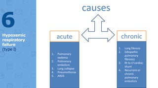
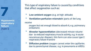
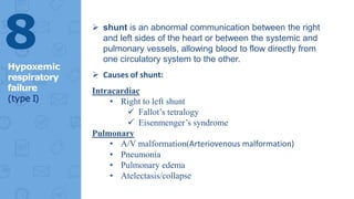
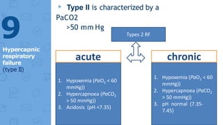
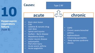
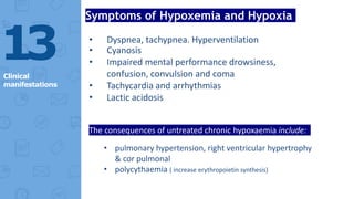
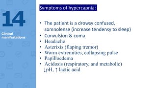
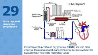
Respiratory
failure
PREPARED BY : ABDULAZIZ MUSLIM SHAHBAZ
SUPERVISED BY : Dr. ABDULAZEEZ SULIMAN SAVO
▸ Background &
introduction
▸ Clinical Presentation
▸ Workup Approach
▸ Treatment & Management
▸ Medication & guidelines
Content
Introduction
Background, pathophysiology and etiology
3
▸ Respiratory failure is a syndrome in
which the respiratory systemfails in
one or both of its gas exchange
functions:
▹oxygenation
▹and carbon dioxide elimination
▸ In practice, it may be classified as
either
 hypoxemic PaO2 <60 mmHg ( < 8,0 kPa)
or
 Hypercapnic PaCO2 >50 mmHg ( > 6,7
kPa)
4
Definition
▸ Type I is characterized by an
arterial oxygen tension (PaO2) <60
mm Hg with a normal or low
arterial carbon dioxide tension
(PaCO2)
▸This is the most common form
of respiratory failure
▸ causes of hypoxemia classified as:
▹V/Q missmatch
▹Impaired diffusion
▹Rt to Lt shunt
5
Hypoxemic
respiratory
failure
(type I)
6
Hypoxemic
respiratory
failure
(type I)
causes
1. Pulmonary
oedema
2. Pulmonary
embolism
3. Lung collapse
4. Pneumothorax
5. ARDS
acute chronic
1. Lung fibrosis
2. (idiopathic
pulmonary
fibrosis)
3. Rt to Lf cardiac
shunt
4. Recurrent or
chronic
pulmonary
embolism
This type of respiratory failure is caused by conditions
that affect oxygenation suchas:
▸ Low ambient oxygen (e.g. at high altitude)
▸ Ventilation-perfusion mismatch (parts of the lung
receive
oxygen but not enough blood to absorb it,e.g. pulmonary
embolism)
▸ Alveolar hypoventilation (decreased minute volume
due to reduced respiratory muscle activity, e.g. in acute
neuromuscular disease); this form can also cause type 2
respiratory failure ifsevere
▸ Diffusion problem (oxygen cannot enter the capillaries
due to parenchymal disease, e.g. in pneumonia or ARDS)
7
Hypoxemic
respiratory
failure
(type I)
 shunt is an abnormal communication between the right
and left sides of the heart or between the systemic and
pulmonary vessels, allowing blood to flow directly from
one circulatory system to the other.
 Causes of shunt:
Intracardiac
• Right to left shunt
 Fallot’s tetralogy
 Eisenmenger’s syndrome
Pulmonary
• A/V malformation(Arteriovenous malformation)
• Pneumonia
• Pulmonary edema
• Atelectasis/collapse
8
Hypoxemic
respiratory
failure
(type I)
▸ Type II is characterized by a
PaCO2
>50 mm Hg
9
Hypercapnic
respiratory
failure
(type II)
Types 2 RF
1. Hypoxemia (PaO2 < 60
mmHg))
2. Hypercapnoea (PaCO2
> 50 mmHg))
3. Acidosis (pH <7.35)
1. Hypoxemia (PaO2 < 60
mmHg))
2. Hypercapnoea (PaCO2
> 50 mmHg))
3. pH normal (7.35-
7.45)
acute chronic
10
Hypercapnic
respiratory
failure
(type II)
Causes: Type 2 RF
• Brain stem lesion
• CVA
• sedative & narcotic drug
over dose
• Spinal cord injuries
• Guillain – Barre disease
• Myasthenia Gravis
• motor neuron disease
• poliomyelitis
• Flail chest injury
• Acute severe asthma
• Acute epiglottitis
• COPD
• Chronic severe bronchial
asthma
• Kyphoscoliosis
• Ankylosing spondyliltis
• Sleep apnoea syndrome
• obesity
acute chronic
acute chronic
Clinical
manifestations
History and physical examination findings
1
1
▸ The diagnosis of acute or chronic
respiratory failure begins with
clinical suspicion of its presence
▸ Confirmation of the diagnosis is basedon
arterial blood gas analysis
▸ Evaluation of an underlying cause must be
initiated early, frequently in the presence of
concurrent treatment for acute respiratory
failure.
▸ The cause of respiratory failure is often
evident after a careful history and physical
examination
1
2
History
13
Clinical
manifestations
Symptoms of Hypoxemia and Hypoxia
• Dyspnea, tachypnea. Hyperventilation
• Cyanosis
• Impaired mental performance drowsiness,
confusion, convulsion and coma
• Tachycardia and arrhythmias
• Lactic acidosis
The consequences of untreated chronic hypoxaemia include:
• pulmonary hypertension, right ventricular hypertrophy
& cor pulmonal
• polycythaemia ( increase erythropoietin synthesis)
14
Clinical
manifestations
Symptoms of hypercapnia:
• The patient is a drowsy confused,
somnolense (increase tendensy to sleep)
• Convulsion & coma
• Headache
• Asterixis (flaping tremor)
• Warm extremities, collapsing pulse
• Papilloedema
• Acidosis (respiratory, and metabolic)
↓pH, ↑ lactic acid
Respiratory
Failure Workup
15
▸ Chest radiography is
essential.
▸ Pulmonary functions tests (PFTs), may
be helpful, although more useful in terms
of defining recovery potential
▸ ECG should be performed to evaluate
the possibility of a cardiovascular cause
of respiratory failure; it also may detect
dysrhythmias resulting from severe
hypoxemia or acidosis
▸ Right-sided heart catheterization
is controversial
16
Approach
Consideration
1. ABG analysis should beperformed to confirm the diagnosis
and to assist in the distinction between acute and chronic
forms
Normal values of arterial blood gases:
-PH = 7.35 – 7.45 mmhg
-PCO2= 35 – 45 mmhg ( 4,7- 6,0 kPa)
-PO2 = 80 – 100 mmhg (10,6-13,3 kPa)
-HCO3 = 22 – 28 meq/L(Milliequivalents Per Litre)
2. CBC may indicate anemia,which can contribute to tissue
hypoxia, whereas polycythemia mayindicate chronic hypoxemic
respiratory failure
1
7
Laboratory
Studies
3. Abnormalities in renal and hepatic
function mayeither provide clues to
the etiology of respiratory failure or
alert the clinician tocomplications
4. Abnormalities in electrolytes
such
as K+, Mg, and phosphate may
aggravate respiratory failure and
other organ function
18
Laboratory
Studies
5. Measuring serum creatine kinase
with fractionation and troponin I
helps exclude recent MI in a patient
with respiratory failure
6. In chronic hypercapnicrespiratory
failure, serum levels of TSH
should be measured to evaluate
the
possibility of hypothyroidism
(a potentially reversible cause of respiratoryfailure)
19
Laboratory
Studies
Chest X-ray
Chest radiography frequently reveals the
cause
20
Bilateral airspace infiltrates on chest radiograph film secondary to acute
respiratory distress syndrome that resulted in respiratory failure
21
Extensive left-lung pneumonia caused respiratory failure; the mechanism of
hypoxia is intrapulmonaryshunting
22
Treatment &
Management
23
▸ The aim to Maintain PaO2 at 55 to 60 mm Hg or
more and SaO2 at 90% or more at the lowest O2
concentration possible without increasing PaCO2 and
acidosis
▸ diagnosis and treatment of the
underlying causes:
• Tracheostomy for laryngeal obstruction
• nebulised bronchodilators in acute severe
asthma
• tube drainage for a tension pneumothorax
• antibiotic for pneumonia
• diuretic for cor pulmonale
2
4
Treatment of
respiratory
failure
▸ Acute on chronic type 11 respiratory
failure:
• The further acute increase in PaCO2 results in acidaemia and
worsening hypercapnia, and may lead to drowsiness and
eventually to coma.
• Main cause is acute exacerbation of COPD
• Controlled oxygen therapy Start with 24% -28% by Venturi
mask is the most important treatment.
• Treating the precipitating condition .
• Frequent physiotherapy ± pharyngeal suction
• Nebulised bronchodilators
• Antibiotic
• If PaCO2 continues to rise or a safe PaO2 cannot be achieved
without severe hypercapnia and acidaemia, mechanical
ventilatory support may be required
25
Treatment
of type 11
RF
26
Correction
of
Hypoxemia
O2 administration
High concentration Low concentration
• such as 40-60% oxygen
• via a high-flow mask
• useful in acute type I
respiratory failure
• pneumonia, asthma or
pulmonary oedema.
• If used for prolonged
periods should be
humidified by passing it
over warm water
• Venturi masks (24% or
28%)
• delivering controlled
oxygen therapy in type II
respiratory failure
• low concentrations of
oxygen (24-28%) may be
needed to avoid
precipitating worsening
respiratory depression
High concentration Low concentration
 Chronic oxygen delivery ( domicilliary home O2
therapy)
LONG-TERM OXYGEN THERAPY (LTOT)
oxygen is often given via a low-concentration mask or
nasal cannulae for > 15 hr /day 24% -28%
Portable oxygen may increase exercise tolerance in some
patients with chronic hypoxic lung disease
Indication
PaO2 ≤ 7.3 kPa (55 mmHg) irrespective of PaCO2 and
FEV1 ≤ 1.5 litres
PaO2 7.3-8 kPa (55-60 mmHg) plus pulmonary
hypertension, peripheral oedema or nocturnal
hypoxaemia
27
Correction
of
Hypoxemia
Adverse effects of O2 therapy:
1-High O2 is irritant & toxic if inhaled for more
than few hours.
2-Retro-lental fibroplasia & blindness in
pre mature infants.
3-Pulmonary oedema in adult if > 40% for
more than 24 hours continuously.
4-Loss of surfactant and atelectasis
5-Convulsion mainly in hyperbaric O2
28
Correction
of
Hypoxemia
Extracorporeal
membrane
oxygenation
29
Extracorporeal membrane oxygenation (ECMO) may be more
effective than conventional management for patients with severe
but potentially reversible respiratoryfailure
Mechanical Ventilation
 Mechanical Ventilation is ventilation of the lungs by artificial
means usually by a ventilator.
Indications for supported ventilation is::
• Failure to respond to initial treatment
• declining conscious level
• Worsening respiratory acidosis (H+ > 50 nmol/L (pH < 7.3),
• PaCO2 > 6.6 kPa (50 mmHg)) on blood gases
 Non- invasive ventillation
 invasive ventillation ( using endotracheal tube)
30
Mechanica
l
Ventilation
Mechanical ventilation is used for
two essential reasons:
1. to increase PaO2 and
2. to lower PaCO2
▸ Mechanical ventilation also rests
the respiratory muscles and is an
appropriate therapy for respiratory
muscle fatigue
31
Mechanical
Ventilation
Respiratory failure

Respiratory failure

  • 1.
    Respiratory failure PREPARED BY :ABDULAZIZ MUSLIM SHAHBAZ SUPERVISED BY : Dr. ABDULAZEEZ SULIMAN SAVO
  • 2.
    ▸ Background & introduction ▸Clinical Presentation ▸ Workup Approach ▸ Treatment & Management ▸ Medication & guidelines Content
  • 3.
  • 4.
    ▸ Respiratory failureis a syndrome in which the respiratory systemfails in one or both of its gas exchange functions: ▹oxygenation ▹and carbon dioxide elimination ▸ In practice, it may be classified as either  hypoxemic PaO2 <60 mmHg ( < 8,0 kPa) or  Hypercapnic PaCO2 >50 mmHg ( > 6,7 kPa) 4 Definition
  • 5.
    ▸ Type Iis characterized by an arterial oxygen tension (PaO2) <60 mm Hg with a normal or low arterial carbon dioxide tension (PaCO2) ▸This is the most common form of respiratory failure ▸ causes of hypoxemia classified as: ▹V/Q missmatch ▹Impaired diffusion ▹Rt to Lt shunt 5 Hypoxemic respiratory failure (type I)
  • 6.
    6 Hypoxemic respiratory failure (type I) causes 1. Pulmonary oedema 2.Pulmonary embolism 3. Lung collapse 4. Pneumothorax 5. ARDS acute chronic 1. Lung fibrosis 2. (idiopathic pulmonary fibrosis) 3. Rt to Lf cardiac shunt 4. Recurrent or chronic pulmonary embolism
  • 7.
    This type ofrespiratory failure is caused by conditions that affect oxygenation suchas: ▸ Low ambient oxygen (e.g. at high altitude) ▸ Ventilation-perfusion mismatch (parts of the lung receive oxygen but not enough blood to absorb it,e.g. pulmonary embolism) ▸ Alveolar hypoventilation (decreased minute volume due to reduced respiratory muscle activity, e.g. in acute neuromuscular disease); this form can also cause type 2 respiratory failure ifsevere ▸ Diffusion problem (oxygen cannot enter the capillaries due to parenchymal disease, e.g. in pneumonia or ARDS) 7 Hypoxemic respiratory failure (type I)
  • 8.
     shunt isan abnormal communication between the right and left sides of the heart or between the systemic and pulmonary vessels, allowing blood to flow directly from one circulatory system to the other.  Causes of shunt: Intracardiac • Right to left shunt  Fallot’s tetralogy  Eisenmenger’s syndrome Pulmonary • A/V malformation(Arteriovenous malformation) • Pneumonia • Pulmonary edema • Atelectasis/collapse 8 Hypoxemic respiratory failure (type I)
  • 9.
    ▸ Type IIis characterized by a PaCO2 >50 mm Hg 9 Hypercapnic respiratory failure (type II) Types 2 RF 1. Hypoxemia (PaO2 < 60 mmHg)) 2. Hypercapnoea (PaCO2 > 50 mmHg)) 3. Acidosis (pH <7.35) 1. Hypoxemia (PaO2 < 60 mmHg)) 2. Hypercapnoea (PaCO2 > 50 mmHg)) 3. pH normal (7.35- 7.45) acute chronic
  • 10.
    10 Hypercapnic respiratory failure (type II) Causes: Type2 RF • Brain stem lesion • CVA • sedative & narcotic drug over dose • Spinal cord injuries • Guillain – Barre disease • Myasthenia Gravis • motor neuron disease • poliomyelitis • Flail chest injury • Acute severe asthma • Acute epiglottitis • COPD • Chronic severe bronchial asthma • Kyphoscoliosis • Ankylosing spondyliltis • Sleep apnoea syndrome • obesity acute chronic acute chronic
  • 11.
  • 12.
    ▸ The diagnosisof acute or chronic respiratory failure begins with clinical suspicion of its presence ▸ Confirmation of the diagnosis is basedon arterial blood gas analysis ▸ Evaluation of an underlying cause must be initiated early, frequently in the presence of concurrent treatment for acute respiratory failure. ▸ The cause of respiratory failure is often evident after a careful history and physical examination 1 2 History
  • 13.
    13 Clinical manifestations Symptoms of Hypoxemiaand Hypoxia • Dyspnea, tachypnea. Hyperventilation • Cyanosis • Impaired mental performance drowsiness, confusion, convulsion and coma • Tachycardia and arrhythmias • Lactic acidosis The consequences of untreated chronic hypoxaemia include: • pulmonary hypertension, right ventricular hypertrophy & cor pulmonal • polycythaemia ( increase erythropoietin synthesis)
  • 14.
    14 Clinical manifestations Symptoms of hypercapnia: •The patient is a drowsy confused, somnolense (increase tendensy to sleep) • Convulsion & coma • Headache • Asterixis (flaping tremor) • Warm extremities, collapsing pulse • Papilloedema • Acidosis (respiratory, and metabolic) ↓pH, ↑ lactic acid
  • 15.
  • 16.
    ▸ Chest radiographyis essential. ▸ Pulmonary functions tests (PFTs), may be helpful, although more useful in terms of defining recovery potential ▸ ECG should be performed to evaluate the possibility of a cardiovascular cause of respiratory failure; it also may detect dysrhythmias resulting from severe hypoxemia or acidosis ▸ Right-sided heart catheterization is controversial 16 Approach Consideration
  • 17.
    1. ABG analysisshould beperformed to confirm the diagnosis and to assist in the distinction between acute and chronic forms Normal values of arterial blood gases: -PH = 7.35 – 7.45 mmhg -PCO2= 35 – 45 mmhg ( 4,7- 6,0 kPa) -PO2 = 80 – 100 mmhg (10,6-13,3 kPa) -HCO3 = 22 – 28 meq/L(Milliequivalents Per Litre) 2. CBC may indicate anemia,which can contribute to tissue hypoxia, whereas polycythemia mayindicate chronic hypoxemic respiratory failure 1 7 Laboratory Studies
  • 18.
    3. Abnormalities inrenal and hepatic function mayeither provide clues to the etiology of respiratory failure or alert the clinician tocomplications 4. Abnormalities in electrolytes such as K+, Mg, and phosphate may aggravate respiratory failure and other organ function 18 Laboratory Studies
  • 19.
    5. Measuring serumcreatine kinase with fractionation and troponin I helps exclude recent MI in a patient with respiratory failure 6. In chronic hypercapnicrespiratory failure, serum levels of TSH should be measured to evaluate the possibility of hypothyroidism (a potentially reversible cause of respiratoryfailure) 19 Laboratory Studies
  • 20.
    Chest X-ray Chest radiographyfrequently reveals the cause 20
  • 21.
    Bilateral airspace infiltrateson chest radiograph film secondary to acute respiratory distress syndrome that resulted in respiratory failure 21
  • 22.
    Extensive left-lung pneumoniacaused respiratory failure; the mechanism of hypoxia is intrapulmonaryshunting 22
  • 23.
  • 24.
    ▸ The aimto Maintain PaO2 at 55 to 60 mm Hg or more and SaO2 at 90% or more at the lowest O2 concentration possible without increasing PaCO2 and acidosis ▸ diagnosis and treatment of the underlying causes: • Tracheostomy for laryngeal obstruction • nebulised bronchodilators in acute severe asthma • tube drainage for a tension pneumothorax • antibiotic for pneumonia • diuretic for cor pulmonale 2 4 Treatment of respiratory failure
  • 25.
    ▸ Acute onchronic type 11 respiratory failure: • The further acute increase in PaCO2 results in acidaemia and worsening hypercapnia, and may lead to drowsiness and eventually to coma. • Main cause is acute exacerbation of COPD • Controlled oxygen therapy Start with 24% -28% by Venturi mask is the most important treatment. • Treating the precipitating condition . • Frequent physiotherapy ± pharyngeal suction • Nebulised bronchodilators • Antibiotic • If PaCO2 continues to rise or a safe PaO2 cannot be achieved without severe hypercapnia and acidaemia, mechanical ventilatory support may be required 25 Treatment of type 11 RF
  • 26.
    26 Correction of Hypoxemia O2 administration High concentrationLow concentration • such as 40-60% oxygen • via a high-flow mask • useful in acute type I respiratory failure • pneumonia, asthma or pulmonary oedema. • If used for prolonged periods should be humidified by passing it over warm water • Venturi masks (24% or 28%) • delivering controlled oxygen therapy in type II respiratory failure • low concentrations of oxygen (24-28%) may be needed to avoid precipitating worsening respiratory depression High concentration Low concentration
  • 27.
     Chronic oxygendelivery ( domicilliary home O2 therapy) LONG-TERM OXYGEN THERAPY (LTOT) oxygen is often given via a low-concentration mask or nasal cannulae for > 15 hr /day 24% -28% Portable oxygen may increase exercise tolerance in some patients with chronic hypoxic lung disease Indication PaO2 ≤ 7.3 kPa (55 mmHg) irrespective of PaCO2 and FEV1 ≤ 1.5 litres PaO2 7.3-8 kPa (55-60 mmHg) plus pulmonary hypertension, peripheral oedema or nocturnal hypoxaemia 27 Correction of Hypoxemia
  • 28.
    Adverse effects ofO2 therapy: 1-High O2 is irritant & toxic if inhaled for more than few hours. 2-Retro-lental fibroplasia & blindness in pre mature infants. 3-Pulmonary oedema in adult if > 40% for more than 24 hours continuously. 4-Loss of surfactant and atelectasis 5-Convulsion mainly in hyperbaric O2 28 Correction of Hypoxemia
  • 29.
    Extracorporeal membrane oxygenation 29 Extracorporeal membrane oxygenation(ECMO) may be more effective than conventional management for patients with severe but potentially reversible respiratoryfailure
  • 30.
    Mechanical Ventilation  MechanicalVentilation is ventilation of the lungs by artificial means usually by a ventilator. Indications for supported ventilation is:: • Failure to respond to initial treatment • declining conscious level • Worsening respiratory acidosis (H+ > 50 nmol/L (pH < 7.3), • PaCO2 > 6.6 kPa (50 mmHg)) on blood gases  Non- invasive ventillation  invasive ventillation ( using endotracheal tube) 30 Mechanica l Ventilation
  • 31.
    Mechanical ventilation isused for two essential reasons: 1. to increase PaO2 and 2. to lower PaCO2 ▸ Mechanical ventilation also rests the respiratory muscles and is an appropriate therapy for respiratory muscle fatigue 31 Mechanical Ventilation

Editor's Notes

  • #25 The aim to Maintain PaO2 at 55 to 60 mm Hg or more and SaO2 at 90% or more at the lowest O2 concentration possible without increasing PaCO2 and acidosis