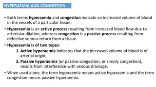
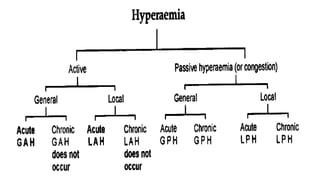
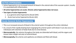
Lecture # 12
• Both terms hyperaemia and congestion indicate an increased volume of blood
in the vessels of a particular tissue.
• Hyperaemia is an active process resulting from increased blood flow due to
arteriolar dilation, whereas congestion is a passive process resulting from
defective venous return from a tissue.
• Hyperaemia is of two types:
1. Active hyperaemia indicates that the increased volume of blood is of
arterial origin,
2. Passive hyperaemia (or passive congestion, or simply congestion),
results from interference with venous drainage.
• When used alone, the term hyperaemia means active hyperaemia and the term
congestion means passive hyperaemia.
HYPERAEMIA AND CONGESTION
• Active hyperaemia is an increased amount of blood in the arterial side of the vascular system. Usually
it is associated with inflammation.
• All active hyperaemias are acute. Chronic active hyperaemia does not occur.
• Two types of active hyperaemia:
1. Acute general active hyperaemia (Acute GAH)
2. Acute local active hyperaemia (Acute LAH)
• This is an increased amount of blood in the arterial system throughout the entire individual.
• As a result of the rapidly beating heart engorging the arterial system with blood. It can also occur in
individuals with retention of body fluids due to renal disease.
• Macroscopically, the arteries throughout the body are distended with blood, and the organs and
tissues have a bright red color of arterial blood.
• Microscopically, the arteries and capillaries are dilated and filled with blood.
ACTIVE HYPERAEMIA
Acute General Active Hyperaemia
• This is an increased amount of blood in the arterial system within a local area (leg, stomach,
or lung) of an individual.
• It is the most common type of hyperaemia. Usually, when the term hyperaemia is used
unqualified, it refers to this type of hyperaemia.
• It is either
1. Physiological
2. and/or pathological.
• Physiological acute local active hyperaemia occurs in:
(1) the stomach and intestine following a meal (2) the lactating mammary gland, (3) the
genital tract during estrus, (4) the organs of locomotion such as muscles during
exercise, and (5) blushing, in humans.
Here, the hyperaemia is induced by neurogenic mechanisms.
Acute Local Active Hyperaemia
• Pathological acute active hyperaemia is the first stage of inflammation.
• Macroscopically, the part is enlarged, swollen, and is heavier than normal due to the
increased amount of blood. The color is bright red.
• Microscopically, when viewed in the living tissues with a magnifying lens, arteries, arterioles,
and capillaries are distended with blood.
Acute Local Active Hyperaemia
• This is an increased amount of blood in the venous side of the vascular system due to some form of
hindrance to the flow of blood from an organ or region.
• It is general if the interference is central, i.e., in the heart or lung,
• and local if in the vein of an organ or part of the body.
• Both general and local passive hyperaemia can be acute or chronic.
• This is an increase in the amount of blood in the venous side of the circulatory system due to a
sudden obstruction to the flow of blood in the heart or lungs.
• The hyperaemia involves the entire venous side of the circulatory system.
• The principal causes are:
1. Degeneration and necrosis of the myocardium.
2. Sudden myocardial accidents, such as myocardial infarction
3. Pneumonia, pulmonary thrombosis and embolism, hydropericardium, haemopericardium, or
pyopericardium, hydrothorax, haemothorax, and pyothorax.
• Macroscopically, organs throughout the body have an intense bluish red cyanotic) color.
• Microscopically, veins and capillaries are distended with blood.
Passive Hyperaemia (or Congestion)
Acute General Passive Hyperaemia
• This is an increase in the amount of blood in the venous side of the circulatory system that
persists for a long period of time and results in permanent alterations (atrophy and
fibrosis) in various tissues and organs throughout the body.
• The lesions have a central location in the heart or lungs.
• Lesions involving heart are:
1. Stenosis of a valvular opening (tricuspid or mitral valve)
2. Valvular insufficiency
3. Myocardial failure
4. Anomalies of the heart (persistent foramen ovale)
5. Constrictive lesions of the pericardium and epicardium, such as traumatic
pericarditis in cattle.
Chronic General Passive Hyperaemia
• Macroscopically, organs throughout the body have an intense bluish red cyanotic color.
• Microscopically, veins and capillaries are distended with blood.
• The lungs, liver, and spleen develop the most noticeable changes.
• Lungs are affected in the left-sided heart failure, that is, due to lesions in the mitral
valve (heart-failure cells)
• Liver is affected in right-sided heart failure, that is, due to lesions in the right atrio-
ventricular valve, or in the lungs (nutmeg liver)
• Spleen is slightly enlarged and slightly cyanotic. Microscopically, sinusoids are markedly
distended.
Chronic General Passive Hyperaemia
• This is an increase in the amount of blood in the veins of an area (foot, tail, kidney, spleen) as
the result of a sudden obstruction to the flow of blood from an organ or region.
• It might be due to:
1. Malposition of the viscera (intussusception, volvulus, and torsion) in which veins are
compressed.
2. External pressure
• Macroscopically, the veins are engorged with blood.
• Microscopically, veins and capillaries are distended with blood. Necrosis and suppurative
inflammation may be present.
Acute Local Passive Hyperaemia
• This is an increase in the amount of blood that persists for a long time in the veins of a
portion, and causes permanent tissue changes (atrophy and fibrosis) in the area.
• Two types of changes produce this form of hyperemia:
• Enlarging neoplasms lymph nodes, and abscesses.
• Obstruction within a vein, such as caused by a thrombus.
• Macroscopically, the affected organ in the beginning is larger than normal. Later, when
scarring (fibrosis) takes place, it becomes smaller.
• Microscopically, veins and capillaries are distended with blood and cells in the involved
tissue are separated by a transudate, which stains faintly pink with eosin.
Chronic Local Passive Hyperaemia
• Hypostatic congestion is the pooling of blood in capillaries and veins in a dependent part due
to the effect of gravity.
• Most commonly seen in sick animals that are in a recumbent position for a long time.
• Hypostatic congestion that occurs at the time of death, when the failing heart is no longer
able to maintain blood pressure, is called agonal congestion. When it is observed after
death, it is known as postmortem congestion.
• Hypostatic congestion indicates the side of the animal that was ventral at the time of
death.
HYPOSTATIC CONGESTION

Lecture # 12 HYPERAEMIA AND CONGESTION.pdf

  • 1.
  • 2.
    • Both termshyperaemia and congestion indicate an increased volume of blood in the vessels of a particular tissue. • Hyperaemia is an active process resulting from increased blood flow due to arteriolar dilation, whereas congestion is a passive process resulting from defective venous return from a tissue. • Hyperaemia is of two types: 1. Active hyperaemia indicates that the increased volume of blood is of arterial origin, 2. Passive hyperaemia (or passive congestion, or simply congestion), results from interference with venous drainage. • When used alone, the term hyperaemia means active hyperaemia and the term congestion means passive hyperaemia. HYPERAEMIA AND CONGESTION
  • 4.
    • Active hyperaemiais an increased amount of blood in the arterial side of the vascular system. Usually it is associated with inflammation. • All active hyperaemias are acute. Chronic active hyperaemia does not occur. • Two types of active hyperaemia: 1. Acute general active hyperaemia (Acute GAH) 2. Acute local active hyperaemia (Acute LAH) • This is an increased amount of blood in the arterial system throughout the entire individual. • As a result of the rapidly beating heart engorging the arterial system with blood. It can also occur in individuals with retention of body fluids due to renal disease. • Macroscopically, the arteries throughout the body are distended with blood, and the organs and tissues have a bright red color of arterial blood. • Microscopically, the arteries and capillaries are dilated and filled with blood. ACTIVE HYPERAEMIA Acute General Active Hyperaemia
  • 5.
    • This isan increased amount of blood in the arterial system within a local area (leg, stomach, or lung) of an individual. • It is the most common type of hyperaemia. Usually, when the term hyperaemia is used unqualified, it refers to this type of hyperaemia. • It is either 1. Physiological 2. and/or pathological. • Physiological acute local active hyperaemia occurs in: (1) the stomach and intestine following a meal (2) the lactating mammary gland, (3) the genital tract during estrus, (4) the organs of locomotion such as muscles during exercise, and (5) blushing, in humans. Here, the hyperaemia is induced by neurogenic mechanisms. Acute Local Active Hyperaemia
  • 6.
    • Pathological acuteactive hyperaemia is the first stage of inflammation. • Macroscopically, the part is enlarged, swollen, and is heavier than normal due to the increased amount of blood. The color is bright red. • Microscopically, when viewed in the living tissues with a magnifying lens, arteries, arterioles, and capillaries are distended with blood. Acute Local Active Hyperaemia
  • 7.
    • This isan increased amount of blood in the venous side of the vascular system due to some form of hindrance to the flow of blood from an organ or region. • It is general if the interference is central, i.e., in the heart or lung, • and local if in the vein of an organ or part of the body. • Both general and local passive hyperaemia can be acute or chronic. • This is an increase in the amount of blood in the venous side of the circulatory system due to a sudden obstruction to the flow of blood in the heart or lungs. • The hyperaemia involves the entire venous side of the circulatory system. • The principal causes are: 1. Degeneration and necrosis of the myocardium. 2. Sudden myocardial accidents, such as myocardial infarction 3. Pneumonia, pulmonary thrombosis and embolism, hydropericardium, haemopericardium, or pyopericardium, hydrothorax, haemothorax, and pyothorax. • Macroscopically, organs throughout the body have an intense bluish red cyanotic) color. • Microscopically, veins and capillaries are distended with blood. Passive Hyperaemia (or Congestion) Acute General Passive Hyperaemia
  • 8.
    • This isan increase in the amount of blood in the venous side of the circulatory system that persists for a long period of time and results in permanent alterations (atrophy and fibrosis) in various tissues and organs throughout the body. • The lesions have a central location in the heart or lungs. • Lesions involving heart are: 1. Stenosis of a valvular opening (tricuspid or mitral valve) 2. Valvular insufficiency 3. Myocardial failure 4. Anomalies of the heart (persistent foramen ovale) 5. Constrictive lesions of the pericardium and epicardium, such as traumatic pericarditis in cattle. Chronic General Passive Hyperaemia
  • 9.
    • Macroscopically, organsthroughout the body have an intense bluish red cyanotic color. • Microscopically, veins and capillaries are distended with blood. • The lungs, liver, and spleen develop the most noticeable changes. • Lungs are affected in the left-sided heart failure, that is, due to lesions in the mitral valve (heart-failure cells) • Liver is affected in right-sided heart failure, that is, due to lesions in the right atrio- ventricular valve, or in the lungs (nutmeg liver) • Spleen is slightly enlarged and slightly cyanotic. Microscopically, sinusoids are markedly distended. Chronic General Passive Hyperaemia
  • 10.
    • This isan increase in the amount of blood in the veins of an area (foot, tail, kidney, spleen) as the result of a sudden obstruction to the flow of blood from an organ or region. • It might be due to: 1. Malposition of the viscera (intussusception, volvulus, and torsion) in which veins are compressed. 2. External pressure • Macroscopically, the veins are engorged with blood. • Microscopically, veins and capillaries are distended with blood. Necrosis and suppurative inflammation may be present. Acute Local Passive Hyperaemia
  • 11.
    • This isan increase in the amount of blood that persists for a long time in the veins of a portion, and causes permanent tissue changes (atrophy and fibrosis) in the area. • Two types of changes produce this form of hyperemia: • Enlarging neoplasms lymph nodes, and abscesses. • Obstruction within a vein, such as caused by a thrombus. • Macroscopically, the affected organ in the beginning is larger than normal. Later, when scarring (fibrosis) takes place, it becomes smaller. • Microscopically, veins and capillaries are distended with blood and cells in the involved tissue are separated by a transudate, which stains faintly pink with eosin. Chronic Local Passive Hyperaemia
  • 12.
    • Hypostatic congestionis the pooling of blood in capillaries and veins in a dependent part due to the effect of gravity. • Most commonly seen in sick animals that are in a recumbent position for a long time. • Hypostatic congestion that occurs at the time of death, when the failing heart is no longer able to maintain blood pressure, is called agonal congestion. When it is observed after death, it is known as postmortem congestion. • Hypostatic congestion indicates the side of the animal that was ventral at the time of death. HYPOSTATIC CONGESTION