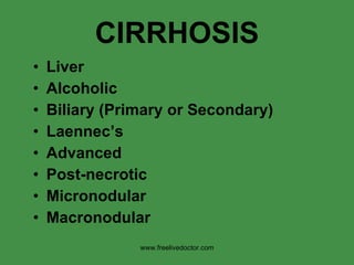
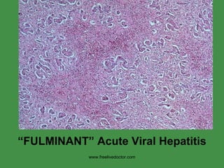
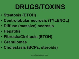
IT INCLUDES ANATOMY, PHYSIOLOGY AND PATHOLOGY OF LIVER .
THE SOURCES ARE:-
THE MEDICAL TEXT BOOK OF ROBBIN'S PATHOLOGY
AND OTHERS
IMAGES SOURCE :- ATLAS BOOKS AND INTERNET
IT INCLUDES ANATOMY, PHYSIOLOGY AND PATHOLOGY OF LIVER .
THE SOURCES ARE:-
THE MEDICAL TEXT BOOK OF ROBBIN'S PATHOLOGY
AND OTHERS
IMAGES SOURCE :- ATLAS BOOKS AND INTERNET
The gall bladder is located in the junction of the right ninth costal cartilage and lateral border of the rectus abdominis.
It is a pear shaped sac lying on the inferior surface of the liver in a fossa between the right and quadrate lobes with a capacity of about 30 to 50 mL.
A chronic, progressive disease characterized by widespread fibrosis(scarring) and nodule formation.
The development of cirrhosis is an insidious, prolonged course, usually after decades of chronic liver disease.
10. www.freelivedoctor.com The FIRST part of the lobule, i.e., portal triad is the FIRST to get blood flow, so it is also the FIRST to get the brunt of general toxic effects, and the LAST to get the brunt of ischemic effects.
60. C LESS common than B (one fourth) LESS dangerous than B in the acute phase MORE likely to go chronic than B MORE closely linked with hepatoma than B www.freelivedoctor.com
91. - “micro-” and “macro-” vesicular steatosis b) necrosis and apoptosis i) centrilobular necrosis c) inflammation i) hepatitis d) regeneration e) fibrosis i) irreversible damage ii) “scar” tissue is referred to as “cirrhosis” www.freelivedoctor.com
92.
93. d) chronic liver failure i) most common route - cirrhosis e) hepatic dysfunction without overt necrosis i) viable without normal function - Reye syndrome - tetracycline toxicity - acute steatosis of pregnancy www.freelivedoctor.com
94. f) clinical: i) jaundice ii) hypoalbuminemia iii) impaired estrogen metabolism - hypogonadism - gynecomastia iv) MSOF susceptibility v) coagulopathy - II, VII, IX and X deficiency of clotting factors vi) death in weeks to months www.freelivedoctor.com
95. vii) 2 grave complications: - hepatic encephalopathy neurotransmission disturbances of CNS and neuromuscular system. - hepatorenal syndrome severe renal failure without any intrinsic disorders. 1. Na + retention 2. H 2 O excretion 3. RBF 4. GFR www.freelivedoctor.com
96.
97. c) cirrhosis essentially irreversible i) main pathogenic processes: - progressive fibrosis - vascular reorganization (blood shunted around parenchyma) ii) collagen deposits (via perisinusoidal satellite cells) - Types I and III d) may be clinically silent e) death: progressive liver failure, portal hypertension, CA www.freelivedoctor.com
101. b) clinical consequences i) ascites ii) portosystemic shunts iii) congestive splenomegaly iv) hepatic encephalopathy 1. ascites a) fluid accumulation of fluid in peritoneum b) Starling forces www.freelivedoctor.com
102. 2. portosystemic shunts a) shunt flow where portal and systemic share common circulation i) rectum (hemorrhoids) ii) cardioesophageal junction - esophageal varices - massive hematemesis iii) periumbilical and abdominal collaterals - caput medusae www.freelivedoctor.com
103.
104. b) bilirubin and bile formation i) bilirubin end product of heme degradation - senescent RBC’s - in spleen, liver and bone marrow - accounts for ~ 0.3 gm/day ii) most remainder of bilirubin - turnover of hepatic heme iii) heme biliverdin (via heme oxidase) bilirubin (via biliverdin reductase) www.freelivedoctor.com
105. iv) bound to albumin (not soluble) - free may in hemolytic diseases v) taken up by liver (carrier mediated), conjugated with glucuronic acid - H 2 O - secreted into bile - degraded by bacteria to urobilinogens - excreted in feces and urine - reab in ileum and colon www.freelivedoctor.com
108. i) small amount may diffuse into brain of infants kernicterus - in hemolytic diseases (erythroblastosis fetalis) c) conjugated is H 2 O soluble i) can be excreted in urine d) normal bilirubin = 0.3-1.2 mg/dl e) jaundice >2-2.5 mg/dl i) imbalance between production and clearance www.freelivedoctor.com
112. iv) Dubin-Johnson syndrome - excretion problem - conjugated bilirubin v) Rotor syndrome - decreased uptake and excretion vi) see table 18-4 www.freelivedoctor.com
113.
114. e) classic lab test is Alk. Phos. i) detergent effects of retained bile salts. ii) must verify hepatic in nature - several isoforms iii) nutritional deficiencies of Vit A, D and K - malabsorption from gut f) extrahepatic (obstruction) i) surgery to correct g) intrahepatic not helped with surgery (see table 18-5) www.freelivedoctor.com
118. 1. hepatic steatosis a) moderate amounts of ETOH intake small lipid droplets (microvesicular) b) chronic etoh intake large lipid globules (macrovesicular) i) compressing and displacing nucleus to periphery of hepatocyte ii) initially centrilobular iii) may involve entire lobe of liver with progression www.freelivedoctor.com
119. Alcoholic liver disease: macrovesicular steatosis, involving most regions of the hepatic lobule. The intracytoplasmic fat is seen as clear vacuoles. Some early fibrosis (stained blue) is present (Masson trichrome). www.freelivedoctor.com
120. The cluster of inflammatory cells marks the site of a necrotic hepatocyte. A Mallory body is present in a second hepatocyte ( arrow ). www.freelivedoctor.com
121. iv) fibrosis with chronic etoh use v) reversible changes with abstinence from etoh 2. alcoholic hepatitis a) swelling of hepatocytes i) single or scattered foci ii) swelling due to fat and H 2 O iii) mild hemosiderin deposits iv) Mallory inclusions - eosinophilic cytoplasm - not specific (also seen in Wilson disease, cholestasis www.freelivedoctor.com
122. 3. alcoholic cirrhosis a) irreversible and final form of alcoholic liver disease b) nodule formation (micro and macro) c) fibrosis d) deranged vascular perfusion www.freelivedoctor.com
123. The characteristic diffuse nodularity of the surface reflects the interplay between nodular regeneration and scarring. The greenish tint of some nodules is due to bile stasis. www.freelivedoctor.com
124. The microscopic view shows nodules of varying sizes entrapped in blue-staining fibrous tissue. www.freelivedoctor.com
136. g) excess iron is directly toxic i) lipid peroxidation - via Fe-induced free radical ii) stimulation of collagen formation iii) O 2 free radicals and iron interact with DNA lethal injury - predisposing to hepatocellular CA iv) removing excess iron is Tx in cells not irreversible damaged www.freelivedoctor.com
137. h) most common 2 o hemochromatosis i) hemolytic anemias associated with ineffective erythropoiesis i) most common sites for hemosiderin deposition (decreasing order) i) liver ii) pancreas iii) myocardium iv) pituitary gland v) adrenals vi) thyroid, parathyroid vii) joints and skin www.freelivedoctor.com
138.
139. d) gene for Wilson disease 13 e) when excretion is defective i) Cu ++ spills over into blood - urinary excretion ii) causes direct toxic effects - hemolysis - organ dysfunction f) rare before 6 yrs. www.freelivedoctor.com
140. g) most common presentation i) acute/chronic liver disease ii) neuropsychiatric (behavioral) iii) Dx urine Cu ++ , serum ceruloplasmin, hepatic Cu ++ iv) plasma Cu ++ NOT useful !! v) “Kayser-Fleischner” rings - green to brown deposits in cornea h) D-penicillamine Tx (Cu ++ chelator) www.freelivedoctor.com
141.
142. d) Most commonly diagnosed genetic liver disease in infants and children e) clinical: i) neonatal hepatitis with cholestatic jaundice 10-20% ii) older children hepatitis or cirrhosis iii) adults emphysema www.freelivedoctor.com
146. c) major features are: i) nonsuppurative inflammation ii) destruction of medium sized bile ducts d) mainly in middle aged women (6:1) i) peak between 40-50 yrs e) initial presentation is pruritis i) jaundice late in course ii) hepatomegaly is typical iii) xanthomas due to cholesterol f) antimitochondrial Ab in > 90% i) against PDH complex E 2 subunit www.freelivedoctor.com
147. g) clinical: i) autoimmune disease ii) sicca complex - dry eyes and mouth iii) etiology remains unclear - lack of “trigger(s)” iv) causes of death: - liver failure - portal hypertension and variceal bleeding - infection www.freelivedoctor.com
148.
149.
150. c) 1 o seen in association with inflammatory bowel disease i) UC - ~75% coexistence ii) only ~4% of patients with UC have have coexistence of 1 o sclerosing cholangitis d) etiology unknown i) association with IBD e) liver transplant is Tx www.freelivedoctor.com
151.
152. b) loss of hepatic artery flow does not always cause infarct i) sufficient collaterals ii) exception is transplanted liver (thrombosis causes infarct) 2 – portal vein a) abdominal pain b) ascites c) similar to portal hypertension i) esophageal varices - prone to rupture www.freelivedoctor.com
154. e) portal vein thrombus NO infarct i) red-blue discolorization which is well demarcated - “ infarct of Zahn” ii) severe hepatocellular atrophy f) neoplastic obstruction 3 – intrahepatic a) most common cause is cirrhosis b) sickle cell disease c) DIC i) eclampsia of pregnancy d) obstructive neoplasms www.freelivedoctor.com
156. 4 – hepatic vein obstruction a) single vein obstruction i) is silent b) 2 or more major hepatic veins (“ Budd-Chiari syndrome ”) i) hepatomegaly ii) pain iii) ascites iv) intrahepatic BP www.freelivedoctor.com
157. c) venous thrombosis associated with i) myeloproliferative disorders ii) inherited disorders of coagulation iii) PNH iv) hepatocellular CA d) BC pills and pregnancy i) with underlying thrombotic disorders e) if untreated mortality is high f) veno-occlusive disease i) following bone marrow trans. www.freelivedoctor.com
158.
159.
160.
161.
162.
163.
164.
Editor's Notes
Like the pancreas, the liver, together with the gallbladder, is considered, also to be derived embryologically, as an outpocketing of the foregut.
Classical anatomic landmarks in the average 1400-1800 gram adult liver.
Relationship of the liver with neighboring organs
The liver is nothing more than an array of cells between the portal and caval venous systems. This shows the direction of flow. The liver gets about 80% of its blood supply fron the portal veins and 20% from the hepatic arterial system.
The IDEAL three-dimensional diagram.
The classical view of liver tissue from a liver biopsy, H&E stained.
What is the direction ov venous blood flow, portal to central, or vice versa?
The FIRST part of the lobule, i.e., portal triad is the FIRST to get blood flow, so it is also the FIRST to get the brunt of general toxic effects, and the LAST to get the brunt of ischemic effects.
The LAST part of the lobule, central vein, VICE VERSA!
The classical classification of diseases!
Heatocyte cytoplasm is “balooned”
Hepatocytes have “feathery” cytoplasm
Fat vacoules are small enough to lie completely WITHIN the hepatocte cytioplasm
Fat vacuoles are large enough to completely REPLACE the hepatocyte cytoplasm. Why is the differential diagnosis of MACRO vesicular steatosis the same as MICRO vesicular steatosis?
Of the three types of common golden pigment found in the body, 1) hemosiderin, 2) melanin, and 3) bile, only hemosiderin stains BLUE with the prussian blue stain. There are special stains to identify melanin and bile however.
Crucially important concept worth repeating. KNOW the difference between an acinus and a lobule.
The “hyalinized” appearing round structures are cells dying from a NORMAL replacement process called apoptosis.
Triads are involved with hepatitis earlier than general sinusoids. Why?
Cirrhosis is the natural END STAGE IRREVERSIBLE disease of ANYTHING which chronically damages the liver!!! Cirrhosis is the natural END STAGE IRREVERSIBLE disease of ANYTHING which chronically damages the liver!!! Cirrhosis is the natural END STAGE IRREVERSIBLE disease of ANYTHING which chronically damages the liver!!! Cirrhosis is the natural END STAGE IRREVERSIBLE disease of ANYTHING which chronically damages the liver!!!
The hepatoma/cirrosis cycle: Hepatomas often, perhaps usually, arise in a cirrhosis background, and can be thought of as regenerating nodules that have lost growth control, ususlly NOT metastasizing, the hepatoma itself causing FURTHER functional liver disease.
Even a blind man would know this liver surface feels “smooth” which generally rules out cirrhosis or tumors even before slicing it for examination.
In a normal liver, ther is connective tissue ONLY in the small portal triad area.
Is this MACROnodfular or MICROdular cirrhosis?
Why are TRICHROME stains recommended for every liver biopsy?
Ther are twokinds of hepatic enzymes: INTRACELLULAR and MEMBRANE ASSOCIATED. This fact forms the basis for hepatic diagnostic enzymology.
Almost all primarilly INTRACELLULAR liver pathologies result in MEMBRANE elevations too, and VICE VERSA!
Sclera and conjunctiva are the best places to see early jaundice, palms too!
A normal liver should NOT have this much bile pigment, in fact bile pigment in a normal liver biopsy should be scarce!
Bile accumulations . Why does it look like a lot is BETWEEN hepatocytes in early stages?
“ swollen” liver?
The inflammation of hepatitis starts in the portal triad areas, with increasing severity it extends to the sinusoids.
“ Fulminant” hepatitis is associated with massive hepatic necrosis and often (usually) results in death.
It would be VERY nice to see Councilman bodies on a liver biopsy to enable the diagnosis of Hepatitis B. Unfortunately, you may not be lucky enough to find them. Does this remind you of the “apoptosis” image? Each “Councilman body” represents apoptotic death of a single liver cell.
Consequences of hepatitis B, surprisingly, most cases of Hepatitis B are SUB-clinical
Consequences of hepatitis C
Laboratory findings in hepatitis B, classical.
The MAIN differential of NON viral “hepatitis”: 1) TOXIC (alcohol the most common), 2) autoimmune, and 3) non-viral infectious!
Many of the classical liver toxins can produce a fairly predictable pattern of liver changes. BUT, they can also be quite UN-predictable and varied as well!
Alpha-1-antitrypsin PROTECTS tissues, especially lung, liver, from HARMFUL NATURAL PROTEASE. Lack of this enzyme, due to a genetic defect, would then be expected to cause destructive changes in these areas.
The liver can be thought of as being a “lymph” node for early metastases of any organ which has a portal circulation!
How would the blind man know this is metastatic cancer, rather than macronodular cirrhosis?
Individually, hepatoma “cells” usually very closely resemble normal hepatocytes!
Cholangiocarcinoma is ALWAYS confused with liver metastases. Why? What feature of this picture would NOT suggest metastases?
Common gallbladder “anomalies” are usually developmental.
Cholecystitis predisposes to cholelithiasis, and VICE VERSA!