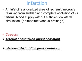
Hemodynamic Changes
Eman Zayed
Hemodynamic Changes
• Hyperaemia
• Thrombosis
• Embolism
• Ischemia and infarct
• Hemorrhage and shock
• Definition:
increased blood volume in a particular tissue or organ, due to
dilatation of the microcirculation resulting from either
increased blood flow (inflow)
or impaired venous drainage (outflow).
• Types:
I. Active hyperemia (hyperemia)
II. Passive hyperemia (congestion)
It occurs when arterial and arteriolar dilatation
produces an increased flow of blood into the
capillary beds with opening of inactive
capillaries.
• Causes: might be
1- Physiological e.g. muscular exercise.
2- Pathological .e.g., acute inflammation.
• Grossly, the affected tissue is red because of
the engorgement with oxygenated blood.
It results from impaired venous drainage (return), and it leads to
accumulation of blood on the venous side of the circulation
• Congestion is almost always pathologic.
• Grossly,The affected tissue is cyanotic (blue- red)
due to accumulation deoxygenated blood (reduced
Hb)
• Examples :
 Leg in deep vein thrombosis
 Lung congestion
Classification and causes of
Congestion
According to its distribution:
• Generalized (systemic)
• Localized
According to onset:
• Acute
• Chronic
I. Localized Venous Congestion:
1. Acute local congestion
• Sudden complete venous obstruction occurs due to: ligature
of a vein, thrombosis of a vein, and mechanical
pressure caused by strangulated hernia, volvulus, and
intussusception.
• Effects: if there is collateral circulation no harm
will occur, but if there is poor collateral
circulation, local venous congestion, edema
and even infarction will occur.
2 .Chronic local congestion
• There is gradual incomplete venous obstruction caused by:
venous compression by a tumour, enlarged lymph node, by
constriction of vein by fibrous tissue (liver cirrhosis or bilharsial
hepatic fibrosis) or obliteration of vein by organizing thrombus
• Effects:
the veins, venules, and capillaries proximal to the obstruction
become dilated and congested resulting in edema. Also it will lead
to gradual opening of the collateral anastomotic veins.
II. Systemic Venous Congestion:
1. Acute generalized venous congestion:
EX,terminal condition in acute
heart failure
2. Chronic generalized venous congestion:
mitral stenosis , or extensive fibrosis of the lung
with resulting obstruction in the pulmonary
circulation.
General effects of venous
congestion:
1- Hypoxia
2- Cyanosis
3- Edema, due to overfilling of venules causing
increased hydrostatic pressure. It might also be
due to venous congestion of the kidney causing
retention of water and electrolytes. Edema
starts in dependent parts where the venous
pressure is greatest especially around the
ankles, and then spreads upwards.
congestion in different organs
• Definition; it is a condition of inadequate
supply of oxygenated blood to a tissue
Types of ischemia:
A. Generalized ischemia:
Generalized decrease in tissue perfusion due to
inadequate cardiac output e.g., shock state.
B. Localized ischemia:
affection of blood supply to a particular organ e.g.,
infarct
2. According to onset and severity
A. Sudden complete ischemia
sudden complete occlusion of arterial
circulation. It might be due to thrombosis, embolism,
surgical ligature of an
artery
B. Gradual ischemia
might be due to atherosclerosis or pressure on an
artery from
outside by enlarged lymph nodes or a growing tumour.
Infarction
• An infarct is a localized area of ischemic necrosis
resulting from sudden and complete occlusion of its
arterial blood supply without sufficient collateral
circulation, (or impaired venous drainage).
• Causes:
 Arterial obstruction (most common)
 Venous obstruction (less common)
Factors that affect the
development of an infarct
• Nature of the blood supply:
• In tissues rich in collateral circulation, blood
flow is not significantly decreased by
occlusion of an artery e.g., lung (double
blood supply: branchial and pulmonary),
fiver)(hepatic artery and portal vein), circle of
Wills (brain).
• In tissues having no collateral circulation,
obstruction of the end artery leads to
infarction e.g., central retinal artery, and
splenic artery.
• The state of collateral circulation (patency
of collateral) e.g., Narrowing of arteries in the
collateral circulation decreases their
effectiveness in maintenance of blood flow
e.g., occlusion of internal carotid artery with
healthy circle of Wills has no effect on blood
flow. Atherosclerotic of these collaterals
leads to infarct when one internal carotid
artery is occluded.
• Rate of development of occlusiohypoxia:
• Slowly developing occlusions are less likely
to cause infarction since they give time to the
development of collateral circulation e.g. In
the heart, occlusion of one of the large
coronary arteries
• The vulnerability of tissue to hypoxia
Neurons are very sensitive to hypoxia, followed
by myocardial muscle fibres
Fibroblasts are resistant to hypoxia.
• Oxygen content of the blood:
Partial flow obstruction of a small blood vessel
in an anemic or cyanotic patient (congestive
heart failure), might lead to tissue infarction
(whereas it would be without effect under
conditions of normal oxygen tension.
Classification of infarction
• The presence or absence of microorganisms
Sterile
Septic
• Septic infarcts are characterized by secondary bacterial infection of the necrotic
tissue.
• Septic infarcts occur:
• When microorganisms are present in thrombus or embolus e.g., emboli of acute
infective endocarditis.
• When infarction occurs in a tissue (intestine) that normally contains bacteria.
• When bacteria from the blood stream cause secondary infection (rare)
May convert infarction into abscess or gangrene
• Microscopic appearance:
• Coagulative necrosis (the most common)
• Liquifactive necrosis (in the brain)
Fate of Infarction:
• A small infarct is healed by fibrous tissue i.e. scar.
• A large infarct is encapsulated by fibrous tissue capsule and undergoes dystrophic
calcification.
• Infection and abscess formation may complicate infarcts.
• Superadded putrifaction, leads to gangrene.
Thrombosis
• Definition:
Process of formation of a solid mass from
the blood constituents
Etiology
• Endothelial lining injury
• Alterations in the normal blood flow This
may be in the form of stasis or turbulence.
• Alterations in blood coagulability
(Hypercoagulability)
Embolism
• the process of impaction of an embolus in
a vessel wall too Small to permit its further
passage, resulting in partial or complete
occlusion of the vessel.
• Embolus: is detached intravascular solid,
liquid or gaseous mass, carried by the
blood to a site distant from its point of
origin.
Types of emboli
• Detached thrombi (99% of all emboli).
• Air emboli.
• Amniotic fluid emboli.
• Fat emboli.
• Tumour emboli
• Miscellaneous emboli such as foreign
body emboli (bullet, silk and tale), parasitic
emboli (worms or ova), and red cell
aggregates especially in infection.
Detached thrombi (thromboembolism)
Depending on the site of origin of these
thromboemboli, they may rest anywhere in
the cardiovascular system. So
thromboemboli are classified according to
whether they lodge in the pulmonary or
systemic circulations.
Systemic thromboembolism
It refers to emboli that travel through the
arterial circulation
• Thrombi in the left heart
• Thrombus on atheromatous lesions on
aorta
• Paradoxical emboli: are venous emboli
that reach the arterial circulation by
crossing right to left heart through
interatrial or interventricular septal defect.
So in this condition, the detached thrombi
bypass the lung and reach directly
Air embolism
Air or gas may gain access to the
circulation:
1-During delivery or abortion
2-Through artificial pneumothorax
3- Injury to the lung or chest wall,
4-Injury to a large neck vein
5-During blood or fluid infusion
• 50-100 c.c. Of air are required to produce
problems, usually they lodge into a major
vessel in the lung leading to sudden death
or acute heart failure as the injected air
reaching the right ventricle and obstructs
the blood flow to the lung by forming
uncompressible column of air-fluid mixture.
Hemorrhage
• Escape of blood outside cardiovascular
system

circulatory disturbance 1.pptx

  • 1.
  • 2.
    Hemodynamic Changes • Hyperaemia •Thrombosis • Embolism • Ischemia and infarct • Hemorrhage and shock
  • 3.
    • Definition: increased bloodvolume in a particular tissue or organ, due to dilatation of the microcirculation resulting from either increased blood flow (inflow) or impaired venous drainage (outflow). • Types: I. Active hyperemia (hyperemia) II. Passive hyperemia (congestion)
  • 4.
    It occurs whenarterial and arteriolar dilatation produces an increased flow of blood into the capillary beds with opening of inactive capillaries. • Causes: might be 1- Physiological e.g. muscular exercise. 2- Pathological .e.g., acute inflammation. • Grossly, the affected tissue is red because of the engorgement with oxygenated blood.
  • 6.
    It results fromimpaired venous drainage (return), and it leads to accumulation of blood on the venous side of the circulation • Congestion is almost always pathologic. • Grossly,The affected tissue is cyanotic (blue- red) due to accumulation deoxygenated blood (reduced Hb) • Examples :  Leg in deep vein thrombosis  Lung congestion
  • 7.
    Classification and causesof Congestion According to its distribution: • Generalized (systemic) • Localized According to onset: • Acute • Chronic
  • 8.
    I. Localized VenousCongestion: 1. Acute local congestion • Sudden complete venous obstruction occurs due to: ligature of a vein, thrombosis of a vein, and mechanical pressure caused by strangulated hernia, volvulus, and intussusception. • Effects: if there is collateral circulation no harm will occur, but if there is poor collateral circulation, local venous congestion, edema and even infarction will occur.
  • 9.
    2 .Chronic localcongestion • There is gradual incomplete venous obstruction caused by: venous compression by a tumour, enlarged lymph node, by constriction of vein by fibrous tissue (liver cirrhosis or bilharsial hepatic fibrosis) or obliteration of vein by organizing thrombus • Effects: the veins, venules, and capillaries proximal to the obstruction become dilated and congested resulting in edema. Also it will lead to gradual opening of the collateral anastomotic veins.
  • 10.
    II. Systemic VenousCongestion: 1. Acute generalized venous congestion: EX,terminal condition in acute heart failure 2. Chronic generalized venous congestion: mitral stenosis , or extensive fibrosis of the lung with resulting obstruction in the pulmonary circulation.
  • 11.
    General effects ofvenous congestion: 1- Hypoxia 2- Cyanosis 3- Edema, due to overfilling of venules causing increased hydrostatic pressure. It might also be due to venous congestion of the kidney causing retention of water and electrolytes. Edema starts in dependent parts where the venous pressure is greatest especially around the ankles, and then spreads upwards.
  • 12.
  • 13.
    • Definition; itis a condition of inadequate supply of oxygenated blood to a tissue
  • 15.
    Types of ischemia: A.Generalized ischemia: Generalized decrease in tissue perfusion due to inadequate cardiac output e.g., shock state. B. Localized ischemia: affection of blood supply to a particular organ e.g., infarct
  • 16.
    2. According toonset and severity A. Sudden complete ischemia sudden complete occlusion of arterial circulation. It might be due to thrombosis, embolism, surgical ligature of an artery B. Gradual ischemia might be due to atherosclerosis or pressure on an artery from outside by enlarged lymph nodes or a growing tumour.
  • 17.
    Infarction • An infarctis a localized area of ischemic necrosis resulting from sudden and complete occlusion of its arterial blood supply without sufficient collateral circulation, (or impaired venous drainage). • Causes:  Arterial obstruction (most common)  Venous obstruction (less common)
  • 18.
    Factors that affectthe development of an infarct • Nature of the blood supply: • In tissues rich in collateral circulation, blood flow is not significantly decreased by occlusion of an artery e.g., lung (double blood supply: branchial and pulmonary), fiver)(hepatic artery and portal vein), circle of Wills (brain). • In tissues having no collateral circulation, obstruction of the end artery leads to infarction e.g., central retinal artery, and splenic artery.
  • 19.
    • The stateof collateral circulation (patency of collateral) e.g., Narrowing of arteries in the collateral circulation decreases their effectiveness in maintenance of blood flow e.g., occlusion of internal carotid artery with healthy circle of Wills has no effect on blood flow. Atherosclerotic of these collaterals leads to infarct when one internal carotid artery is occluded.
  • 20.
    • Rate ofdevelopment of occlusiohypoxia: • Slowly developing occlusions are less likely to cause infarction since they give time to the development of collateral circulation e.g. In the heart, occlusion of one of the large coronary arteries • The vulnerability of tissue to hypoxia Neurons are very sensitive to hypoxia, followed by myocardial muscle fibres Fibroblasts are resistant to hypoxia.
  • 21.
    • Oxygen contentof the blood: Partial flow obstruction of a small blood vessel in an anemic or cyanotic patient (congestive heart failure), might lead to tissue infarction (whereas it would be without effect under conditions of normal oxygen tension.
  • 22.
  • 23.
    • The presenceor absence of microorganisms Sterile Septic • Septic infarcts are characterized by secondary bacterial infection of the necrotic tissue. • Septic infarcts occur: • When microorganisms are present in thrombus or embolus e.g., emboli of acute infective endocarditis. • When infarction occurs in a tissue (intestine) that normally contains bacteria. • When bacteria from the blood stream cause secondary infection (rare) May convert infarction into abscess or gangrene
  • 24.
    • Microscopic appearance: •Coagulative necrosis (the most common) • Liquifactive necrosis (in the brain)
  • 25.
    Fate of Infarction: •A small infarct is healed by fibrous tissue i.e. scar. • A large infarct is encapsulated by fibrous tissue capsule and undergoes dystrophic calcification. • Infection and abscess formation may complicate infarcts. • Superadded putrifaction, leads to gangrene.
  • 26.
    Thrombosis • Definition: Process offormation of a solid mass from the blood constituents
  • 27.
  • 28.
    • Endothelial lininginjury • Alterations in the normal blood flow This may be in the form of stasis or turbulence. • Alterations in blood coagulability (Hypercoagulability)
  • 30.
    Embolism • the processof impaction of an embolus in a vessel wall too Small to permit its further passage, resulting in partial or complete occlusion of the vessel. • Embolus: is detached intravascular solid, liquid or gaseous mass, carried by the blood to a site distant from its point of origin.
  • 31.
    Types of emboli •Detached thrombi (99% of all emboli). • Air emboli. • Amniotic fluid emboli. • Fat emboli. • Tumour emboli • Miscellaneous emboli such as foreign body emboli (bullet, silk and tale), parasitic emboli (worms or ova), and red cell aggregates especially in infection.
  • 32.
    Detached thrombi (thromboembolism) Dependingon the site of origin of these thromboemboli, they may rest anywhere in the cardiovascular system. So thromboemboli are classified according to whether they lodge in the pulmonary or systemic circulations.
  • 33.
    Systemic thromboembolism It refersto emboli that travel through the arterial circulation
  • 34.
    • Thrombi inthe left heart • Thrombus on atheromatous lesions on aorta • Paradoxical emboli: are venous emboli that reach the arterial circulation by crossing right to left heart through interatrial or interventricular septal defect. So in this condition, the detached thrombi bypass the lung and reach directly
  • 35.
    Air embolism Air orgas may gain access to the circulation: 1-During delivery or abortion 2-Through artificial pneumothorax 3- Injury to the lung or chest wall, 4-Injury to a large neck vein 5-During blood or fluid infusion
  • 36.
    • 50-100 c.c.Of air are required to produce problems, usually they lodge into a major vessel in the lung leading to sudden death or acute heart failure as the injected air reaching the right ventricle and obstructs the blood flow to the lung by forming uncompressible column of air-fluid mixture.
  • 37.
    Hemorrhage • Escape ofblood outside cardiovascular system