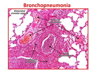
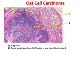
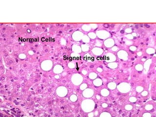
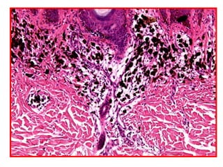
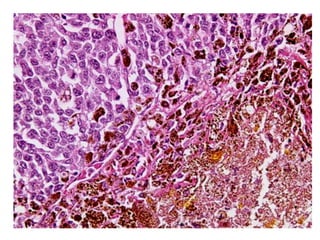
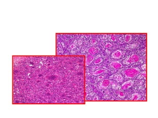
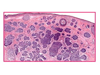
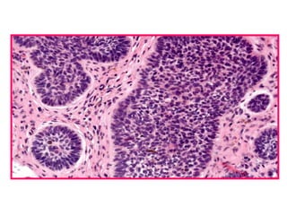
Allergic Nasal Polyp is characterized by:
1) Hyperplastic respiratory epithelium
2) Edematous stroma
3) Dilated congested capillaries
The document contains diagrams and photomicrographs of various pathological conditions affecting multiple organ systems. The sections describe microscopic findings, histological features, and pathological changes seen in conditions such as pneumonia, emphysema, infarction, cancer, glomerulonephritis, bilharziasis, appendicitis, liver diseases, and others.
This document discusses patterns of lymph node hyperplasia. It begins by describing the normal anatomy of a lymph node and then discusses various causes of lymph node hyperplasia including infectious, immune, and neoplastic causes. Various patterns of lymph node hyperplasia are classified including follicular, paracortical, sinus, diffuse, and mixed patterns. Specific types of follicular, paracortical, sinus, and mantle/marginal zone hyperplasia are described in detail. The document compares the architectural and cytological features of follicular hyperplasia versus follicular lymphoma. References for further information are also provided.
This document discusses various histopathological patterns seen in tissue samples. It defines terms like trabeculae, syncytium, alveolus, herringbone, storiform, fascicle, cribriform, tubule, papillae, Indian file, hobnail, rossette, microcystic, and follicle. Examples of diseases that exhibit each pattern are provided. The objectives are to help differentiate patterns in histopathology. Key patterns include trabecular, syncytial, alveolar, herringbone, storiform, fascicular, cribriform, tubular, papillary, micropapillary, Indian file, hobnail, and rossette
Lymph nodes are bean-shaped organs found throughout the body that filter lymph and house immune cells. A lymph node contains a fibrous capsule enclosing compartments of connective tissue and lymphocytes. The parenchyma is divided into an outer cortex and inner medulla. A normal lymph node contains mature lymphocytes, plasma cells, centrocytes, centroblasts, and immunoblasts. Lymphadenopathy refers to enlarged lymph nodes, which can be caused by infection, inflammation, autoimmune disease, or cancer metastasis. Physical examination of lymph nodes considers location, number, size, consistency, tenderness, and mobility to evaluate causes of lymphadenopathy.
The document discusses various pathologies of the prostate, including:
- Benign prostatic hyperplasia (BPH), which is a noncancerous enlargement of the prostate due to aging. Histologic features include glandular and stromal hyperplasia.
- Prostatic intraepithelial neoplasia (PIN), which is considered a precursor lesion to prostate cancer. It is graded as low or high grade.
- Prostate adenocarcinoma, which microscopically appears as small, simple glandular structures. The Gleason grading system is used to characterize prostate cancers.
- Other rare pathologies that can involve the prostate such as lymphoma, small cell carcinoma,
This document discusses the classification and characteristics of various types of non-Hodgkin lymphoma (NHL). It describes the historical classifications of NHL from the 1940s to the current 2008 WHO classification. It then provides details on specific NHL subtypes, including small lymphocytic lymphoma/chronic lymphocytic leukemia, follicular lymphoma, mantle cell lymphoma, and marginal zone B-cell lymphoma. For each subtype, it discusses immunophenotype, genetic abnormalities, clinical features, histopathology, immunostaining patterns, and differential diagnosis.
The document summarizes the International Academy of Cytology (IAC) System for classifying breast malignancy based on fine-needle aspiration cytology (FNAC) results. The system was developed at a meeting in Yokohama in 2016. It aims to standardize breast cytology reporting to improve diagnosis and patient management. The system categorizes FNAC results as insufficient, benign, atypical, suspicious of malignancy, or malignant, with associated risks of malignancy. Cytological features and management recommendations are provided for each category. The goal is to link cytology reports to optimal breast care.
An array of presentation of lymphoma spillover in the peripheral smear and bone marrow. All types of lymphomas are discussed along with a bouquet of HPE pictures
This document provides an overview of lymph nodes and lymphomas. It discusses the anatomy, histology, embryology and functions of lymph nodes. It also examines various pathological conditions of lymph nodes including infections, reactive hyperplasias and lymphomas. The document summarizes different classification systems for lymphomas and describes some of the major lymphoid neoplasms that can involve lymph nodes, such as mantle cell lymphoma. Clinical features and techniques like fine needle aspiration cytology for evaluating lymphomas are also outlined.
The document provides an outline and overview of a presentation on cytopathology of the breast. It discusses the normal breast anatomy and cells seen on fine needle aspiration (FNA). It covers patient workup, techniques for FNA, and considerations for interpreting results. Inflammatory conditions, benign and malignant breast tumors are addressed. The accuracy and limitations of FNA are summarized. Reporting categories for breast FNA results are also outlined.
The document provides an overview of thyroid gland embryology, anatomy, common pathologies, and the steps for grossing a thyroid specimen in the laboratory. The thyroid gland develops from an endodermal outpouching in the embryo and normally descends to the anterior neck. Common pathologies include Hashimoto's thyroiditis, Graves' disease, multinodular goiter, and various thyroid cancers. When grossing a thyroid specimen, the pathologist examines, describes, weighs, measures, and sections the gland to identify any lesions for histological analysis.
The document discusses effusion cytology. It begins by describing the anatomy of serous cavities and membranes that line them, producing serous fluid. Any excess fluid is an effusion, indicating a pathological process. Effusions can be classified as hydrostatic, infectious, inflammatory, or malignant. Samples are collected and prepared as smears for staining.
Normal components in effusions include mesothelial cells, histiocytes, lymphocytes, and other inflammatory cells. Reactive mesothelial cells can appear atypical but maintain a uniform appearance. Malignant effusions result from direct extension or metastasis of cancers. Identifying malignant cells involves comparing size, shape and number to determine the primary tumor type and origin. The most
This document discusses tumors of infancy and childhood. It begins by describing tumor-like lesions such as hamartomas and choristomas. It then discusses common benign tumors including hemangiomas, lymphangiomas, and sacrococcygeal teratomas. Malignant tumors that are discussed include leukemias, lymphomas, brain tumors, liver tumors, kidney tumors, soft tissue sarcomas, and bone tumors. Specific malignant tumors that are common in different age groups are also outlined. The document concludes by discussing characteristics of common childhood cancers like leukemia, lymphomas, brain tumors, and others.
- The document discusses the interpretation of endoscopic biopsies, including technical aspects, common findings, and pitfalls. It covers biopsies of the esophagus, stomach, and small intestine.
- Key points include descriptions of normal histology, as well as abnormalities seen in conditions like esophagitis, gastritis, celiac disease, infections, and more. Specific features of various diseases are outlined at both low and high power.
- Accurate biopsy interpretation requires correlation of histological findings with clinical information and endoscopic features. Multiple biopsy samples are often needed to make a definitive diagnosis.
Gastric Cancer - Deifinition , epidemiology , histological types and molecular genetics and WHO update
Reference - WHO Classificiation of tumors of Digestive system
Rosai and Ackermann
For undergradutes
Revise structure of lymph node and spleen
Classify non-neoplastic lesions
Various histological patterns
Etiologies of each lesion / pattern
Small round cell tumors are a group of highly aggressive cancers composed of small, undifferentiated cells. The diagnostic approach involves clinical findings, imaging, pathology, and molecular genetics testing. Key small round cell tumors in pediatric patients include Ewing sarcoma, neuroblastoma, nephroblastoma, rhabdomyosarcoma, medulloblastoma, retinoblastoma, and lymphoblastic lymphoma. Immunohistochemistry and genetic testing are used to determine the specific tumor type to help guide treatment.
This document discusses soft tissue tumors. It defines soft tissue and describes its composition. It notes that soft tissue tumors can be caused by radiation, burns, trauma, viruses, or genetic syndromes. The document outlines various tumor types including liposarcomas, fibrosarcomas, and malignant fibrous histiocytomas. It describes histologic patterns seen in different tumors and discusses grading systems. Pseudosarcomas like nodular fasciitis are also summarized.
This slide presentation summarizes a cytology case involving a 38-year-old female patient. A cervical smear shows atypical cells with dense, scanty cytoplasm and enlarged hyperchromatic nuclei throughout the smear. Background shows sheets of neutrophils, coccobacilli and hemorrhage. The specimen is categorized as an epithelial cell abnormality and interpreted as a high-grade squamous intra-epithelial lesion. Colposcopic examination is suggested. The presentation provides further details on cytology techniques, classifications of abnormal cervical cells, screening guidelines, and characteristics of low and high-grade squamous lesions.
This document discusses the pathology of gastrointestinal tract lesions. It describes the gross and microscopic features of peptic ulcers, carcinoma of the stomach, and carcinoma of the colon. For peptic ulcers, it outlines the four histologic zones seen microscopically. For carcinoma of the stomach, it notes the common flat, infiltrating growth pattern and varying degrees of tumor cell differentiation. Carcinoma of the colon is described as having a right-sided fungating growth pattern or left-sided napkin ring configuration microscopically.
This document summarizes key information about renal biopsies and nephrectomies:
1) Renal biopsies are typically performed under ultrasound guidance by a nephrologist and radiologist to evaluate renal function abnormalities. Adequate biopsy specimens contain at least 10 glomeruli and two arteries. Tissue is processed for light microscopy, immunofluorescence, and electron microscopy.
2) Nephrectomies are performed for renal tumors, nonfunctioning transplants, or native kidneys. Radical nephrectomies remove the entire kidney and surrounding tissues while partial nephrectomies resect tumors. Specimens are examined grossly and microscopically, with sections
This is a presentation on the topic of cytology of the breast, prepared by Dr Ashish Jawarkar, he is MD in pathology and a teacher at Parul institute of Medical sciences and research Vadodara.
The bethesda system for reporting thyroid cytopathology dhanya89
The Bethesda System for Reporting Thyroid Cytopathology document outlines the standardized categories for reporting thyroid fine needle aspiration biopsy results. The categories include: Nondiagnostic/Unsatisfactory, Benign, Atypia of Undetermined Significance/Follicular Lesion of Undetermined Significance, Follicular Neoplasm/Suspicious for Follicular Neoplasm, Suspicious for Malignancy, and Malignant. Each category has specific criteria for cellularity, architecture, and nuclear features to provide consistent terminology for thyroid FNA interpretation and patient management.
This document summarizes key information about major and minor salivary glands including their location and cell types. It describes common benign and malignant epithelial tumors of the salivary glands such as pleomorphic adenoma, Warthin's tumor, oncocytoma, and mucoepidermoid carcinoma. For each tumor, the clinical features, microscopic appearance, differential diagnosis, and important histological characteristics are outlined. The document provides an overview of salivary gland anatomy, histology, and the spectrum of tumors that can arise in these glands.
The document discusses myeloproliferative disorders (MPDs), which are clonal stem cell disorders characterized by increased blood cell counts and enlarged spleen and bone marrow. It focuses on chronic myeloid leukemia (CML), describing it as a MPD caused by a genetic mutation that results in uncontrolled white blood cell growth. CML progresses through chronic, accelerated, and blast phases, with symptoms ranging from fatigue to organ enlargement. Diagnosis involves blood and bone marrow tests detecting elevated white and platelet counts and the Philadelphia chromosome genetic abnormality associated with CML.
This document summarizes various congenital abnormalities, diseases, and conditions that can affect the stomach. It discusses pyloric stenosis, diaphragmatic hernias, acute and chronic gastritis, peptic ulcer disease, gastric carcinoma, and other benign tumors and miscellaneous conditions. For many of these, it describes the pathogenesis, clinical presentation, diagnosis, and treatment.
This document discusses various histopathological patterns seen under the microscope. It defines terms like trabecular, syncytial, alveolar, herringbone, stromiform, fascicular, glandular, cribriform, tubular, papillary, micropapillary, Indian file, hobnail, and follicular patterns. It also explains structures like rossettes, microcysts, and different types of rossettes seen in various tumors. Examples of tumors showing each pattern are provided. The document aims to help differentiate between the most common histopathological patterns.
The thyroid gland is located in the neck and is composed of two lobes connected by an isthmus. It contains follicles lined by follicular cells that secrete thyroid hormones. Parafollicular cells secrete calcitonin. Fine needle aspiration cytology is used to evaluate thyroid nodules and can identify normal thyroid tissue, non-malignant conditions like goiter and thyroiditis, and malignant tumors including follicular neoplasms, Hurthle cell neoplasms, papillary carcinoma, medullary carcinoma, anaplastic carcinoma, and insular carcinoma based on cellular appearance and arrangement.
Giant cell lesions of bone include both reactive and neoplastic conditions characterized by the presence of multinucleated giant cells. Reactive giant cell lesions include giant cell reparative granuloma and brown tumor of hyperparathyroidism. Benign neoplastic giant cell lesions include giant cell tumor and aneurysmal bone cyst. Giant cell tumor is the most common, occurring most frequently in long bones of the extremities in young and middle aged adults. Histologically it is characterized by uniformly distributed osteoclast-like giant cells and mononuclear stromal cells that express RANKL.
This document discusses the process of tissue processing in histology and histopathology laboratories. [1] Tissue samples are obtained from biopsies and autopsies and undergo histotechniques to prepare them for microscopic examination. [2] The key steps include fixation, processing, embedding in paraffin wax, sectioning, staining, and mounting. [3] Automated equipment is now commonly used to improve efficiency at many steps such as tissue processing, sectioning, and staining.
This document contains summaries of various medical conditions in 3 sentences or less. It includes summaries of lobar pneumonia, bronchopneumonia, acute diffuse glomerulonephritis, rapidly progressive glomerulonephritis, intestinal adenoma, biliary cirrhosis, endometrial hyperplasia, fibrocystic changes of the breast, prostatic hyperplasia, acute suppurative appendicitis, atheroma, chronic venous congestion of the lung, colloid goitre, fatty change of the liver, renal cell carcinoma, rhinoscleroma, rheumatic myocarditis, chondrosarcoma, and biliary cirrhosis. Each summary briefly describes the histological findings and key
The document provides an overview of the department of histopathology and its various benches. It describes histopathology as the microscopic examination of tissue to study disease manifestations. The key benches mentioned are processing, gross sectioning, tissue processing, embedding, cutting, staining including H&E, immunohistochemistry, special stains, cytology, cytogenetics, and semen analysis. The roles of each bench are briefly outlined.
Secondary osteoarthritis can occur at any age in a previously damaged or congenitally abnormal joint. It is caused by "wear and tear" and most commonly affects the vertebrae, hips, and knees. There are two main theories for the cause of osteoarthritis - the biomechanical theory, which states that wear and tear leads to cartilage breakdown and bone changes, and the biochemical theory, which maintains that aging causes reduced cartilage maintenance and an excess of enzymes that degrade the cartilage. The primary symptom of osteoarthritis is thinning and fragmentation of the articular cartilage, which leads to joint stiffness, pain, and disability.
This document discusses tuberculosis (TB), sepsis, and their classifications. It describes the causative agents of TB, including Mycobacterium tuberculosis, and how it is transmitted. TB has several classifications including primary TB occurring from an initial infection, hematogenous TB which spreads through the bloodstream from a latent infection, and secondary TB developing from a pre-existing lesion. Sepsis is a serious infection that can affect the entire body and be caused by many pathogens. It spreads locally from an initial septic focus through the lymphatic or circulatory systems. Sepsis is classified based on causative agent, site of the initial infection, and clinical and pathological features.
This document contains a list of medical terms organized by order number and description. The terms include descriptions of conditions and pathological findings such as hemorrhagic pulmonary infarction, ischemic renal infarction, phlegmonous inflammation, epidemic encephalitis type B with findings of neuronophagia and satellitosis, pulmonary abscess, mild viral hepatitis, and amebiasis.
This e book in pdf format will serve as a rapid reference book for undergraduate and postgraduate students of pathology during pathology practical classes and also during exams.
The document discusses various lung pathologies including:
1. Congenital anomalies such as cystic diseases and tracheal/bronchial anomalies.
2. Traumas from mechanical injuries or bone fractures caused by traffic accidents or firearms.
3. Vascular diseases such as pulmonary congestion and edema from left heart failure, acute respiratory distress syndrome from diffuse capillary damage, and pulmonary embolism from deep vein thrombosis.
Purulent inflammation, also known as suppurative inflammation, results from bacterial infection and is characterized by large amounts of pus consisting of neutrophils, dead cells, and fluid. Abscesses form when pus accumulates in enclosed tissue spaces. Examples given include suppurative appendicitis, pyelonephritis, and purulent meningitis. Suppurative appendicitis specifically involves obstruction of the appendix leading to bacterial infection, swelling, and eventual rupture and abscess formation. Pyelonephritis is a urinary tract infection of the kidneys that can cause interstitial abscesses and suppuration if severe. Purulent meningitis is an infectious inflammation of the meninges often involving the brain and
A hydatidiform mole is a benign tumor of the placenta that can develop after conception. It occurs more frequently in Asian women and women over age 45. A complete mole contains no embryo and results from fertilization by a sperm that duplicates the paternal chromosomes. A partial mole contains some normal placental tissue but also abnormal molar areas, resulting from fertilization by two sperm. Diagnosis is based on an elevated hCG level and ultrasound findings, and treatment involves surgical evacuation of the uterus. Follow up hCG testing is needed to monitor for choriocarcinoma, a rare type of cancer that can develop after a molar pregnancy.
Tuberculosis is caused by the bacterium Mycobacterium tuberculosis. It affects many parts of the body and has a variable clinical presentation ranging from no symptoms to severe symptoms like fever, cough, and weight loss. Diagnosis involves tests like the tuberculin skin test and imaging tests. Treatment requires a long course of multiple anti-tuberculosis medications.
This document describes a case of a 17-year-old Thai woman diagnosed with a molar pregnancy. It provides background on molar pregnancies including risk factors, types (complete vs. partial), clinical presentation, diagnosis, management including termination of pregnancy and follow up. Molar pregnancies are abnormal pregnancies characterized by trophoblastic proliferation and edema of the chorionic villi. Complete moles have higher malignant potential and are usually diploid and paternal in origin, while partial moles have lower risk and are typically triploid. Diagnosis involves history, exam, ultrasound and hCG levels. Management involves termination of pregnancy, usually by suction curettage, followed by hCG monitoring and contraception to prevent recurrence.
This document discusses tuberculosis (TB), caused by the bacterium Mycobacterium tuberculosis. It spreads through the air and affects the lungs and other organs. Poverty, malnutrition and overcrowding increase the risk. Primary TB occurs via inhaled bacteria forming lesions, while secondary TB results from reactivation of dormant bacteria. Symptoms include cough, fever and weight loss. Diagnosis involves staining samples to identify bacteria and culturing sputum. Treatment requires long-term antibiotic therapy.
Gross anatomy & histology of ileum, jejunumAbdul Ansari
This document provides an overview of the gross anatomy and histology of parts of the small and large intestines. It discusses the learning objectives which are to identify the segments of the small intestines, gross features of the large intestines, and histological features of the ileum, jejunum, appendix and colon. For each section, it describes the key anatomical structures and features. Diagrams and ultrasound images are also included to illustrate aspects of the gross anatomy.
The document describes the histological features of various oral lesions seen on hematoxylin and eosin staining of biopsy specimens. These include premalignant lesions like mild and moderate epithelial dysplasia and carcinoma in situ. Other conditions described are lichen planus, oral submucous fibrosis, salivary gland diseases like necrotizing sialometaplasia and chronic sialadenitis, benign salivary gland tumors like pleomorphic adenoma, Warthin's tumor and oncocytoma. Malignant salivary gland tumors discussed include mucoepidermoid carcinoma and adenoid cystic carcinoma. Features of perineural invasion are also highlighted. Finally benign and malignant oral neoplasms like
Bronchiectasis is defined as abnormal irreversibly dilated and thick-walled bronchi resulting from destruction of the bronchial wall. Its pathogenesis involves defects in mucociliary clearance, cellular immunity, or associated conditions. High-resolution CT is helpful for diagnosis by showing features like tram lines or honeycombing. Additional tests may be needed to identify underlying causes. Microbiology of infected airways guides antimicrobial therapy for managing the vicious cycle of infection and inflammation that can progress the disease.
RESPIRATORY SYSTEM: PATHOLOGY OF PNEUMONIASVijay Shankar
Dr. Vijay Shankar presents a case of a 35-year-old male with a 2-day cough producing green-yellow sputum, along with fever, chills, and dyspnea. Examination reveals abnormal breath sounds and dullness in the right lower lung. Gram stain of sputum shows gram-positive cocci and neutrophils. The document then discusses pneumonia as a leading killer of children worldwide and outlines World Pneumonia Day. It proceeds to describe pathology of pneumonia, summarizing types of pneumonia including lobar, bronchial, and interstitial. Defense mechanisms, etiology, pathogenesis, clinical features and complications of pneumonia are explored in detail.
The document contains a multiple choice pathology question test with 54 questions covering various topics in pathology including inflammation, neoplasia, pulmonary diseases, gastrointestinal diseases, and liver diseases. The questions assess knowledge of topics like the cellular and molecular mechanisms of acute and chronic inflammation, characteristics of different tumor types, features of diseases of various organ systems, and clinical presentations and histopathological findings associated with various diseases.
This document provides information about myocardial infarction including:
- Myocardial infarction is caused by interruption of blood supply to the heart muscle, usually due to blockage of a coronary artery.
- Risk factors that can lead to coronary artery blockage include hypertension, hyperlipidemia, diabetes, and smoking.
- A thrombus or embolism in a coronary artery cuts off the blood supply, causing cell death in the affected heart muscle area.
- The location and size of the infarction depends on which coronary artery is blocked. Transmural infarcts that penetrate the full heart wall thickness are more likely to cause complications than smaller subendocardial infarcts.
Hypoxic ischemic encephalopathy (HIE) results from global reduction in blood flow, oxygen, or glucose to the brain. It depends on gestational age, duration of insult, and collateral circulation. In term infants, injury occurs in cortical and subcortical watershed zones, while preterm infants experience injury in deep periventricular white matter. Imaging findings include infarction, hemorrhage, edema, and in severe cases, multicystic encephalomalacia. Outcomes range from full recovery to death, with preterm infants having a worse prognosis. Treatment focuses on supportive care, seizure control, and managing brain edema. HIE remains an important cause of neonatal mortality and long-term neurological impairments
The document describes various types of inflammatory cells and patterns of inflammation seen in different disease processes. It discusses granulomatous inflammation, noting the presence of epithelioid macrophages surrounded by lymphocytes. Examples of giant cells seen in tuberculosis and foreign body reactions are provided. The stages of repair from granulation tissue to scar formation are outlined. Features of gastric ulcers, liver cirrhosis, and alcoholic cirrhosis are summarized with examples of histological findings.
This document contains numerous pathology slides and descriptions of different types of cell and tissue changes seen microscopically. Some key points summarized:
1) Slides show examples of necrosis, inflammation, repair processes like granulation tissue and scarring, and various neoplastic conditions including benign tumors like lipomas and malignant tumors with areas of necrosis, abnormal mitosis, and anaplasia.
2) Specific examples include liver necrosis showing pale hepatocytes without nuclei, acute bronchopneumonia with neutrophils filling alveoli, and granulation tissue forming during repair.
3) Benign tumors discussed include pleomorphic adenomas, fibroadenomas, and mature teratomas, while malignant tumors shown
Detailed presentation on Varicose veins, examination and management
Detailed presentation on Deep Vein Thrombosis, categories, staging and scoring systems and management.
Management also includes Endovascular and Surgical techniques.
Short notes made on IVC filters
The document discusses the anatomy, structure, blood supply, lymphatic drainage, and clinical features of conjunctivitis. Some key points:
- The conjunctiva lines the eyelids and eyeball and contains epithelium, an adenoid layer, and a fibrous layer. It has different structures in different regions.
- Trachoma is a chronic conjunctivitis caused by Chlamydia trachomatis. It presents with follicles, papillae, pannus, scarring, and can lead to entropion and trichiasis.
- Clinical features of conjunctivitis include redness, discharge, follicles, membranes, and papillae
PMU third/fourth year Clinical pathonanatomy Part 1BenjiH
Part one of the full clinical anatomy micro and macroscopic slides.
this part covers the first 32 microscopic slide topics. For medical students Plovdiv medical university.
The document discusses edema and fluid balance in the body. It describes the normal circulation of fluid between blood and tissues, mediated by hydrostatic and oncotic pressures. Edema occurs when there is increased hydrostatic pressure, decreased oncotic pressure, increased capillary permeability, or impaired lymphatic drainage. Specific types of edema discussed include cardiac, hepatic, pulmonary, cerebral, and lymphatic edema. Sites of common edema and clinical significance are also reviewed.
Chronic venous insufficiency (CVI) refers to functional changes that may occur in the lower extremity due to persistent elevation of venous pressures, most commonly resulting from venous reflux due to faulty valve function. CVI affects about 7% of the population and prevalence of venous leg ulcers ranges from 1% to 2%. Diagnosis involves clinical examination showing signs such as varicose veins, edema, skin changes, as well as noninvasive testing including venous duplex imaging and air plethysmography and invasive testing such as contrast venography. Pathophysiology involves retrograde blood flow due to valve incompetence leading to increased hydrostatic pressures, edema, skin changes, and ulceration through mechanisms such as venous microangiop
1. The liver and biliary ducts arise from a foregut diverticulum, with the cranial part forming the liver, caudal part forming the ventral pancreas, and intermediate bud forming the gallbladder.
2. The liver has 4 lobes and 8 segments, each supplied by its own branches of the hepatic artery, portal vein and bile duct, allowing surgical resection of individual segments.
3. Liver injuries are most often caused by blunt trauma or penetrating injuries, and may involve lacerations, hematomas or ruptures requiring laparotomy, suturing, and sometimes lobectomy or segmentectomy.
Glomerulonephritis refers to inflammation of the glomeruli in the kidney. It can be classified as either nephritic or nephrotic based on urine findings and the parts of the glomerulus affected. Nephritic glomerulonephritis presents with active urine sediment and involvement of endothelial cells and mesangial cells. Nephrotic glomerulonephritis presents with proteinuria, hyaline casts, and lipiduria with involvement of podocytes and the glomerular basement membrane. Minimal change disease is a common cause of nephrotic syndrome characterized by effacement and fusion of podocytes without inflammatory changes. Treatment involves corticosteroids with a high rate of remission
1) The document discusses various types of inflammation, benign and malignant tumors found in different organs of the body. It provides microscopic images and descriptions of slides showing examples like acute appendicitis, chronic skin inflammation, myocardial scarring, etc.
2) Key pathological entities summarized from the slides include acute suppurative appendicitis, chronic skin inflammation, fatty change of the liver, recent thrombus, atherosclerosis, lobar pneumonia, nasal polyp, bronchogenic carcinoma, squamous cell papilloma, fibroadenoma, fibroma, and fibrosarcoma.
3) The document provides detailed summaries of the histological features seen in slides of various common benign and malignant epithelial and mesen
The lymphatic system consists of lymph vessels, lymph nodes, the spleen, thymus, and bone marrow. It works with the immune and cardiovascular systems. Lymph is formed from excess interstitial fluid drained by lymphatic capillaries. It flows through lymph vessels and nodes which filter the lymph before returning it to blood circulation. The thymus and bone marrow are primary lymphatic organs where immune cells mature and multiply. Secondary organs like lymph nodes, spleen and tonsils initiate immune responses against pathogens.
The document summarizes the structure and functions of the vascular endothelium. It discusses how endothelial cells form a single layer lining the interior of blood vessels, acting as a permeability barrier and performing important roles in coagulation, immune response, angiogenesis, and regulation of vascular tone. Dysfunction of the endothelium is implicated in many vascular diseases such as atherosclerosis, hypertension, diabetes, and sepsis. The integrity of the endothelial layer is essential for organ health.
Urinary system overview, it's functional histology and it's congenital diseasesAttique Hassan
This assignment is all about urinary system of animals.
It covers overview of urinary system with perfect pictures of different animal's kidney.
General histology of each part of system and each part of nephron.
At last a brief overview of important congenital diseases with pics....
The document provides an overview of the urinary system and kidney anatomy and histology. It describes the key functions and components of the kidneys, including the cortex and medulla. It explains the nephron is the functional unit of the kidney, consisting of the renal corpuscle and uriniferous tubule. The renal corpuscle contains the glomerulus and Bowman's capsule. Filtration occurs across the glomerular capillary endothelium, glomerular basement membrane, and podocytes within Bowman's capsule.
The digestive glands include the salivary glands, liver, and pancreas. The salivary glands secrete saliva to aid digestion in the oral cavity. The liver secretes bile and produces proteins to aid digestion in the duodenum. The pancreas secretes digestive enzymes through its exocrine function and hormones through its endocrine function to regulate digestion in the duodenum.
This document discusses circulatory disturbances and disturbances of blood and body fluids. It covers topics such as hyperaemia and congestion, thrombosis, and venous thrombosis.
Hyperaemia is an increase in blood flow due to vessel dilation, while congestion is passive hyperaemia due to blood engorgement in veins. Thrombosis is the formation of a blood clot within a vessel. It can be caused by endothelial injury, abnormal blood flow, and hypercoagulability. Venous thrombosis commonly occurs in the lower limbs and is due to stasis, hypercoagulability, and endothelial damage. Migratory thrombophlebitis is associated with disseminated cancers. Capillary thrombosis
This presentation was provided by Steph Pollock of The American Psychological Association’s Journals Program, and Damita Snow, of The American Society of Civil Engineers (ASCE), for the initial session of NISO's 2024 Training Series "DEIA in the Scholarly Landscape." Session One: 'Setting Expectations: a DEIA Primer,' was held June 6, 2024.
ISO/IEC 27001, ISO/IEC 42001, and GDPR: Best Practices for Implementation and...PECB
Denis is a dynamic and results-driven Chief Information Officer (CIO) with a distinguished career spanning information systems analysis and technical project management. With a proven track record of spearheading the design and delivery of cutting-edge Information Management solutions, he has consistently elevated business operations, streamlined reporting functions, and maximized process efficiency.
Certified as an ISO/IEC 27001: Information Security Management Systems (ISMS) Lead Implementer, Data Protection Officer, and Cyber Risks Analyst, Denis brings a heightened focus on data security, privacy, and cyber resilience to every endeavor.
His expertise extends across a diverse spectrum of reporting, database, and web development applications, underpinned by an exceptional grasp of data storage and virtualization technologies. His proficiency in application testing, database administration, and data cleansing ensures seamless execution of complex projects.
What sets Denis apart is his comprehensive understanding of Business and Systems Analysis technologies, honed through involvement in all phases of the Software Development Lifecycle (SDLC). From meticulous requirements gathering to precise analysis, innovative design, rigorous development, thorough testing, and successful implementation, he has consistently delivered exceptional results.
Throughout his career, he has taken on multifaceted roles, from leading technical project management teams to owning solutions that drive operational excellence. His conscientious and proactive approach is unwavering, whether he is working independently or collaboratively within a team. His ability to connect with colleagues on a personal level underscores his commitment to fostering a harmonious and productive workplace environment.
Date: May 29, 2024
Tags: Information Security, ISO/IEC 27001, ISO/IEC 42001, Artificial Intelligence, GDPR
-------------------------------------------------------------------------------
Find out more about ISO training and certification services
Training: ISO/IEC 27001 Information Security Management System - EN | PECB
ISO/IEC 42001 Artificial Intelligence Management System - EN | PECB
General Data Protection Regulation (GDPR) - Training Courses - EN | PECB
Webinars: https://pecb.com/webinars
Article: https://pecb.com/article
-------------------------------------------------------------------------------
For more information about PECB:
Website: https://pecb.com/
LinkedIn: https://www.linkedin.com/company/pecb/
Facebook: https://www.facebook.com/PECBInternational/
Slideshare: http://www.slideshare.net/PECBCERTIFICATION
The simplified electron and muon model, Oscillating Spacetime: The Foundation...RitikBhardwaj56
Discover the Simplified Electron and Muon Model: A New Wave-Based Approach to Understanding Particles delves into a groundbreaking theory that presents electrons and muons as rotating soliton waves within oscillating spacetime. Geared towards students, researchers, and science buffs, this book breaks down complex ideas into simple explanations. It covers topics such as electron waves, temporal dynamics, and the implications of this model on particle physics. With clear illustrations and easy-to-follow explanations, readers will gain a new outlook on the universe's fundamental nature.
This presentation includes basic of PCOS their pathology and treatment and also Ayurveda correlation of PCOS and Ayurvedic line of treatment mentioned in classics.
How to Build a Module in Odoo 17 Using the Scaffold MethodCeline George
Odoo provides an option for creating a module by using a single line command. By using this command the user can make a whole structure of a module. It is very easy for a beginner to make a module. There is no need to make each file manually. This slide will show how to create a module using the scaffold method.
Thinking of getting a dog? Be aware that breeds like Pit Bulls, Rottweilers, and German Shepherds can be loyal and dangerous. Proper training and socialization are crucial to preventing aggressive behaviors. Ensure safety by understanding their needs and always supervising interactions. Stay safe, and enjoy your furry friends!
How to Add Chatter in the odoo 17 ERP ModuleCeline George
In Odoo, the chatter is like a chat tool that helps you work together on records. You can leave notes and track things, making it easier to talk with your team and partners. Inside chatter, all communication history, activity, and changes will be displayed.
Executive Directors Chat Leveraging AI for Diversity, Equity, and InclusionTechSoup
Let’s explore the intersection of technology and equity in the final session of our DEI series. Discover how AI tools, like ChatGPT, can be used to support and enhance your nonprofit's DEI initiatives. Participants will gain insights into practical AI applications and get tips for leveraging technology to advance their DEI goals.
How to Fix the Import Error in the Odoo 17Celine George
An import error occurs when a program fails to import a module or library, disrupting its execution. In languages like Python, this issue arises when the specified module cannot be found or accessed, hindering the program's functionality. Resolving import errors is crucial for maintaining smooth software operation and uninterrupted development processes.
বাংলাদেশের অর্থনৈতিক সমীক্ষা ২০২৪ [Bangladesh Economic Review 2024 Bangla.pdf] কম্পিউটার , ট্যাব ও স্মার্ট ফোন ভার্সন সহ সম্পূর্ণ বাংলা ই-বুক বা pdf বই " সুচিপত্র ...বুকমার্ক মেনু 🔖 ও হাইপার লিংক মেনু 📝👆 যুক্ত ..
আমাদের সবার জন্য খুব খুব গুরুত্বপূর্ণ একটি বই ..বিসিএস, ব্যাংক, ইউনিভার্সিটি ভর্তি ও যে কোন প্রতিযোগিতা মূলক পরীক্ষার জন্য এর খুব ইম্পরট্যান্ট একটি বিষয় ...তাছাড়া বাংলাদেশের সাম্প্রতিক যে কোন ডাটা বা তথ্য এই বইতে পাবেন ...
তাই একজন নাগরিক হিসাবে এই তথ্য গুলো আপনার জানা প্রয়োজন ...।
বিসিএস ও ব্যাংক এর লিখিত পরীক্ষা ...+এছাড়া মাধ্যমিক ও উচ্চমাধ্যমিকের স্টুডেন্টদের জন্য অনেক কাজে আসবে ...
Macroeconomics- Movie Location
This will be used as part of your Personal Professional Portfolio once graded.
Objective:
Prepare a presentation or a paper using research, basic comparative analysis, data organization and application of economic information. You will make an informed assessment of an economic climate outside of the United States to accomplish an entertainment industry objective.
21. Acute Diffuse Glomerulonephritis
3
1
2
1) This glomerulus is enlarged, hypercellular and capillary loops are poorly defined.
2) Increased numbers of epithelial, endothelial, and mesangial cells as well as
neutrophils in and around the glomerular capillary loops.
3) Tubule shows intraluminal casts
75. Proliferative Phase
Glands are regular, round or elongated,
Lined by columnar epithelium
No secretion
Stroma is dense and cellular
76. Secretory Phase
Glands are dilated and tortuous
Lined with col. Or cuboidal epith.
Sub- or supranuclear vacuoles
Luminal secretion
Stroma is edematous
Cells are plump