Downloaded 722 times
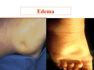
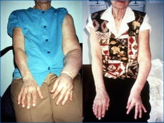
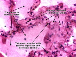
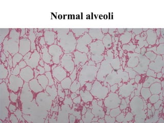
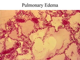
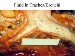
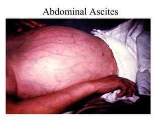
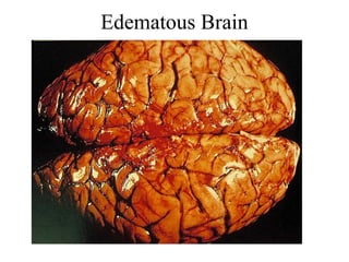
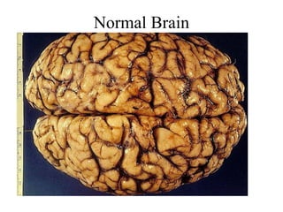
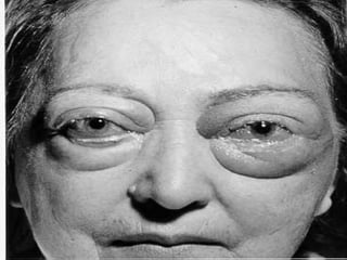
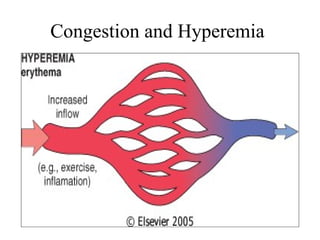
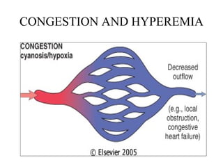
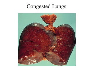
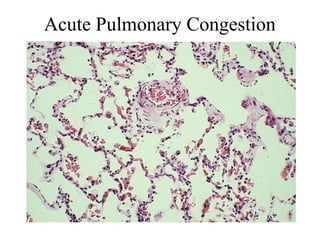
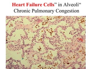
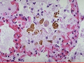
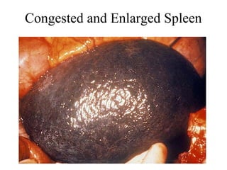
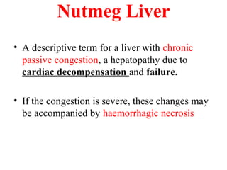
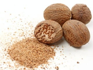

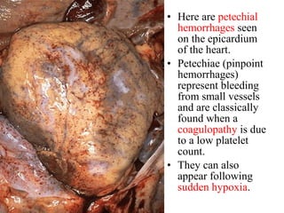
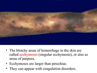
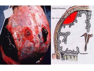
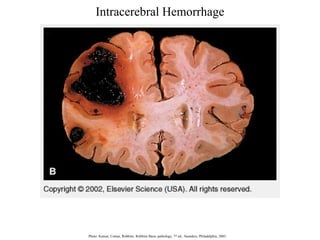
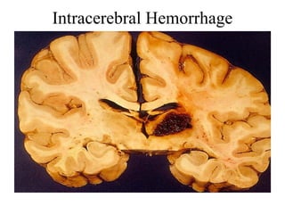
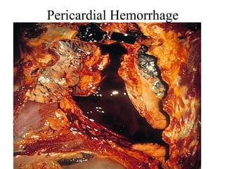
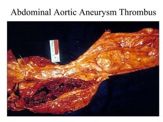
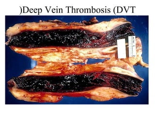
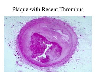
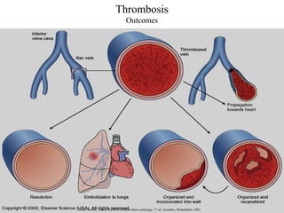
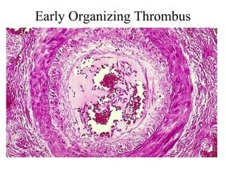
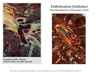
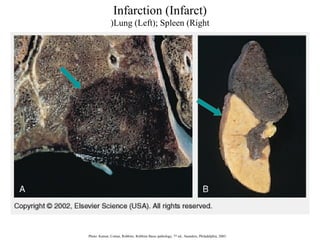
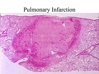
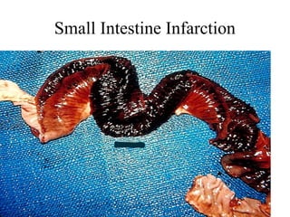
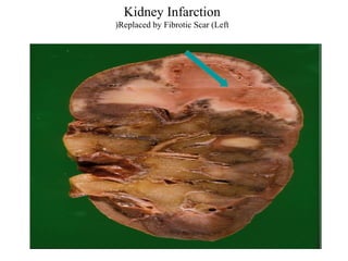
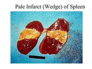


This document discusses hemodynamic disorders and thrombosis. It covers several topics including edema, congestion, hemorrhage, thrombosis, embolism, and infarction. Edema is an accumulation of fluid in tissues and organs, and can occur in the lungs (pulmonary edema), abdomen (ascites), and brain. Congestion and hyperemia involve increased blood volume in organs and tissues, seen in conditions like heart failure and liver disease. Thrombosis is the formation of a clot (thrombus) in a blood vessel. Key factors in thrombosis are described by Virchow's triad. Thrombi can embolize and block vessels in other organs, potentially leading to infarction or tissue death.


![Prac ex'cises 3[1].5](https://cdn.slidesharecdn.com/ss_thumbnails/pracexcises31-5-130213071026-phpapp01-thumbnail.jpg?width=640&height=640&fit=bounds)


![Prac excises 3[1].5](https://cdn.slidesharecdn.com/ss_thumbnails/pracexcises31-150331131154-conversion-gate01-thumbnail.jpg?width=640&height=640&fit=bounds)


![ONFH[AVN HIP] -TRIPLE REGIME -A NOVAL SURGICAL CONCEPT .pptx](https://cdn.slidesharecdn.com/ss_thumbnails/onfhavnhip2026koaconcalicutdrgokuldevdrmashraf-260210064517-213ec005-thumbnail.jpg?width=640&height=640&fit=bounds)


