Edema
General
• Approximately 60% of lean body weight is water
• Two thirds of which is intracellular and one third is extracellular.
• Extracellular is further divided into intravascular , interstitial and trans
vascular
Total body water
Extracellular
1/3
Interstitial
3/4
Intravascular
¼
Trans vascular
Intracellular
2/3
Normal Fluid Circulation
- Fluid movement between the vascular and interstitial spaces is governed mainly by two opposing
forces—the vascular hydrostatic pressure and the colloid osmotic pressure produced by plasma proteins.
Hydrostatic pressure is more at the arteriolar end whereas osmotic pressure is more at the venous end.
Exit of fluid from arteriolar end is balanced by inflow at venular end of microcirculation.
Small residual amount of excess interstitial fluid is drained by lymphatics.
HPc = hydrostatic pressure of the capillary
OPc = osmotic pressure of the capillary
venulararteriolar
Capillary
What is Edema?
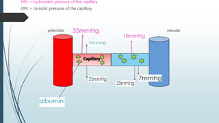
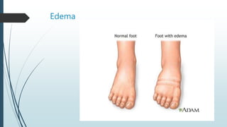
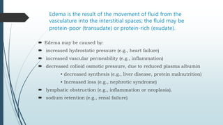
Edema is an abnormal accumulation of fluid in the interstitium,
located beneath the skin and in the cavities of the body.
Edema is a normal response of the body to inflammation or injury.
For example, a twisted ankle , a bee sting , or a skin infection will
all result in edema in the involved area.
In some cases , such as in an infection, this may be beneficial.
Increased fluid from the blood vessels allows more infection-
fighting white blood cells to enter the affected area.
Edema
Edema is the result of the movement of fluid from the
vasculature into the interstitial spaces; the fluid may be
protein-poor (transudate) or protein-rich (exudate).
 Edema may be caused by:
 increased hydrostatic pressure (e.g., heart failure)
 increased vascular permeability (e.g., inflammation)
 decreased colloid osmotic pressure, due to reduced plasma albumin
• decreased synthesis (e.g., liver disease, protein malnutrition)
• Increased loss (e.g., nephrotic syndrome)
 lymphatic obstruction (e.g., inflammation or neoplasia).
 sodium retention (e.g., renal failure)
Treatment
 When possible, treatment involves resolving the underlying cause.
Treatment may also involve positioning the affected body parts to
improve drainage. For example, swelling in feet or ankles may be
reduced by having the person lie down in bed or sit with the feet
propped up on cushions. Intermittent pneumatic compression can be
used to pressurize tissue in a limb, forcing fluids – both blood and
lymph – to flow out of the pressurized area. Treatment of persistent
edemas, such as idiopathic bilateral lymphedema, may also use
manual lymphatic drainage, a low-pressure massage technique
designed to pump lymphatic fluid by a similar mechanism
CONGESTIONHyperemia and
Hyperemia and congestion both refer to an
increase in blood volume within a tissue but
they have different underlying mechanisms.
Hyperemia
is an active process
resulting from arteriolar dilation and increased
blood inflow,
as occurs at sites of inflammation or in exercising
skeletal muscle.
Hyperemic tissues are redder than normal because
of
engorgement with oxygenated blood.
Congestion
is a passive process
resulting from impaired outflow of venous blood from a tissue. It can
occur systemically, as in cardiac failure, or locally as a consequence of
an isolated venous obstruction.
Congested tissues have an abnormal blue-red color (cyanosis)
that stems from the accumulation of deoxygenated hemoglobin in the
affected area.
hyperemia
Active
(arterial)
physiological
-skeletal muscle during
exercise
-glands during secretion
pathological Acute inflammation
Passive
(venal)
Acute
Ex. Venous occlusion by
thrombosis
Portal hypertension
HEMORRAHGE
 Is escape of blood from a blood vessel
bleeding may occur externally or internally into serious cavities
(e.g hemothorax- hemoperitonium- hemopericardium)
or it’s called revealed hemorrhage which starts internal
The reveals it self e.g. vomiting blood
 This form of blood loss can be large and sudden (Acute)
 Or small repeated amount of blood loss over a period of time (Chronic)
Causes of Hemorrhage
 Trauma to vessel wall – penetrating wound.
 Spontaneous Hemorrhage – Rapture of aneurysm, acute anemia, scurvy.
 Inflammatory lesion of vessel wall – Bleeding from chronic peptic ulcer,
polyarteritis nodosa.
 Neoplastic invasion – following vascular invasion in carcinoma (of tongue).
 Vascular Diseases – Atherosclerosis.
 Elevated pressure within vessels – Retinal hemorrhage in systemic hypertension.
Types of hemorrhage
Arterial
Venous
Capillary
Arterial hemorrhage
 Bright red
 Emitted as spurting jet which rises and falls in
time with the pulse.
 Can lead to severe blood loss
 Blood becomes watery if too much fluids
given in protracted bleed.
Venous hemorrhage
 Darker red
 Steady and copious flow
 Color becomes further darker with oxygen desaturation
 Bleed from jugular, femoral, portal veins and esophageal
varices is fatal if not controlled
Capillary hemorrhage
 Bright red
 Rapid and oozing
 Blood loss becomes serious if continues for hours
 Common in haemophiliacs.
Types of hemorrhage
Primary
Reactionary
Secondary
Primary hemorrhage
 Occurs at the time of injury or operation.
 Cause is injury to vessels.
 May be arterial, venous or capillary.
 More common in surgery on malignancies.
Reactionary hemorrhage
 Bleeding within 24 hours ( usually 4-6 hrs ) of primary hemorrhage
 Cause is slipping of ligature, dislodgement of clot or cessation of reflex
vasospasm
 Bleed starts when there is a rise in the arterial or venous pressure.
Secondary hemorrhage
 Occurs after 7-14 days of primary hemorrhage
 Cause is sloughing of vessel due to infection
 1st a warning stain followed by a sudden severe bleed
 Common after hemorrhoids surgery, GI surgery & amputations.
Cutaneous Hemorrhage
1. Petechiae:
A tiny, purplish-red spot on the skin(1 to 2 mm in
diameter). Caused by the leakage of a bit of blood out of a vessel and into
the skin, mucous membranes, or serosal surfaces
 Causes:
 Physical trauma (coughing, sunburn)
 Non-infectious agents
• low platelet counts(thrombocytopenia)
• defective platelet function
• and loss of vascular wall support, as in vitamin C deficiency
 Infectious agents
• Typhus
2. Purpura:
 a small hemorrhage in the skin, mucous membrane, or se
sal surface.
 Slightly larger than petechiae (3-5 mm in diameter)
 The color is first red, gradually darkens to purple, fades to
brownish yellow, and usually disappears in 2-3 weeks
 Causes:
can result from the same disorders that cause petechiae, as
as
1) trauma
2) vascular inflammation (vasculitis)
3) increased vascular fragility.
3. Ecchymoses:
 bluish discoloration of an area of skin or mucous membrane caus
d by the extravasation of blood into the subcutaneous
tissues as a result of trauma to the underlying blood vessels or fra
ility of the vessel walls. Also called bruise.
 (1-2 cm)
 The characteristic color changes of a bruise are due to the
enzymatic conversion of
Hemoglobin (red-blue color)
Bilirubin (blue-green color)
Hemosiderin(golden-brown).
Clinical significance of hemorrhage
 The clinical significance of any particular hemorrhage depends on the volume of blood lost
and the rate of bleeding.
 Loss of 20% of blood volume = little clinical effect due to compensatory mechanism
 Sudden loss of 33% of blood volume may cause death
 50% loss of blood volume over longer period of time (24 hours) may not be fatal
 However chronic blood loss generally produce iron deficiency anaemia, where as acute
hemorrhage may lead immediate consequences like HYPOVOLMIC SHOCK

Edema, congestion and hemorrage

  • 2.
  • 3.
    General • Approximately 60%of lean body weight is water • Two thirds of which is intracellular and one third is extracellular. • Extracellular is further divided into intravascular , interstitial and trans vascular Total body water Extracellular 1/3 Interstitial 3/4 Intravascular ¼ Trans vascular Intracellular 2/3
  • 4.
    Normal Fluid Circulation -Fluid movement between the vascular and interstitial spaces is governed mainly by two opposing forces—the vascular hydrostatic pressure and the colloid osmotic pressure produced by plasma proteins. Hydrostatic pressure is more at the arteriolar end whereas osmotic pressure is more at the venous end. Exit of fluid from arteriolar end is balanced by inflow at venular end of microcirculation. Small residual amount of excess interstitial fluid is drained by lymphatics.
  • 5.
    HPc = hydrostaticpressure of the capillary OPc = osmotic pressure of the capillary venulararteriolar Capillary
  • 6.
    What is Edema? Edemais an abnormal accumulation of fluid in the interstitium, located beneath the skin and in the cavities of the body. Edema is a normal response of the body to inflammation or injury. For example, a twisted ankle , a bee sting , or a skin infection will all result in edema in the involved area. In some cases , such as in an infection, this may be beneficial. Increased fluid from the blood vessels allows more infection- fighting white blood cells to enter the affected area.
  • 7.
  • 8.
    Edema is theresult of the movement of fluid from the vasculature into the interstitial spaces; the fluid may be protein-poor (transudate) or protein-rich (exudate).  Edema may be caused by:  increased hydrostatic pressure (e.g., heart failure)  increased vascular permeability (e.g., inflammation)  decreased colloid osmotic pressure, due to reduced plasma albumin • decreased synthesis (e.g., liver disease, protein malnutrition) • Increased loss (e.g., nephrotic syndrome)  lymphatic obstruction (e.g., inflammation or neoplasia).  sodium retention (e.g., renal failure)
  • 9.
    Treatment  When possible,treatment involves resolving the underlying cause. Treatment may also involve positioning the affected body parts to improve drainage. For example, swelling in feet or ankles may be reduced by having the person lie down in bed or sit with the feet propped up on cushions. Intermittent pneumatic compression can be used to pressurize tissue in a limb, forcing fluids – both blood and lymph – to flow out of the pressurized area. Treatment of persistent edemas, such as idiopathic bilateral lymphedema, may also use manual lymphatic drainage, a low-pressure massage technique designed to pump lymphatic fluid by a similar mechanism
  • 10.
    CONGESTIONHyperemia and Hyperemia andcongestion both refer to an increase in blood volume within a tissue but they have different underlying mechanisms.
  • 11.
    Hyperemia is an activeprocess resulting from arteriolar dilation and increased blood inflow, as occurs at sites of inflammation or in exercising skeletal muscle. Hyperemic tissues are redder than normal because of engorgement with oxygenated blood. Congestion is a passive process resulting from impaired outflow of venous blood from a tissue. It can occur systemically, as in cardiac failure, or locally as a consequence of an isolated venous obstruction. Congested tissues have an abnormal blue-red color (cyanosis) that stems from the accumulation of deoxygenated hemoglobin in the affected area.
  • 12.
    hyperemia Active (arterial) physiological -skeletal muscle during exercise -glandsduring secretion pathological Acute inflammation Passive (venal) Acute Ex. Venous occlusion by thrombosis Portal hypertension
  • 13.
    HEMORRAHGE  Is escapeof blood from a blood vessel bleeding may occur externally or internally into serious cavities (e.g hemothorax- hemoperitonium- hemopericardium) or it’s called revealed hemorrhage which starts internal The reveals it self e.g. vomiting blood  This form of blood loss can be large and sudden (Acute)  Or small repeated amount of blood loss over a period of time (Chronic)
  • 14.
    Causes of Hemorrhage Trauma to vessel wall – penetrating wound.  Spontaneous Hemorrhage – Rapture of aneurysm, acute anemia, scurvy.  Inflammatory lesion of vessel wall – Bleeding from chronic peptic ulcer, polyarteritis nodosa.  Neoplastic invasion – following vascular invasion in carcinoma (of tongue).  Vascular Diseases – Atherosclerosis.  Elevated pressure within vessels – Retinal hemorrhage in systemic hypertension.
  • 15.
  • 16.
    Arterial hemorrhage  Brightred  Emitted as spurting jet which rises and falls in time with the pulse.  Can lead to severe blood loss  Blood becomes watery if too much fluids given in protracted bleed.
  • 17.
    Venous hemorrhage  Darkerred  Steady and copious flow  Color becomes further darker with oxygen desaturation  Bleed from jugular, femoral, portal veins and esophageal varices is fatal if not controlled
  • 18.
    Capillary hemorrhage  Brightred  Rapid and oozing  Blood loss becomes serious if continues for hours  Common in haemophiliacs.
  • 19.
  • 20.
    Primary hemorrhage  Occursat the time of injury or operation.  Cause is injury to vessels.  May be arterial, venous or capillary.  More common in surgery on malignancies.
  • 21.
    Reactionary hemorrhage  Bleedingwithin 24 hours ( usually 4-6 hrs ) of primary hemorrhage  Cause is slipping of ligature, dislodgement of clot or cessation of reflex vasospasm  Bleed starts when there is a rise in the arterial or venous pressure.
  • 22.
    Secondary hemorrhage  Occursafter 7-14 days of primary hemorrhage  Cause is sloughing of vessel due to infection  1st a warning stain followed by a sudden severe bleed  Common after hemorrhoids surgery, GI surgery & amputations.
  • 23.
    Cutaneous Hemorrhage 1. Petechiae: Atiny, purplish-red spot on the skin(1 to 2 mm in diameter). Caused by the leakage of a bit of blood out of a vessel and into the skin, mucous membranes, or serosal surfaces  Causes:  Physical trauma (coughing, sunburn)  Non-infectious agents • low platelet counts(thrombocytopenia) • defective platelet function • and loss of vascular wall support, as in vitamin C deficiency  Infectious agents • Typhus
  • 24.
    2. Purpura:  asmall hemorrhage in the skin, mucous membrane, or se sal surface.  Slightly larger than petechiae (3-5 mm in diameter)  The color is first red, gradually darkens to purple, fades to brownish yellow, and usually disappears in 2-3 weeks  Causes: can result from the same disorders that cause petechiae, as as 1) trauma 2) vascular inflammation (vasculitis) 3) increased vascular fragility.
  • 25.
    3. Ecchymoses:  bluishdiscoloration of an area of skin or mucous membrane caus d by the extravasation of blood into the subcutaneous tissues as a result of trauma to the underlying blood vessels or fra ility of the vessel walls. Also called bruise.  (1-2 cm)  The characteristic color changes of a bruise are due to the enzymatic conversion of Hemoglobin (red-blue color) Bilirubin (blue-green color) Hemosiderin(golden-brown).
  • 27.
    Clinical significance ofhemorrhage  The clinical significance of any particular hemorrhage depends on the volume of blood lost and the rate of bleeding.  Loss of 20% of blood volume = little clinical effect due to compensatory mechanism  Sudden loss of 33% of blood volume may cause death  50% loss of blood volume over longer period of time (24 hours) may not be fatal  However chronic blood loss generally produce iron deficiency anaemia, where as acute hemorrhage may lead immediate consequences like HYPOVOLMIC SHOCK