SlideShare a Scribd company logo
1 of 25
Slide Title Slide Number
Osteology of Elbow Complex Slide 2
Review of Superficial Veins in Arm Slide 3
Arm: Introduction Slide 4
Arm: Anterior Compartment Muscles Slide 5
Arm: Posterior Compartment Muscles Slide 6
Cubital Fossa Slide 7
Brachial Artery Slide 8
Brachial Artery Pulse Slide 9
Bicipital Aponeurosis Slide 10
Musculocutaneous Nerve Slide 11
Ulnar Nerve Slide 12
Ulnar Nerve Lesion at Elbow Slide 13
Ulnar Nerve Lesion at Wrist Slide 14
Median Nerve Slide 15
Slide Title Slide Number
Supracondylar Fractures Slide 16
Radial Head Fracture Slide 17
Median Nerve Lesion at Elbow Slide 18
Radial Nerve Slide 19
Humeral Shaft Fracture Slide 20
Medial Cutaneous Nerve of Arm Slide 21
Elbow Joint Complex Slide 22
Elbow Capsule & Ligaments Slide 23
Nursemaid’s Elbow Slide 24
Olecranon Bursitis (Student’s Bursitis) Slide 25
PowerPoint Handout: Lab 10, Arm, Cubital Fossa, and Elbow Joint
To adequately review the learning objectives covering osteology of the distal humerus, radius, and ulna, view the Lower Limb Osteology and Medical Imaging Guide.
Osteology of Elbow Complex
Review of Superficial Veins in Arm
The cephalic and basilic veins are the main superficial
veins of the upper limb. They originate from the dorsal
venous network on the dorsum of the hand.
• The cephalic vein ascends along the anterolateral
aspect of the forearm and arm. It then follows the
superior border of the pectoralis major muscle to enter
the deltopectoral triangle. It ultimately joins the
axillary vein after passing through the clavipectoral
fascia.
• The basilic vein ascends along the medial forearm and
the arm. In the arm, it passes deep to the brachial
fascia where it courses in close proximity to the
brachial artery and medial cutaneous nerve of the
forearm along its path into the axilla. In the axilla, it
joins with venae comitantes (accompanying axillary
artery) to form the axillary vein.
• The median cubital vein is a branch of the cephalic
vein that passes obliquely across the anterior elbow
region (cubital fossa) to join with the basilic vein.
CLINICAL ANATOMY: Veins in the dorsal venous
network are commonly used for long-term
introduction of fluids.
Arm: Introduction
The arm consists of the humerus, which articulates proximally
and distally.
• Proximally, the humerus articulates with the scapula at the
glenohumeral (shoulder) joint.
• Distally, the humerus articulates with the forearm at the
elbow joint
The fascia of the arm separates the arm’s muscles into two
compartments.
• Anterior: The anterior compartment of the arm contains
primarily flexors of the shoulder and elbow.
• The muscles of the anterior compartment are
innervated by the musculocutaneous nerve (motor
and sensory).
• Blood supply is from the brachial artery.
• Posterior: The posterior compartment of the arm contains
primarily extensors of the shoulder and elbow.
• The muscles of the posterior compartment are
innervated by the radial nerve (motor and sensory).
• Blood supply is from the deep brachial artery.
Glenohumeral (Shoulder) Joint
Elbow Joint
• Humeroulnar Joint
• Humeroradial Joint
• Radioulnar Joint
Arm: Anterior Compartment Muscles
MUSCLE INNERVATION BLOOD SUPPLY ACTION
Biceps brachii Musculocutaneous n Brachial a Flexes and supinates forearm
Coracobrachialis Musculocutaneous n Brachial a Adducts and flexes arm
Brachialis Musculocutaneous n Brachial a Flexes forearm
https://3d4medic.al/2gPqFlNq https://3d4medic.al/PPKGOOIE
FUNCTIONAL ANATOMY: Because the biceps brachii muscle inserts on the radial tuberosity it is capable of supinating the
forearm when the elbow is flexed. In this position, the biceps brachii is the most powerful supinator of the forearm.
MUSCLE INNERVATION BLOOD SUPPLY ACTION
Triceps brachii
• Medial Head: Radial n.
• Lateral Head: Radial n.
• Long Head: Radial n. (in addition to radial,
sometimes innervation by axillary n)
Deep Brachial a Extends forearm
Arm: Posterior Compartment Muscles
https://3d4medic.al/7Z6xn1C2 https://3d4medic.al/4pXmeeTR
The boundaries of the cubital fossa are listed below
• Lateral: brachioradialis muscle
• Medial: pronator teres muscle
• Superior: an imaginary line connecting the
epicondyles of the humerus
• Roof: the bicipital aponeurosis
• Floor: brachialis muscle (proximally) supinator
muscle (distally)
The cubital fossa is a depression on the anterior side of the elbow that is a transition area between the arm and the forearm.
The contents of the cubital fossa are listed below from lateral
to medial.
• Bicipital tendon
• Brachial artery
• Median nerve
• (Radial nerve: Technically , the radial nerve isn’t considered
to be a structure within the cubital fossa, but courses close
by as it passes along the deep surface of the brachioradialis
muscle. In this area, it bifurcates into the superficial and
deep radial nerves.)
https://3d4medic.al/CmWeGhiV
Cubital Fossa
Brachial Artery
The brachial artery is a continuation of the axillary artery after it crosses the tendon of the inferior border of the teres major muscle in the arm.
• The profunda brachii artery (deep artery of the arm or deep brachial artery) is the first branch of the brachial artery in the arm. After branching from the
brachial artery, it courses posteriorly to pass through the triceps hiatus along with the radial nerve to supply the posterior compartment of the arm.
• The brachial artery courses through the arm in the medial bicipital groove along its path to the cubital fossa where it typically terminates by bifurcating into
the radial and ulnar arteries.
• It supplies blood to structures in the anterior compartment of the arm
• At the elbow it gives off several collateral branches that supply the elbow joint. The elbow joint is also supplied by recurrent arteries that branch from
the ulnar and radial arteries.
https://3d4medic.al/4pXmeeTR
https://3d4medic.al/Fhm3HUJE
CLINICAL ANATOMY: In approximately 3% of limbs, the bifurcation of the brachial artery occurs in the arm. When it does, the ulnar artery may course
superficial to the superficial group of flexor muscles, where it can be mistaken for a superficial vein. A quick check for a pulse prevents such a mishap.
https://3d4medic.al/sQCe946a
CLINICAL ANATOMY: The best place to compress the brachial artery to control hemorrhage (bleeding) is in the middle of the arm, in what is known
anatomically as the medial bicipital groove. In the proximal portion of the medial bicipital groove, the brachial artery is coursing between the biceps
brachii and and the triceps brachii. In the distal part of the medial bicipital groove the brachial artery courses between brachialis and biceps brachii.
The brachial pulse can be palpated easily in the proximal medial bicipital groove by pushing the biceps brachii muscle anteriorly to compress the
brachial artery against the humerus.
Brachial Artery Pulse
CLINICAL ANATOMY: The bicipital aponeurosis is located between the more
superficial median cubital vein and the brachial artery, which is deep.
Because of this location, the brachial artery is protected when blood is
drawn from the median cubital vein during venipuncture.
The bicipital aponeurosis (an aponeurosis is a broad, flat tendon) fuses with deep
fascia of the proximal, medial forearm. The biceps brachii tendon crosses the
cubital fossa deep to the bicipital aponeurosis on its path to its attachment on
the radial tuberosity.
Bicipital Aponeurosis
https://3d4medic.al/hszyWLgA
The musculocutaneous nerve pierces coracobrachialis and descends through the arm by passing between the biceps brachii and brachialis muscles. Ultimately,
it emerges from between the biceps brachii, pierces the deep fascia, and continues into the forearm as the lateral antebrachial cutaneous nerve.
• Motor innervation
• Coracobrachialis
• Biceps brachii
• Brachialis
• Sensory innervation via lateral cutaneous nerve of forearm
• Anterior lateral forearm
CLINICAL ANATOMY: The musculocutaneous nerve is rarely injured because of its protected
position beneath the biceps brachii muscle. If it is injured high up in the arm, this results in
weakness of supination (biceps brachii) and forearm flexion (brachialis and biceps brachii)
https://3d4medic.al/idCSLm3n
Musculocutaneous Nerve
In the arm, the Ulnar nerve pierces the medial intermuscular septum to course
on the anterior surface of the medial head of the triceps brachii. It then passes
posterior to the medial epicondyle of the humerus to enter the cubital tunnel,
which is a fibro-osseous passage along the ulnar groove of the medial
epicondyle of the humerus. It doesn’t give off any branches in the arm.
The tunnel is bounded by the following structures:
• Roof: humero-ulnar arcade (arcuate ligament of Osborne)
• Floor: elbow joint capsule
• Medial border: medial epicondyle
• Lateral border: olecranon
It enters the anterior compartment of the forearm by passing between the two
heads of the flexor carpi ulnaris muscle.
https://3d4medic.al/DDP9bPKH
Ulnar Nerve
CLINICAL ANATOMY:
• The most common site of ulnar nerve entrapment is at or near the elbow, especially in the
the cubital tunnel. Cubital tunnel syndrome results from a narrowing of the cubital tunnel,
which is reduced in size when the elbow is flexed. This reduction in size, increases pressure on
the ulnar nerve and results in an ulnar neuropathy. The arcuate ligament of Osborne is thought
to be the point of maximum compression in this condition. Cubital tunnel syndrome is
diagnosed based on signs and symptoms of ulnar neuropathy.
• Common symptoms include:
• Pain and numbness in the elbow
• Paresthesia/numbness on palmar and dorsal aspects of ulnar (medial) half of ring
finger and all of little finger
• More severe symptoms can include:
• Weakened flexion of wrist (hand will deviate towards radial side during flexion)
• Inability to flex MCP joints and extend PIP and DIP joints of ring and little finger
• Inability to abduct and adduct the digits
Ulnar Nerve Lesion at Elbow
The second most likely site for ulnar nerve entrapment is at or near the wrist, especially in the area of the anatomic structure called the ulnar tunnel (canal of
Guyon), which will be studied in detail in the next lab. However, it makes sense to compare an ulnar lesion at the elbow to a lesion at the wrist at this point in
time.
• If an ulnar nerve lesion occurs BELOW the elbow BEYOND the point at which the flexor digitorum profundus receives its ulnar innervation, ulnar claw
hand can occur. Ulnar claw hand describes the position of the hand when at REST (This is an important distinction from Hand of Benediction in which the
examiner is asking the patient to make a fist.)
• The 4th and 5th MP joints are extended due to the unopposed action of extensor digitorum. The extensor digitorum is normally opposed by
the actions of the lumbricals and interossei flexing the MP joints.
• The 4th and 5th IP joints are flexed due to the unopposed action of flexor digitorum profundus. Normally the flexor digitorum is opposed by
the actions of lumbricals extending the IP joints.
For a nice overview of “Ulnar Claw Hand” and how it
differs from “Hand of Benediction,” visit Dr. Nabil
Ebraheim’s YouTube video:
https://www.youtube.com/watch?v=GyqaKGg3HmM
Ulnar Nerve Lesion at Wrist
• On the medial side of the arm, the median nerve courses with brachial artery in a groove between the biceps brachii and brachialis. Along its
path in the arm, it doesn’t give off any branches.
• It enters the cubital fossa by passing inferior to the bicipital aponeurosis. After entering the cubital fossa, it passes between the two heads of
the pronator teres muscle to enter the anterior compartment of the forearm. In the anterior compartment of the forearm, it begins
innervating muscles, beginning with the pronator teres muscle. The anterior interosseous nerve branches from the median nerve soon after
passing between the heads of the pronator teres muscle.
https://3d4medic.al/JzuRZ0kq
https://3d4medic.al/DDP9bPKH
Median Nerve
CLINICAL ANATOMY: Elbow fractures can occur in direct falls on the elbow or when Falling On
an Outstretched Hand (the clinical acronym is FOOSH). In children (esp 5-8-years-old), such falls may
result in a supracondylar fracture of the distal humerus. If displacement of the distal segment occurs at
the fracture site, nerves traversing the elbow and the brachial artery are at risk of injury.
Depending upon which direction the distal segment displaces, different structures are at risk of injury.
• Due to its location deep within the cubital fossa, the median nerve (and/or its anterior
interosseous branch in cases where this nerve leaves the nerve superiorly) is at risk of injury
in supracondylar fracture when the distal segment displaces posterolaterally. In addition, the
brachial artery is at risk in this location.
• A posteromedial displacement of the distal segment, puts the radial nerve at risk of injury.
Supracondylar Fractures
CLINICAL ANATOMY: Radial head and neck fractures are the most common elbow
fractures in adults, comprising approximately 33%–50% of elbow fractures, and
are seen in roughly 20% of elbow trauma cases (Figures 1 and 2). The majority of
radial head fractures result from a fall on an outstretched hand (FOOSH), but may
also result from direct impact on the elbow, a twisting injury, or dislocation. A
fracture of the radial head results in localized pain at the radial head that is worse
during supination. Localized edema due to hemarthrosis is usually present along
with limited passive motion of the elbow. Fracture of the capitellum may occur
simultaneously.
Extra information if you are interested… (Figure 3)
Type I: The fracture consists of a simple split-wedge fragment which may be
displaced or non-displaced. It is also called a chisel fracture.
Type II: In this fracture pattern, part of the head and neck remain intact. The
portion involved in the fracture is tilted and impacted.
Comminution is variable.
Type III: A severely comminuted fracture. The hallmark of this fracture is that no
portion of the head or neck remains in continuity.
Type IV: Fracture of the radial head with dislocation of the elbow joint.
Figure 1 Figure 2
Figure 3
Radial Head Fracture
Median Nerve Lesion at Elbow
• A lesion of the median nerve at the elbow (supracondylar fracture) can result in the following deficits.
• Motor
• Weakened pronation of forearm (loss of pronator quadratus)
• Weakened flexion at wrist along with hand deviation to ulnar side during flexion (loss flexor carpi radialis, but flexor carpi ulnaris still intact)
• Hand of Benediction: Active flexion of the digits (making a fist) results in ONLY digits 4 and 5 flexing at MCP, DIP, and PIP.
• Inability to flex index and middle finger at MCP, PIP and DIP joints (loss of flexor digitorum superficialis, flexor pollicis longus, radial
½ of flexor digitorum profundus)
• Inability to abduct, oppose and flex the thumb (loss of flexor pollicis longus, flexor pollicis brevis, opponens pollicis, abductor pollicis)
• Atrophy of the thenar muscles
• Ape Hand Deformity: loss of thenar muscles causes the thumb to fall into the same plane as the other digits
• Thumb is adducted due to unopposed action of the adductor pollicis muscle.
• Sensory
• Loss of sensation on palmar and dorsal aspects of index, middle, and lateral half of ring fingers
• Loss of sensation to palmar aspect of thumb
Ape Hand
Thenar Atrophy
Thumb in same
plane as fingers
Ask patient to
make a fist.
Hand of Benediction
Video that clearly describes Ulnar Claw Hand, Ape Hand, and Hand of
Benediction: https://www.youtube.com/watch?v=0AAligXLJ1A
The Radial nerve exits the axilla by passing through the triceps hiatus (triangular interval)
along with the deep brachial artery. Both structures pass between the medial and long heads
of the triceps brachii to enter the radial groove of the humerus. After exiting the radial
groove, it courses between the brachialis and brachioradialis muscles. Ultimately it enters the
forearm by crossing the anterior to the capsule of the elbow joint.
• Motor innervation: triceps brachii (all 3 heads) NOTE: The long head of triceps brachii can
also be innervated by the axillary nerve
• Cutaneous innervation: cutaneous branches to posterior arm and forearm.
CLINICAL ANATOMY: Because of their relationship to the posterior surface of the humerus,
the radial nerve and profunda brachii artery are at risk of injury in fractures of the
humeral shaft. Compression of the radial nerve against the humerus for extended periods,
such as when falling asleep with the back of the arm compressed against a solid object
(“Saturday night palsy”, “honeymoon palsy”) or when fitted improperly for crutches (“crutch
palsy”) results in a temporary mononeuropathy characterized by numbness of the back of
the hand and digits, and an inability to extend the wrist and digits.
Radial Nerve
Humeral Shaft Fracture
CLINICAL ANATOMY: Midshaft humerus fractures can involve injury to the radial nerve and is the
most common peripheral nerve injury associated with long bone fractures. The radial nerve is most
likely to be damaged in humerus fractures that have a lateral displacement of the distal fracture
segment because the nerve is tethered to the bone and cannot withstand the forces applied to it as a
result of the displacement.
On physical exam, patients with a radial nerve injury may have the following signs/symptoms.
• wrist drop (loss or weakness of wrist extensors)
• loss or weakness of finger extension
• decreased or absent sensation to the posterior forearm, digits 1 to 3, and the radial half of the
fourth digit.
Up to 90% of patients with a closed humeral fracture with radial nerve injury will have a resolution of
neuropraxia within three to four months following the injury.
Medial Cutaneous nerve of Arm: The medial cutaneous nerve
of the arm branches from the medial cord of the brachial
plexus and supplies the anteromedial skin of the arm.
Medial Cutaneous Nerve of Arm
In comparison to the shoulder, motion at the elbow joint is
relatively restricted. The elbow joint consists of three
articulations.
1. Humeroulnar articulation
• Joint between the trochlea of the humerus and
the trochlear notch of the ulna
• movements: flexion and extension
2. Humeroradial articulation
• Joint between the capitulum of the humerus and
the head of the radius
• Movements: flexion and extension
3. Proximal radioulnar articulation: This articulation is also
enclosed within the elbow joint capsule.
• Joint between the head of the radius and a notch
on the ulna called the radial notch. The radial head
is held in place by the annular ligament, which
forms a collar-like ring around the joint
• Movements: supination and pronation
(i.e., rotation around the long axis of the radius)
The radius and ulna also articulate near the wrist. This joint is
called the distal radio-ulnar joint.
Elbow Joint Complex
Proximal Radioulnar
Joint
Humeroulnar
Joint
Humeroradial
Joint
Proximal Radioulnar Joint
Distal Radioulnar
Joint
Capsule: A single capsule encloses all three joints of the elbow joint complex. The capsule of the elbow joint is loose, which accommodates a large degree of
flexion and extension. However, the capsule is reinforced by ligaments.
Elbow Complex Ligaments
• The ulnar collateral ligament spans from the medial epicondyle of the humerus to the proximal ulna. It functions to provide medial stability to the joint.
• The radial collateral ligament spans from the lateral epicondyle of the humerus to the annular ligament and the olecranon process of the ulna. It
functions to provide lateral stability to the joint.
• The annular ligament is attached to the anterior and posterior surfaces of the radial notch. Along its path from one attachment to the next, it forms a
ring around the radial head.
https://3d4medic.al/FHJS4mq6
https://3d4medic.al/3RVlCiHz
Elbow Capsule & Ligaments
CLINICAL ANATOMY: Subluxation and/or dislocation of the proximal radio-ulnar joint, so-called nursemaid's elbow,
may occur with excessive force with sudden pull upward on a child's arm. It is a common injury in children 5- to 7-
years-old, but with increasing age, the ligaments become stronger and the risk of such injury is reduced.
Nursemaid’s Elbow
CLINICAL ANATOMY: Elbow (olecranon) bursitis involving the
subcutaneous bursa (student’s bursitis) results from repeated excessive
pressure on the elbow. Bursitis of the subtendinous bursa is due to
excessive friction between the tendon and the olecranon. Pain from the
latter form increases substantially during elbow flexion due to
compression of the bursa by the triceps tendon.
Olecranon Bursitis (Student’s Bursitis)

More Related Content

Similar to PowerPointHandout_ArmCubitalFossaElbow.pptx

Blood supply of upper limb by Dr-Ismail Khan
Blood supply of upper limb by Dr-Ismail KhanBlood supply of upper limb by Dr-Ismail Khan
Blood supply of upper limb by Dr-Ismail KhanDr-Ismail Khan
 
musclesofthelimbs-170212083605.pdf
musclesofthelimbs-170212083605.pdfmusclesofthelimbs-170212083605.pdf
musclesofthelimbs-170212083605.pdfssuserbf4af22
 
Blood supply of upper limb
Blood supply of upper limbBlood supply of upper limb
Blood supply of upper limbIdris Siddiqui
 
Lecture 5.1 Axilla and Brachial Plexus Moodle version.pdf
Lecture 5.1 Axilla and Brachial Plexus Moodle version.pdfLecture 5.1 Axilla and Brachial Plexus Moodle version.pdf
Lecture 5.1 Axilla and Brachial Plexus Moodle version.pdfakshayabatti
 
Anatomy pectoral arm02122010
Anatomy pectoral arm02122010Anatomy pectoral arm02122010
Anatomy pectoral arm02122010Lawrence James
 
4 arm region and cubital
4  arm region and cubital4  arm region and cubital
4 arm region and cubitalmehrdad asgari
 
Arm & Cubital Fossa.pptx
Arm & Cubital Fossa.pptxArm & Cubital Fossa.pptx
Arm & Cubital Fossa.pptxShrutiYadav335673
 
Arm and cubital fossa- Dr.K.S.Ravi
Arm and cubital fossa- Dr.K.S.RaviArm and cubital fossa- Dr.K.S.Ravi
Arm and cubital fossa- Dr.K.S.RaviDr.Kumar Satish Ravi
 
Blood supply & innervation of upper limb
Blood supply & innervation of upper limbBlood supply & innervation of upper limb
Blood supply & innervation of upper limbEneutron
 
Axilla and brachial plexus abba
Axilla and brachial plexus abbaAxilla and brachial plexus abba
Axilla and brachial plexus abbakazibwe kazibwe
 
Arterial Supply of Upper Limb
Arterial Supply of Upper LimbArterial Supply of Upper Limb
Arterial Supply of Upper LimbAsish Rajak
 
15 Vascular anatomy of the upper limb2010 (2).ppt
15 Vascular anatomy of the upper limb2010 (2).ppt15 Vascular anatomy of the upper limb2010 (2).ppt
15 Vascular anatomy of the upper limb2010 (2).pptAmanuelIbrahim
 
Anatomy upper limb scapulohumeral 24112010
Anatomy upper limb scapulohumeral  24112010Anatomy upper limb scapulohumeral  24112010
Anatomy upper limb scapulohumeral 24112010Lawrence James
 
MUSCLES of the limbs.ppt
MUSCLES of the limbs.pptMUSCLES of the limbs.ppt
MUSCLES of the limbs.pptDaniyar19
 
Anatomy of the arm (detailed)
Anatomy of the arm (detailed)Anatomy of the arm (detailed)
Anatomy of the arm (detailed)drjumarasekh
 
Axilla of human body
Axilla of human bodyAxilla of human body
Axilla of human bodyCity university
 
Anatomy axilla 25112010
Anatomy axilla 25112010Anatomy axilla 25112010
Anatomy axilla 25112010Lawrence James
 

Similar to PowerPointHandout_ArmCubitalFossaElbow.pptx (20)

Muscles of arm
Muscles of armMuscles of arm
Muscles of arm
 
Blood supply of upper limb by Dr-Ismail Khan
Blood supply of upper limb by Dr-Ismail KhanBlood supply of upper limb by Dr-Ismail Khan
Blood supply of upper limb by Dr-Ismail Khan
 
musclesofthelimbs-170212083605.pdf
musclesofthelimbs-170212083605.pdfmusclesofthelimbs-170212083605.pdf
musclesofthelimbs-170212083605.pdf
 
Blood supply of upper limb
Blood supply of upper limbBlood supply of upper limb
Blood supply of upper limb
 
Lecture 5.1 Axilla and Brachial Plexus Moodle version.pdf
Lecture 5.1 Axilla and Brachial Plexus Moodle version.pdfLecture 5.1 Axilla and Brachial Plexus Moodle version.pdf
Lecture 5.1 Axilla and Brachial Plexus Moodle version.pdf
 
Anatomy pectoral arm02122010
Anatomy pectoral arm02122010Anatomy pectoral arm02122010
Anatomy pectoral arm02122010
 
4 arm region and cubital
4  arm region and cubital4  arm region and cubital
4 arm region and cubital
 
Arm & Cubital Fossa.pptx
Arm & Cubital Fossa.pptxArm & Cubital Fossa.pptx
Arm & Cubital Fossa.pptx
 
Arm and cubital fossa- Dr.K.S.Ravi
Arm and cubital fossa- Dr.K.S.RaviArm and cubital fossa- Dr.K.S.Ravi
Arm and cubital fossa- Dr.K.S.Ravi
 
Blood supply & innervation of upper limb
Blood supply & innervation of upper limbBlood supply & innervation of upper limb
Blood supply & innervation of upper limb
 
Axilla and brachial plexus abba
Axilla and brachial plexus abbaAxilla and brachial plexus abba
Axilla and brachial plexus abba
 
Arterial Supply of Upper Limb
Arterial Supply of Upper LimbArterial Supply of Upper Limb
Arterial Supply of Upper Limb
 
Arm.pptx
Arm.pptxArm.pptx
Arm.pptx
 
15 Vascular anatomy of the upper limb2010 (2).ppt
15 Vascular anatomy of the upper limb2010 (2).ppt15 Vascular anatomy of the upper limb2010 (2).ppt
15 Vascular anatomy of the upper limb2010 (2).ppt
 
Anatomy upper limb scapulohumeral 24112010
Anatomy upper limb scapulohumeral  24112010Anatomy upper limb scapulohumeral  24112010
Anatomy upper limb scapulohumeral 24112010
 
Shoulder region
Shoulder regionShoulder region
Shoulder region
 
MUSCLES of the limbs.ppt
MUSCLES of the limbs.pptMUSCLES of the limbs.ppt
MUSCLES of the limbs.ppt
 
Anatomy of the arm (detailed)
Anatomy of the arm (detailed)Anatomy of the arm (detailed)
Anatomy of the arm (detailed)
 
Axilla of human body
Axilla of human bodyAxilla of human body
Axilla of human body
 
Anatomy axilla 25112010
Anatomy axilla 25112010Anatomy axilla 25112010
Anatomy axilla 25112010
 

More from Shivani Bhardwaj

Classification of diseases in gyneacology and obs.pptx
Classification of diseases in gyneacology and obs.pptxClassification of diseases in gyneacology and obs.pptx
Classification of diseases in gyneacology and obs.pptxShivani Bhardwaj
 
Eating disorders in early infancy and childhood.pptx
Eating disorders in early infancy and childhood.pptxEating disorders in early infancy and childhood.pptx
Eating disorders in early infancy and childhood.pptxShivani Bhardwaj
 
Obg and Gynaecology notes and examination in hospital
Obg and Gynaecology notes and examination in hospitalObg and Gynaecology notes and examination in hospital
Obg and Gynaecology notes and examination in hospitalShivani Bhardwaj
 
final rheumatoid arthritis medical .ppt
final rheumatoid arthritis medical  .pptfinal rheumatoid arthritis medical  .ppt
final rheumatoid arthritis medical .pptShivani Bhardwaj
 
_var_www_html_wp-content_uploads_2021_02_23788_adhd-comorbidity-childhood.pdf
_var_www_html_wp-content_uploads_2021_02_23788_adhd-comorbidity-childhood.pdf_var_www_html_wp-content_uploads_2021_02_23788_adhd-comorbidity-childhood.pdf
_var_www_html_wp-content_uploads_2021_02_23788_adhd-comorbidity-childhood.pdfShivani Bhardwaj
 
Special Education importance in autism .pptx
Special Education importance in autism .pptxSpecial Education importance in autism .pptx
Special Education importance in autism .pptxShivani Bhardwaj
 

More from Shivani Bhardwaj (6)

Classification of diseases in gyneacology and obs.pptx
Classification of diseases in gyneacology and obs.pptxClassification of diseases in gyneacology and obs.pptx
Classification of diseases in gyneacology and obs.pptx
 
Eating disorders in early infancy and childhood.pptx
Eating disorders in early infancy and childhood.pptxEating disorders in early infancy and childhood.pptx
Eating disorders in early infancy and childhood.pptx
 
Obg and Gynaecology notes and examination in hospital
Obg and Gynaecology notes and examination in hospitalObg and Gynaecology notes and examination in hospital
Obg and Gynaecology notes and examination in hospital
 
final rheumatoid arthritis medical .ppt
final rheumatoid arthritis medical  .pptfinal rheumatoid arthritis medical  .ppt
final rheumatoid arthritis medical .ppt
 
_var_www_html_wp-content_uploads_2021_02_23788_adhd-comorbidity-childhood.pdf
_var_www_html_wp-content_uploads_2021_02_23788_adhd-comorbidity-childhood.pdf_var_www_html_wp-content_uploads_2021_02_23788_adhd-comorbidity-childhood.pdf
_var_www_html_wp-content_uploads_2021_02_23788_adhd-comorbidity-childhood.pdf
 
Special Education importance in autism .pptx
Special Education importance in autism .pptxSpecial Education importance in autism .pptx
Special Education importance in autism .pptx
 

Recently uploaded

Gurgaon iffco chowk 🔝 Call Girls Service 🔝 ( 8264348440 ) unlimited hard sex ...
Gurgaon iffco chowk 🔝 Call Girls Service 🔝 ( 8264348440 ) unlimited hard sex ...Gurgaon iffco chowk 🔝 Call Girls Service 🔝 ( 8264348440 ) unlimited hard sex ...
Gurgaon iffco chowk 🔝 Call Girls Service 🔝 ( 8264348440 ) unlimited hard sex ...soniya singh
 
pOOJA sexy Call Girls In Sector 49,9999965857 Young Female Escorts Service In...
pOOJA sexy Call Girls In Sector 49,9999965857 Young Female Escorts Service In...pOOJA sexy Call Girls In Sector 49,9999965857 Young Female Escorts Service In...
pOOJA sexy Call Girls In Sector 49,9999965857 Young Female Escorts Service In...Call Girls Noida
 
VIP Call Girl Sector 25 Gurgaon Just Call Me 9899900591
VIP Call Girl Sector 25 Gurgaon Just Call Me 9899900591VIP Call Girl Sector 25 Gurgaon Just Call Me 9899900591
VIP Call Girl Sector 25 Gurgaon Just Call Me 9899900591adityaroy0215
 
Basics of Anatomy- Language of Anatomy.pptx
Basics of Anatomy- Language of Anatomy.pptxBasics of Anatomy- Language of Anatomy.pptx
Basics of Anatomy- Language of Anatomy.pptxAyush Gupta
 
Russian Call Girls in Chandigarh Ojaswi ❤️🍑 9907093804 👄🫦 Independent Escort ...
Russian Call Girls in Chandigarh Ojaswi ❤️🍑 9907093804 👄🫦 Independent Escort ...Russian Call Girls in Chandigarh Ojaswi ❤️🍑 9907093804 👄🫦 Independent Escort ...
Russian Call Girls in Chandigarh Ojaswi ❤️🍑 9907093804 👄🫦 Independent Escort ...High Profile Call Girls Chandigarh Aarushi
 
No Advance 9053900678 Chandigarh Call Girls , Indian Call Girls For Full Ni...
No Advance 9053900678 Chandigarh  Call Girls , Indian Call Girls  For Full Ni...No Advance 9053900678 Chandigarh  Call Girls , Indian Call Girls  For Full Ni...
No Advance 9053900678 Chandigarh Call Girls , Indian Call Girls For Full Ni...Vip call girls In Chandigarh
 
Escorts in Gurgaon Aarohi 9711199171 VIP Call Girl in Gurgaon Personal Number
Escorts in Gurgaon Aarohi 9711199171 VIP Call Girl in Gurgaon Personal NumberEscorts in Gurgaon Aarohi 9711199171 VIP Call Girl in Gurgaon Personal Number
Escorts in Gurgaon Aarohi 9711199171 VIP Call Girl in Gurgaon Personal NumberCall Girls Service Gurgaon
 
Call Girls in Hyderabad Lavanya 9907093804 Independent Escort Service Hyderabad
Call Girls in Hyderabad Lavanya 9907093804 Independent Escort Service HyderabadCall Girls in Hyderabad Lavanya 9907093804 Independent Escort Service Hyderabad
Call Girls in Hyderabad Lavanya 9907093804 Independent Escort Service Hyderabaddelhimodelshub1
 
Russian Call Girls in Goa Samaira 7001305949 Independent Escort Service Goa
Russian Call Girls in Goa Samaira 7001305949 Independent Escort Service GoaRussian Call Girls in Goa Samaira 7001305949 Independent Escort Service Goa
Russian Call Girls in Goa Samaira 7001305949 Independent Escort Service Goanarwatsonia7
 
Call Girls Hyderabad Kirti 9907093804 Independent Escort Service Hyderabad
Call Girls Hyderabad Kirti 9907093804 Independent Escort Service HyderabadCall Girls Hyderabad Kirti 9907093804 Independent Escort Service Hyderabad
Call Girls Hyderabad Kirti 9907093804 Independent Escort Service Hyderabaddelhimodelshub1
 
Gurgaon Sector 90 Call Girls ( 9873940964 ) Book Hot And Sexy Girls In A Few ...
Gurgaon Sector 90 Call Girls ( 9873940964 ) Book Hot And Sexy Girls In A Few ...Gurgaon Sector 90 Call Girls ( 9873940964 ) Book Hot And Sexy Girls In A Few ...
Gurgaon Sector 90 Call Girls ( 9873940964 ) Book Hot And Sexy Girls In A Few ...ggsonu500
 
Call Girl Hyderabad Madhuri 9907093804 Independent Escort Service Hyderabad
Call Girl Hyderabad Madhuri 9907093804 Independent Escort Service HyderabadCall Girl Hyderabad Madhuri 9907093804 Independent Escort Service Hyderabad
Call Girl Hyderabad Madhuri 9907093804 Independent Escort Service Hyderabaddelhimodelshub1
 
Call Girl Gurgaon Saloni 9711199012 Independent Escort Service Gurgaon
Call Girl Gurgaon Saloni 9711199012 Independent Escort Service GurgaonCall Girl Gurgaon Saloni 9711199012 Independent Escort Service Gurgaon
Call Girl Gurgaon Saloni 9711199012 Independent Escort Service GurgaonCall Girls Service Gurgaon
 
VIP Call Girls Sector 67 Gurgaon Just Call Me 9711199012
VIP Call Girls Sector 67 Gurgaon Just Call Me 9711199012VIP Call Girls Sector 67 Gurgaon Just Call Me 9711199012
VIP Call Girls Sector 67 Gurgaon Just Call Me 9711199012Call Girls Service Gurgaon
 

Recently uploaded (20)

College Call Girls Dehradun Kavya 🔝 7001305949 🔝 📍 Independent Escort Service...
College Call Girls Dehradun Kavya 🔝 7001305949 🔝 📍 Independent Escort Service...College Call Girls Dehradun Kavya 🔝 7001305949 🔝 📍 Independent Escort Service...
College Call Girls Dehradun Kavya 🔝 7001305949 🔝 📍 Independent Escort Service...
 
Gurgaon iffco chowk 🔝 Call Girls Service 🔝 ( 8264348440 ) unlimited hard sex ...
Gurgaon iffco chowk 🔝 Call Girls Service 🔝 ( 8264348440 ) unlimited hard sex ...Gurgaon iffco chowk 🔝 Call Girls Service 🔝 ( 8264348440 ) unlimited hard sex ...
Gurgaon iffco chowk 🔝 Call Girls Service 🔝 ( 8264348440 ) unlimited hard sex ...
 
pOOJA sexy Call Girls In Sector 49,9999965857 Young Female Escorts Service In...
pOOJA sexy Call Girls In Sector 49,9999965857 Young Female Escorts Service In...pOOJA sexy Call Girls In Sector 49,9999965857 Young Female Escorts Service In...
pOOJA sexy Call Girls In Sector 49,9999965857 Young Female Escorts Service In...
 
VIP Call Girl Sector 25 Gurgaon Just Call Me 9899900591
VIP Call Girl Sector 25 Gurgaon Just Call Me 9899900591VIP Call Girl Sector 25 Gurgaon Just Call Me 9899900591
VIP Call Girl Sector 25 Gurgaon Just Call Me 9899900591
 
Russian Call Girls in Dehradun Komal 🔝 7001305949 🔝 📍 Independent Escort Serv...
Russian Call Girls in Dehradun Komal 🔝 7001305949 🔝 📍 Independent Escort Serv...Russian Call Girls in Dehradun Komal 🔝 7001305949 🔝 📍 Independent Escort Serv...
Russian Call Girls in Dehradun Komal 🔝 7001305949 🔝 📍 Independent Escort Serv...
 
Basics of Anatomy- Language of Anatomy.pptx
Basics of Anatomy- Language of Anatomy.pptxBasics of Anatomy- Language of Anatomy.pptx
Basics of Anatomy- Language of Anatomy.pptx
 
Russian Call Girls in Chandigarh Ojaswi ❤️🍑 9907093804 👄🫦 Independent Escort ...
Russian Call Girls in Chandigarh Ojaswi ❤️🍑 9907093804 👄🫦 Independent Escort ...Russian Call Girls in Chandigarh Ojaswi ❤️🍑 9907093804 👄🫦 Independent Escort ...
Russian Call Girls in Chandigarh Ojaswi ❤️🍑 9907093804 👄🫦 Independent Escort ...
 
No Advance 9053900678 Chandigarh Call Girls , Indian Call Girls For Full Ni...
No Advance 9053900678 Chandigarh  Call Girls , Indian Call Girls  For Full Ni...No Advance 9053900678 Chandigarh  Call Girls , Indian Call Girls  For Full Ni...
No Advance 9053900678 Chandigarh Call Girls , Indian Call Girls For Full Ni...
 
Call Girl Lucknow Gauri 🔝 8923113531 🔝 🎶 Independent Escort Service Lucknow
Call Girl Lucknow Gauri 🔝 8923113531  🔝 🎶 Independent Escort Service LucknowCall Girl Lucknow Gauri 🔝 8923113531  🔝 🎶 Independent Escort Service Lucknow
Call Girl Lucknow Gauri 🔝 8923113531 🔝 🎶 Independent Escort Service Lucknow
 
Escorts in Gurgaon Aarohi 9711199171 VIP Call Girl in Gurgaon Personal Number
Escorts in Gurgaon Aarohi 9711199171 VIP Call Girl in Gurgaon Personal NumberEscorts in Gurgaon Aarohi 9711199171 VIP Call Girl in Gurgaon Personal Number
Escorts in Gurgaon Aarohi 9711199171 VIP Call Girl in Gurgaon Personal Number
 
Call Girls in Hyderabad Lavanya 9907093804 Independent Escort Service Hyderabad
Call Girls in Hyderabad Lavanya 9907093804 Independent Escort Service HyderabadCall Girls in Hyderabad Lavanya 9907093804 Independent Escort Service Hyderabad
Call Girls in Hyderabad Lavanya 9907093804 Independent Escort Service Hyderabad
 
Russian Call Girls in Goa Samaira 7001305949 Independent Escort Service Goa
Russian Call Girls in Goa Samaira 7001305949 Independent Escort Service GoaRussian Call Girls in Goa Samaira 7001305949 Independent Escort Service Goa
Russian Call Girls in Goa Samaira 7001305949 Independent Escort Service Goa
 
Call Girls Hyderabad Kirti 9907093804 Independent Escort Service Hyderabad
Call Girls Hyderabad Kirti 9907093804 Independent Escort Service HyderabadCall Girls Hyderabad Kirti 9907093804 Independent Escort Service Hyderabad
Call Girls Hyderabad Kirti 9907093804 Independent Escort Service Hyderabad
 
Russian Call Girls South Delhi 9711199171 discount on your booking
Russian Call Girls South Delhi 9711199171 discount on your bookingRussian Call Girls South Delhi 9711199171 discount on your booking
Russian Call Girls South Delhi 9711199171 discount on your booking
 
Gurgaon Sector 90 Call Girls ( 9873940964 ) Book Hot And Sexy Girls In A Few ...
Gurgaon Sector 90 Call Girls ( 9873940964 ) Book Hot And Sexy Girls In A Few ...Gurgaon Sector 90 Call Girls ( 9873940964 ) Book Hot And Sexy Girls In A Few ...
Gurgaon Sector 90 Call Girls ( 9873940964 ) Book Hot And Sexy Girls In A Few ...
 
Call Girl Hyderabad Madhuri 9907093804 Independent Escort Service Hyderabad
Call Girl Hyderabad Madhuri 9907093804 Independent Escort Service HyderabadCall Girl Hyderabad Madhuri 9907093804 Independent Escort Service Hyderabad
Call Girl Hyderabad Madhuri 9907093804 Independent Escort Service Hyderabad
 
Call Girl Gurgaon Saloni 9711199012 Independent Escort Service Gurgaon
Call Girl Gurgaon Saloni 9711199012 Independent Escort Service GurgaonCall Girl Gurgaon Saloni 9711199012 Independent Escort Service Gurgaon
Call Girl Gurgaon Saloni 9711199012 Independent Escort Service Gurgaon
 
Model Call Girl in Subhash Nagar Delhi reach out to us at 🔝9953056974🔝
Model Call Girl in Subhash Nagar Delhi reach out to us at 🔝9953056974🔝Model Call Girl in Subhash Nagar Delhi reach out to us at 🔝9953056974🔝
Model Call Girl in Subhash Nagar Delhi reach out to us at 🔝9953056974🔝
 
VIP Call Girls Sector 67 Gurgaon Just Call Me 9711199012
VIP Call Girls Sector 67 Gurgaon Just Call Me 9711199012VIP Call Girls Sector 67 Gurgaon Just Call Me 9711199012
VIP Call Girls Sector 67 Gurgaon Just Call Me 9711199012
 
Call Girl Guwahati Aashi 👉 7001305949 👈 🔝 Independent Escort Service Guwahati
Call Girl Guwahati Aashi 👉 7001305949 👈 🔝 Independent Escort Service GuwahatiCall Girl Guwahati Aashi 👉 7001305949 👈 🔝 Independent Escort Service Guwahati
Call Girl Guwahati Aashi 👉 7001305949 👈 🔝 Independent Escort Service Guwahati
 

PowerPointHandout_ArmCubitalFossaElbow.pptx

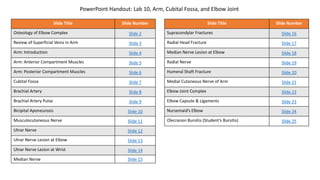
  • 1. Slide Title Slide Number Osteology of Elbow Complex Slide 2 Review of Superficial Veins in Arm Slide 3 Arm: Introduction Slide 4 Arm: Anterior Compartment Muscles Slide 5 Arm: Posterior Compartment Muscles Slide 6 Cubital Fossa Slide 7 Brachial Artery Slide 8 Brachial Artery Pulse Slide 9 Bicipital Aponeurosis Slide 10 Musculocutaneous Nerve Slide 11 Ulnar Nerve Slide 12 Ulnar Nerve Lesion at Elbow Slide 13 Ulnar Nerve Lesion at Wrist Slide 14 Median Nerve Slide 15 Slide Title Slide Number Supracondylar Fractures Slide 16 Radial Head Fracture Slide 17 Median Nerve Lesion at Elbow Slide 18 Radial Nerve Slide 19 Humeral Shaft Fracture Slide 20 Medial Cutaneous Nerve of Arm Slide 21 Elbow Joint Complex Slide 22 Elbow Capsule & Ligaments Slide 23 Nursemaid’s Elbow Slide 24 Olecranon Bursitis (Student’s Bursitis) Slide 25 PowerPoint Handout: Lab 10, Arm, Cubital Fossa, and Elbow Joint
  • 2. To adequately review the learning objectives covering osteology of the distal humerus, radius, and ulna, view the Lower Limb Osteology and Medical Imaging Guide. Osteology of Elbow Complex
  • 3. Review of Superficial Veins in Arm The cephalic and basilic veins are the main superficial veins of the upper limb. They originate from the dorsal venous network on the dorsum of the hand. • The cephalic vein ascends along the anterolateral aspect of the forearm and arm. It then follows the superior border of the pectoralis major muscle to enter the deltopectoral triangle. It ultimately joins the axillary vein after passing through the clavipectoral fascia. • The basilic vein ascends along the medial forearm and the arm. In the arm, it passes deep to the brachial fascia where it courses in close proximity to the brachial artery and medial cutaneous nerve of the forearm along its path into the axilla. In the axilla, it joins with venae comitantes (accompanying axillary artery) to form the axillary vein. • The median cubital vein is a branch of the cephalic vein that passes obliquely across the anterior elbow region (cubital fossa) to join with the basilic vein. CLINICAL ANATOMY: Veins in the dorsal venous network are commonly used for long-term introduction of fluids.
  • 4. Arm: Introduction The arm consists of the humerus, which articulates proximally and distally. • Proximally, the humerus articulates with the scapula at the glenohumeral (shoulder) joint. • Distally, the humerus articulates with the forearm at the elbow joint The fascia of the arm separates the arm’s muscles into two compartments. • Anterior: The anterior compartment of the arm contains primarily flexors of the shoulder and elbow. • The muscles of the anterior compartment are innervated by the musculocutaneous nerve (motor and sensory). • Blood supply is from the brachial artery. • Posterior: The posterior compartment of the arm contains primarily extensors of the shoulder and elbow. • The muscles of the posterior compartment are innervated by the radial nerve (motor and sensory). • Blood supply is from the deep brachial artery. Glenohumeral (Shoulder) Joint Elbow Joint • Humeroulnar Joint • Humeroradial Joint • Radioulnar Joint
  • 5. Arm: Anterior Compartment Muscles MUSCLE INNERVATION BLOOD SUPPLY ACTION Biceps brachii Musculocutaneous n Brachial a Flexes and supinates forearm Coracobrachialis Musculocutaneous n Brachial a Adducts and flexes arm Brachialis Musculocutaneous n Brachial a Flexes forearm https://3d4medic.al/2gPqFlNq https://3d4medic.al/PPKGOOIE FUNCTIONAL ANATOMY: Because the biceps brachii muscle inserts on the radial tuberosity it is capable of supinating the forearm when the elbow is flexed. In this position, the biceps brachii is the most powerful supinator of the forearm.
  • 6. MUSCLE INNERVATION BLOOD SUPPLY ACTION Triceps brachii • Medial Head: Radial n. • Lateral Head: Radial n. • Long Head: Radial n. (in addition to radial, sometimes innervation by axillary n) Deep Brachial a Extends forearm Arm: Posterior Compartment Muscles https://3d4medic.al/7Z6xn1C2 https://3d4medic.al/4pXmeeTR
  • 7. The boundaries of the cubital fossa are listed below • Lateral: brachioradialis muscle • Medial: pronator teres muscle • Superior: an imaginary line connecting the epicondyles of the humerus • Roof: the bicipital aponeurosis • Floor: brachialis muscle (proximally) supinator muscle (distally) The cubital fossa is a depression on the anterior side of the elbow that is a transition area between the arm and the forearm. The contents of the cubital fossa are listed below from lateral to medial. • Bicipital tendon • Brachial artery • Median nerve • (Radial nerve: Technically , the radial nerve isn’t considered to be a structure within the cubital fossa, but courses close by as it passes along the deep surface of the brachioradialis muscle. In this area, it bifurcates into the superficial and deep radial nerves.) https://3d4medic.al/CmWeGhiV Cubital Fossa
  • 8. Brachial Artery The brachial artery is a continuation of the axillary artery after it crosses the tendon of the inferior border of the teres major muscle in the arm. • The profunda brachii artery (deep artery of the arm or deep brachial artery) is the first branch of the brachial artery in the arm. After branching from the brachial artery, it courses posteriorly to pass through the triceps hiatus along with the radial nerve to supply the posterior compartment of the arm. • The brachial artery courses through the arm in the medial bicipital groove along its path to the cubital fossa where it typically terminates by bifurcating into the radial and ulnar arteries. • It supplies blood to structures in the anterior compartment of the arm • At the elbow it gives off several collateral branches that supply the elbow joint. The elbow joint is also supplied by recurrent arteries that branch from the ulnar and radial arteries. https://3d4medic.al/4pXmeeTR https://3d4medic.al/Fhm3HUJE CLINICAL ANATOMY: In approximately 3% of limbs, the bifurcation of the brachial artery occurs in the arm. When it does, the ulnar artery may course superficial to the superficial group of flexor muscles, where it can be mistaken for a superficial vein. A quick check for a pulse prevents such a mishap. https://3d4medic.al/sQCe946a
  • 9. CLINICAL ANATOMY: The best place to compress the brachial artery to control hemorrhage (bleeding) is in the middle of the arm, in what is known anatomically as the medial bicipital groove. In the proximal portion of the medial bicipital groove, the brachial artery is coursing between the biceps brachii and and the triceps brachii. In the distal part of the medial bicipital groove the brachial artery courses between brachialis and biceps brachii. The brachial pulse can be palpated easily in the proximal medial bicipital groove by pushing the biceps brachii muscle anteriorly to compress the brachial artery against the humerus. Brachial Artery Pulse
  • 10. CLINICAL ANATOMY: The bicipital aponeurosis is located between the more superficial median cubital vein and the brachial artery, which is deep. Because of this location, the brachial artery is protected when blood is drawn from the median cubital vein during venipuncture. The bicipital aponeurosis (an aponeurosis is a broad, flat tendon) fuses with deep fascia of the proximal, medial forearm. The biceps brachii tendon crosses the cubital fossa deep to the bicipital aponeurosis on its path to its attachment on the radial tuberosity. Bicipital Aponeurosis https://3d4medic.al/hszyWLgA
  • 11. The musculocutaneous nerve pierces coracobrachialis and descends through the arm by passing between the biceps brachii and brachialis muscles. Ultimately, it emerges from between the biceps brachii, pierces the deep fascia, and continues into the forearm as the lateral antebrachial cutaneous nerve. • Motor innervation • Coracobrachialis • Biceps brachii • Brachialis • Sensory innervation via lateral cutaneous nerve of forearm • Anterior lateral forearm CLINICAL ANATOMY: The musculocutaneous nerve is rarely injured because of its protected position beneath the biceps brachii muscle. If it is injured high up in the arm, this results in weakness of supination (biceps brachii) and forearm flexion (brachialis and biceps brachii) https://3d4medic.al/idCSLm3n Musculocutaneous Nerve
  • 12. In the arm, the Ulnar nerve pierces the medial intermuscular septum to course on the anterior surface of the medial head of the triceps brachii. It then passes posterior to the medial epicondyle of the humerus to enter the cubital tunnel, which is a fibro-osseous passage along the ulnar groove of the medial epicondyle of the humerus. It doesn’t give off any branches in the arm. The tunnel is bounded by the following structures: • Roof: humero-ulnar arcade (arcuate ligament of Osborne) • Floor: elbow joint capsule • Medial border: medial epicondyle • Lateral border: olecranon It enters the anterior compartment of the forearm by passing between the two heads of the flexor carpi ulnaris muscle. https://3d4medic.al/DDP9bPKH Ulnar Nerve
  • 13. CLINICAL ANATOMY: • The most common site of ulnar nerve entrapment is at or near the elbow, especially in the the cubital tunnel. Cubital tunnel syndrome results from a narrowing of the cubital tunnel, which is reduced in size when the elbow is flexed. This reduction in size, increases pressure on the ulnar nerve and results in an ulnar neuropathy. The arcuate ligament of Osborne is thought to be the point of maximum compression in this condition. Cubital tunnel syndrome is diagnosed based on signs and symptoms of ulnar neuropathy. • Common symptoms include: • Pain and numbness in the elbow • Paresthesia/numbness on palmar and dorsal aspects of ulnar (medial) half of ring finger and all of little finger • More severe symptoms can include: • Weakened flexion of wrist (hand will deviate towards radial side during flexion) • Inability to flex MCP joints and extend PIP and DIP joints of ring and little finger • Inability to abduct and adduct the digits Ulnar Nerve Lesion at Elbow
  • 14. The second most likely site for ulnar nerve entrapment is at or near the wrist, especially in the area of the anatomic structure called the ulnar tunnel (canal of Guyon), which will be studied in detail in the next lab. However, it makes sense to compare an ulnar lesion at the elbow to a lesion at the wrist at this point in time. • If an ulnar nerve lesion occurs BELOW the elbow BEYOND the point at which the flexor digitorum profundus receives its ulnar innervation, ulnar claw hand can occur. Ulnar claw hand describes the position of the hand when at REST (This is an important distinction from Hand of Benediction in which the examiner is asking the patient to make a fist.) • The 4th and 5th MP joints are extended due to the unopposed action of extensor digitorum. The extensor digitorum is normally opposed by the actions of the lumbricals and interossei flexing the MP joints. • The 4th and 5th IP joints are flexed due to the unopposed action of flexor digitorum profundus. Normally the flexor digitorum is opposed by the actions of lumbricals extending the IP joints. For a nice overview of “Ulnar Claw Hand” and how it differs from “Hand of Benediction,” visit Dr. Nabil Ebraheim’s YouTube video: https://www.youtube.com/watch?v=GyqaKGg3HmM Ulnar Nerve Lesion at Wrist
  • 15. • On the medial side of the arm, the median nerve courses with brachial artery in a groove between the biceps brachii and brachialis. Along its path in the arm, it doesn’t give off any branches. • It enters the cubital fossa by passing inferior to the bicipital aponeurosis. After entering the cubital fossa, it passes between the two heads of the pronator teres muscle to enter the anterior compartment of the forearm. In the anterior compartment of the forearm, it begins innervating muscles, beginning with the pronator teres muscle. The anterior interosseous nerve branches from the median nerve soon after passing between the heads of the pronator teres muscle. https://3d4medic.al/JzuRZ0kq https://3d4medic.al/DDP9bPKH Median Nerve
  • 16. CLINICAL ANATOMY: Elbow fractures can occur in direct falls on the elbow or when Falling On an Outstretched Hand (the clinical acronym is FOOSH). In children (esp 5-8-years-old), such falls may result in a supracondylar fracture of the distal humerus. If displacement of the distal segment occurs at the fracture site, nerves traversing the elbow and the brachial artery are at risk of injury. Depending upon which direction the distal segment displaces, different structures are at risk of injury. • Due to its location deep within the cubital fossa, the median nerve (and/or its anterior interosseous branch in cases where this nerve leaves the nerve superiorly) is at risk of injury in supracondylar fracture when the distal segment displaces posterolaterally. In addition, the brachial artery is at risk in this location. • A posteromedial displacement of the distal segment, puts the radial nerve at risk of injury. Supracondylar Fractures
  • 17. CLINICAL ANATOMY: Radial head and neck fractures are the most common elbow fractures in adults, comprising approximately 33%–50% of elbow fractures, and are seen in roughly 20% of elbow trauma cases (Figures 1 and 2). The majority of radial head fractures result from a fall on an outstretched hand (FOOSH), but may also result from direct impact on the elbow, a twisting injury, or dislocation. A fracture of the radial head results in localized pain at the radial head that is worse during supination. Localized edema due to hemarthrosis is usually present along with limited passive motion of the elbow. Fracture of the capitellum may occur simultaneously. Extra information if you are interested… (Figure 3) Type I: The fracture consists of a simple split-wedge fragment which may be displaced or non-displaced. It is also called a chisel fracture. Type II: In this fracture pattern, part of the head and neck remain intact. The portion involved in the fracture is tilted and impacted. Comminution is variable. Type III: A severely comminuted fracture. The hallmark of this fracture is that no portion of the head or neck remains in continuity. Type IV: Fracture of the radial head with dislocation of the elbow joint. Figure 1 Figure 2 Figure 3 Radial Head Fracture
  • 18. Median Nerve Lesion at Elbow • A lesion of the median nerve at the elbow (supracondylar fracture) can result in the following deficits. • Motor • Weakened pronation of forearm (loss of pronator quadratus) • Weakened flexion at wrist along with hand deviation to ulnar side during flexion (loss flexor carpi radialis, but flexor carpi ulnaris still intact) • Hand of Benediction: Active flexion of the digits (making a fist) results in ONLY digits 4 and 5 flexing at MCP, DIP, and PIP. • Inability to flex index and middle finger at MCP, PIP and DIP joints (loss of flexor digitorum superficialis, flexor pollicis longus, radial ½ of flexor digitorum profundus) • Inability to abduct, oppose and flex the thumb (loss of flexor pollicis longus, flexor pollicis brevis, opponens pollicis, abductor pollicis) • Atrophy of the thenar muscles • Ape Hand Deformity: loss of thenar muscles causes the thumb to fall into the same plane as the other digits • Thumb is adducted due to unopposed action of the adductor pollicis muscle. • Sensory • Loss of sensation on palmar and dorsal aspects of index, middle, and lateral half of ring fingers • Loss of sensation to palmar aspect of thumb Ape Hand Thenar Atrophy Thumb in same plane as fingers Ask patient to make a fist. Hand of Benediction Video that clearly describes Ulnar Claw Hand, Ape Hand, and Hand of Benediction: https://www.youtube.com/watch?v=0AAligXLJ1A
  • 19. The Radial nerve exits the axilla by passing through the triceps hiatus (triangular interval) along with the deep brachial artery. Both structures pass between the medial and long heads of the triceps brachii to enter the radial groove of the humerus. After exiting the radial groove, it courses between the brachialis and brachioradialis muscles. Ultimately it enters the forearm by crossing the anterior to the capsule of the elbow joint. • Motor innervation: triceps brachii (all 3 heads) NOTE: The long head of triceps brachii can also be innervated by the axillary nerve • Cutaneous innervation: cutaneous branches to posterior arm and forearm. CLINICAL ANATOMY: Because of their relationship to the posterior surface of the humerus, the radial nerve and profunda brachii artery are at risk of injury in fractures of the humeral shaft. Compression of the radial nerve against the humerus for extended periods, such as when falling asleep with the back of the arm compressed against a solid object (“Saturday night palsy”, “honeymoon palsy”) or when fitted improperly for crutches (“crutch palsy”) results in a temporary mononeuropathy characterized by numbness of the back of the hand and digits, and an inability to extend the wrist and digits. Radial Nerve
  • 20. Humeral Shaft Fracture CLINICAL ANATOMY: Midshaft humerus fractures can involve injury to the radial nerve and is the most common peripheral nerve injury associated with long bone fractures. The radial nerve is most likely to be damaged in humerus fractures that have a lateral displacement of the distal fracture segment because the nerve is tethered to the bone and cannot withstand the forces applied to it as a result of the displacement. On physical exam, patients with a radial nerve injury may have the following signs/symptoms. • wrist drop (loss or weakness of wrist extensors) • loss or weakness of finger extension • decreased or absent sensation to the posterior forearm, digits 1 to 3, and the radial half of the fourth digit. Up to 90% of patients with a closed humeral fracture with radial nerve injury will have a resolution of neuropraxia within three to four months following the injury.
  • 21. Medial Cutaneous nerve of Arm: The medial cutaneous nerve of the arm branches from the medial cord of the brachial plexus and supplies the anteromedial skin of the arm. Medial Cutaneous Nerve of Arm
  • 22. In comparison to the shoulder, motion at the elbow joint is relatively restricted. The elbow joint consists of three articulations. 1. Humeroulnar articulation • Joint between the trochlea of the humerus and the trochlear notch of the ulna • movements: flexion and extension 2. Humeroradial articulation • Joint between the capitulum of the humerus and the head of the radius • Movements: flexion and extension 3. Proximal radioulnar articulation: This articulation is also enclosed within the elbow joint capsule. • Joint between the head of the radius and a notch on the ulna called the radial notch. The radial head is held in place by the annular ligament, which forms a collar-like ring around the joint • Movements: supination and pronation (i.e., rotation around the long axis of the radius) The radius and ulna also articulate near the wrist. This joint is called the distal radio-ulnar joint. Elbow Joint Complex Proximal Radioulnar Joint Humeroulnar Joint Humeroradial Joint Proximal Radioulnar Joint Distal Radioulnar Joint
  • 23. Capsule: A single capsule encloses all three joints of the elbow joint complex. The capsule of the elbow joint is loose, which accommodates a large degree of flexion and extension. However, the capsule is reinforced by ligaments. Elbow Complex Ligaments • The ulnar collateral ligament spans from the medial epicondyle of the humerus to the proximal ulna. It functions to provide medial stability to the joint. • The radial collateral ligament spans from the lateral epicondyle of the humerus to the annular ligament and the olecranon process of the ulna. It functions to provide lateral stability to the joint. • The annular ligament is attached to the anterior and posterior surfaces of the radial notch. Along its path from one attachment to the next, it forms a ring around the radial head. https://3d4medic.al/FHJS4mq6 https://3d4medic.al/3RVlCiHz Elbow Capsule & Ligaments
  • 24. CLINICAL ANATOMY: Subluxation and/or dislocation of the proximal radio-ulnar joint, so-called nursemaid's elbow, may occur with excessive force with sudden pull upward on a child's arm. It is a common injury in children 5- to 7- years-old, but with increasing age, the ligaments become stronger and the risk of such injury is reduced. Nursemaid’s Elbow
  • 25. CLINICAL ANATOMY: Elbow (olecranon) bursitis involving the subcutaneous bursa (student’s bursitis) results from repeated excessive pressure on the elbow. Bursitis of the subtendinous bursa is due to excessive friction between the tendon and the olecranon. Pain from the latter form increases substantially during elbow flexion due to compression of the bursa by the triceps tendon. Olecranon Bursitis (Student’s Bursitis)