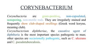
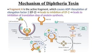
GRAM POSITIVE BACILLI
CORYNEBACTERIUM DIPHTHERIAE
Prabhakar Singh Patel
(Assistant Lecturer)
SINPS, Lucknow
All Paramedical, Nursing and MBBS Courses
CORYNEBACTERIUM
Corynebacteria are gram-positive, non-capsulated,
nonsporing, non-motile rods. They are irregularly stained and
frequently show club-shaped swellings (Greek word koryne,
meaning club).
Corynebacterium diphtheriae, the causative agent of
diphtheria is the most important species pathogenic to man;
other species are occasionally pathogenic, such as C. ulcerans
and C. pseudotuberculosis.
CORYNEBACTERIUM DIPHTHERIAE
Corynebacterium diphtheriae is club-shaped, irregularly stained gram-
positive pleomorphic rod that typically shows two characteristic
features:-
1. Chinese letter or cuneiform arrangement: They appear as V- or
L-shaped in smear, because the bacterial cells divide and daughter
cells tend to lie at acute angles to each other. This type of cell
division is called snapping type of division.
2. Metachromatic granules: They are present at ends or poles of the
bacilli (also called polar bodies or Babes-Ernst bodies or volutin
granules). They are storage granules of the organism, composed of
polymetaphosphates. Granules are well developed on enriched media,
such as blood agar or Loeffler's serum slope.
VIRULENCE FACTORS
CORYNEBACTERIUM DIPHTHERIAE
Diphtheria Toxin:-
Diphtheria toxin (DT) is the primary virulence factor responsible for
diphtheria.
 The toxin is synthesized in precursor form of molecular weight 58,700
Da (Daltons) containing a polypeptide made up of 535 amino acids.
 Toxin has two fragments; A (active) and B (binding) of molecular
weight 21,500 and 37,200 Da respectively.
 Fragment B is the binding fragment which binds to the host cell
receptors (such as epidermal growth factor) and helps in entry of
fragment A.
 Fragment A is the active fragment, gets internalized into the cell and
then acts by the mechanism given below.
Mechanism of Diphtheria Toxin
• Fragment A is the active fragment, which causes ADP ribosylation of
elongation factor 2 (EF-2) → leads to inhibition of EF-2 → leads to
inhibition of translation step of protein synthesis.
Factors Regulating Toxin Production
• Phage coded
• Iron concentration
• DT repressor gene
• Biotypes
• Other species- C. ulcerans and C. pseudotuberculosis.
Pathogenicity and Clinical Manifestations
CORYNEBACTERIUM DIPHTHERIAE
Pathogenesis of diphtheria is toxin mediated.
Diphtheria is toxemia but never a bacteremia.
Bacilli are noninvasive, present only at local site (pharynx), secrete the
toxin which spreads via bloodstream to various organs.
It is the toxin which is responsible for all types of manifestations including
local (respiratory) and systemic complications (except the skin lesions,
which is caused due to the organism, not toxin).
Respiratory Diphtheria
Cutaneous Diphtheria
Systemic Complications
Respiratory Diphtheria
• This is the most common form of diphtheria. Tonsil and pharynx (faucial
diphtheria) are the most common sites followed by nose and larynx.
Incubation period is about 3-4 days.
Faucial diphtheria
Extension of pseudomembrane
Bull-neck appearance
Faucial Diphtheria
Diphtheria toxin elicits an inflammatory response, that leads to
necrosis of the epithelium and exudate formation
• This leads to formation of mucosal ulcers, lined by a tough leathery
greyish white pseudomembrane coat; composed of an inner band of
fibrin surrounded by neutrophils, RBCs and bacteria.
• It is so named as it is adherent to the
mucosal base and bleeds on removal,
in contrast to the true membrane which
can be easily separated.
Extension of Pseudomembrane
In severe cases, it may extend into the larynx and bronchial airways,
which may result in fatal airway obstruction leading to asphyxia. This
mandates immediate tracheostomy.
Bull-Neck Appearance
It is characterized by massive tonsillar swelling and neck edema.
Patients present with foul breath, thick speech, and stridor (noisy
breathing).
Cutaneous Diphtheria
It presents as punched-out ulcerative lesions with necrosis, or rarely
pseudomembrane formation; most commonly occurs on the extremities.
Systemic Complications
Polyneuropathy and myocarditis are the late toxic manifestations of
diphtheria, occurring after weeks of infection. Other complications of
diphtheria include pneumonia, renal failure, encephalitis, cerebral infarction,
and pulmonary embolism.
Neurologic manifestations: It is a toxin mediated non-inflammatory
demyelinating disorder; presented with-
• Cranial nerve involvement
• Peripheral neuropathy
• Ciliary paralysis
CILIARY PARALYSIS
ARRHYTHMIAS DILATED CARDIOMYOPATHY
Myocarditis: It is typically associated with arrhythmias and
dilated cardiomyopathy.
LABORATORY DIAGNOSIS
Specimen Collection:-
A PORTION OF MEMBRANE THROAT SWAB
DIRECT SMEAR
Gram-stain: Club shaped
gram-positive bacilli with
Chinese letter arrangement.
Albert's stain: Green bacilli
with bluish black metachromatic
granules.
CULTURE MEDIA
Enriched Medium:-
Loeffler's Serum Slope
Chocolate Agar
Blood Agar
Selective Medium:
Potassium Tellurite Agar Tinsdale Medium
BIOCHEMICAL TESTS
 Hiss's serum sugar media: Ferments glucose, maltose and /
or starch
 Urease test negative
Urease test
Diphtheria Toxin Demonstration
In Vivo Tests (Guinea Pig Inoculation): Subcutaneous and
Intracutaneous Tests
In vitro tests
Elek's gel precipitation test Detection of tox gene-by PCR
Detection of toxin-by ELISA or ICT Cytotoxicity on cell lines
TREATMENT
Treatment should be started immediately on clinical suspicion of
diphtheria.
Antidiphtheritic serum or ADS (antitoxin)
Antibiotics:-
 Penicillin or Erythromycin is the drug of choice.
Vaccination
Active immunization is done with diphtheria toxoid as it induces antitoxin
production in the body.
Types of Vaccine:-
Single Vaccine: Diphtheria toxoid (alum or formal precipitated)
Combined vaccine: Various vaccines available are:
 DPT: Contains DT (diphtheria toxoid), Pertussis. (whole cell) and TT
(tetanus toxoid}
 DaPT: Contains DT, TT and acellular pertussis (Ap)
 DT: Contains DT and TT
 dT: Contains TT and adult dose diphtheria toxoid (d)
 Pentavalent Vaccine: DPT can also be given along with hepatitis B and
Haemophilus influenzae type b.
Diphtheria Vaccines
Reference
1. J. G. Collee, James Elvins McCartney, and Thomas J. Mackie (1996); Mackie &
Mccartney Practical Medical Microbiology.
2. Apurva S Sastry, Sandhya Bhatt (2019); Essential of medical microbiology.
3. Apurva S Sastry, Sandhya Bhatt (2022); Essential of applied microbiology for nurses.
4. Anmol Chaudhary; Shivlal Pandey (2023); Corynebacterium Diphtheriae,
https://www.ncbi.nlm.nih.gov/books/NBK559015/
5. National Immunization Schedule
CORYNEBACTERIUM DIPHTHERIAE (Diphtheria) GPB.pptx

CORYNEBACTERIUM DIPHTHERIAE (Diphtheria) GPB.pptx

  • 1.
    GRAM POSITIVE BACILLI CORYNEBACTERIUMDIPHTHERIAE Prabhakar Singh Patel (Assistant Lecturer) SINPS, Lucknow All Paramedical, Nursing and MBBS Courses
  • 2.
    CORYNEBACTERIUM Corynebacteria are gram-positive,non-capsulated, nonsporing, non-motile rods. They are irregularly stained and frequently show club-shaped swellings (Greek word koryne, meaning club). Corynebacterium diphtheriae, the causative agent of diphtheria is the most important species pathogenic to man; other species are occasionally pathogenic, such as C. ulcerans and C. pseudotuberculosis.
  • 3.
    CORYNEBACTERIUM DIPHTHERIAE Corynebacterium diphtheriaeis club-shaped, irregularly stained gram- positive pleomorphic rod that typically shows two characteristic features:- 1. Chinese letter or cuneiform arrangement: They appear as V- or L-shaped in smear, because the bacterial cells divide and daughter cells tend to lie at acute angles to each other. This type of cell division is called snapping type of division.
  • 4.
    2. Metachromatic granules:They are present at ends or poles of the bacilli (also called polar bodies or Babes-Ernst bodies or volutin granules). They are storage granules of the organism, composed of polymetaphosphates. Granules are well developed on enriched media, such as blood agar or Loeffler's serum slope.
  • 5.
    VIRULENCE FACTORS CORYNEBACTERIUM DIPHTHERIAE DiphtheriaToxin:- Diphtheria toxin (DT) is the primary virulence factor responsible for diphtheria.  The toxin is synthesized in precursor form of molecular weight 58,700 Da (Daltons) containing a polypeptide made up of 535 amino acids.  Toxin has two fragments; A (active) and B (binding) of molecular weight 21,500 and 37,200 Da respectively.  Fragment B is the binding fragment which binds to the host cell receptors (such as epidermal growth factor) and helps in entry of fragment A.  Fragment A is the active fragment, gets internalized into the cell and then acts by the mechanism given below.
  • 6.
    Mechanism of DiphtheriaToxin • Fragment A is the active fragment, which causes ADP ribosylation of elongation factor 2 (EF-2) → leads to inhibition of EF-2 → leads to inhibition of translation step of protein synthesis.
  • 7.
    Factors Regulating ToxinProduction • Phage coded • Iron concentration • DT repressor gene • Biotypes • Other species- C. ulcerans and C. pseudotuberculosis.
  • 8.
    Pathogenicity and ClinicalManifestations CORYNEBACTERIUM DIPHTHERIAE Pathogenesis of diphtheria is toxin mediated. Diphtheria is toxemia but never a bacteremia. Bacilli are noninvasive, present only at local site (pharynx), secrete the toxin which spreads via bloodstream to various organs. It is the toxin which is responsible for all types of manifestations including local (respiratory) and systemic complications (except the skin lesions, which is caused due to the organism, not toxin). Respiratory Diphtheria Cutaneous Diphtheria Systemic Complications
  • 9.
    Respiratory Diphtheria • Thisis the most common form of diphtheria. Tonsil and pharynx (faucial diphtheria) are the most common sites followed by nose and larynx. Incubation period is about 3-4 days. Faucial diphtheria Extension of pseudomembrane Bull-neck appearance
  • 10.
    Faucial Diphtheria Diphtheria toxinelicits an inflammatory response, that leads to necrosis of the epithelium and exudate formation • This leads to formation of mucosal ulcers, lined by a tough leathery greyish white pseudomembrane coat; composed of an inner band of fibrin surrounded by neutrophils, RBCs and bacteria. • It is so named as it is adherent to the mucosal base and bleeds on removal, in contrast to the true membrane which can be easily separated.
  • 11.
    Extension of Pseudomembrane Insevere cases, it may extend into the larynx and bronchial airways, which may result in fatal airway obstruction leading to asphyxia. This mandates immediate tracheostomy.
  • 12.
    Bull-Neck Appearance It ischaracterized by massive tonsillar swelling and neck edema. Patients present with foul breath, thick speech, and stridor (noisy breathing).
  • 13.
    Cutaneous Diphtheria It presentsas punched-out ulcerative lesions with necrosis, or rarely pseudomembrane formation; most commonly occurs on the extremities.
  • 14.
    Systemic Complications Polyneuropathy andmyocarditis are the late toxic manifestations of diphtheria, occurring after weeks of infection. Other complications of diphtheria include pneumonia, renal failure, encephalitis, cerebral infarction, and pulmonary embolism. Neurologic manifestations: It is a toxin mediated non-inflammatory demyelinating disorder; presented with- • Cranial nerve involvement • Peripheral neuropathy • Ciliary paralysis CILIARY PARALYSIS
  • 15.
    ARRHYTHMIAS DILATED CARDIOMYOPATHY Myocarditis:It is typically associated with arrhythmias and dilated cardiomyopathy.
  • 16.
    LABORATORY DIAGNOSIS Specimen Collection:- APORTION OF MEMBRANE THROAT SWAB
  • 17.
    DIRECT SMEAR Gram-stain: Clubshaped gram-positive bacilli with Chinese letter arrangement. Albert's stain: Green bacilli with bluish black metachromatic granules.
  • 18.
    CULTURE MEDIA Enriched Medium:- Loeffler'sSerum Slope Chocolate Agar Blood Agar
  • 19.
  • 20.
    BIOCHEMICAL TESTS  Hiss'sserum sugar media: Ferments glucose, maltose and / or starch  Urease test negative Urease test
  • 21.
    Diphtheria Toxin Demonstration InVivo Tests (Guinea Pig Inoculation): Subcutaneous and Intracutaneous Tests
  • 22.
    In vitro tests Elek'sgel precipitation test Detection of tox gene-by PCR
  • 23.
    Detection of toxin-byELISA or ICT Cytotoxicity on cell lines
  • 24.
    TREATMENT Treatment should bestarted immediately on clinical suspicion of diphtheria. Antidiphtheritic serum or ADS (antitoxin) Antibiotics:-  Penicillin or Erythromycin is the drug of choice.
  • 25.
    Vaccination Active immunization isdone with diphtheria toxoid as it induces antitoxin production in the body. Types of Vaccine:- Single Vaccine: Diphtheria toxoid (alum or formal precipitated) Combined vaccine: Various vaccines available are:  DPT: Contains DT (diphtheria toxoid), Pertussis. (whole cell) and TT (tetanus toxoid}  DaPT: Contains DT, TT and acellular pertussis (Ap)  DT: Contains DT and TT  dT: Contains TT and adult dose diphtheria toxoid (d)  Pentavalent Vaccine: DPT can also be given along with hepatitis B and Haemophilus influenzae type b.
  • 26.
  • 28.
    Reference 1. J. G.Collee, James Elvins McCartney, and Thomas J. Mackie (1996); Mackie & Mccartney Practical Medical Microbiology. 2. Apurva S Sastry, Sandhya Bhatt (2019); Essential of medical microbiology. 3. Apurva S Sastry, Sandhya Bhatt (2022); Essential of applied microbiology for nurses. 4. Anmol Chaudhary; Shivlal Pandey (2023); Corynebacterium Diphtheriae, https://www.ncbi.nlm.nih.gov/books/NBK559015/ 5. National Immunization Schedule