SlideShare a Scribd company logo
1 of 54
Enterobacteriaceae
Gram-Negative Rods
Basic bacteriology
2021/1/23
Outlines
• Enterobacteriaceae : Overview
• I. Escherichia coli : General characteristics
• II. Salmonella : General characteristics
• III. Shigella : General characteristics
• IV. OTHER ENTEROBACTERIACEAE
• General Diagnosis
Enterobacteriaceae : Overview
1. Are a large heterogeneous group of Gram-negative rods.
2. Natural habitat is the human or animal intestinal.
3. Many also have alternative habitats in soil or water.
4. The family includes many genera.
5. Are facultative anaerobes or aerobes.
6. Ferment a wide range of carbohydrates.
7. possess a complex antigenic Structure, and produce a variety of
toxins.
Enterobacteriaceae : Overview
8. Many as part of the normal flora.
9. Grow well on MacConkey agar.
10. Catalase positive.
11. Oxidase negative (except for Plesiomonas).
12. Reduce nitrate to nitrite.
13. Non spore forming.
14. Motile And Non-Motile.
15. Some has capsule.
16. Some are nosocomial infections.
Enterobacteriaceae Disease Overview.
Enterobacteriaceae: Classification
Gram-negative rods related to the enteric tract include a
large number of genera. These genera have therefore been
divided into three groups depending on the major anatomic location of
disease, namely,
1. pathogens both within and outside the enteric tract.
2. pathogens primarily within the enteric tract.
3. pathogens outside the enteric tract.
Source of Site of Infection Enteric tract Genus
1. Both within and outside Escherichia, Salmonella
2. Primarily within Shigella, Vibrio, Campylobacter, Helicobacter
3. Outside only Klebsiella–Enterobacter–Serratia group, Proteus-
Pseudomonas, Bacteroides, Prevotella, Fusobacterium
Providencia–Morganella group
CHAPTER 18-levinson
Classification according biochemical characteristics
• Ewing grouped bacterial species with similar biochemical
characteristics. Within the tribes.
Enterobacteriaceae: Classification
Lippincot-ch12
Lactose fermented rapidly
Escherichia coli: metallic sheen on differential media; motile; flat,
nonviscous colonies
Enterobacter aerogenes: raised colonies, no metallic sheen; often
motile; more viscous growth
Enterobacter cloacae: similar to Enterobacter aerogenes
Klebsiella pneumoniae: very viscous, mucoid growth; nonmotile
Lactose fermented slowly
Edwardsiella, Serratia, Citrobacter, Salmonella Arizona subgroup,
Erwinia, some strains of Shigella sonnei (on extended incubation)
Lactose not fermented
Shigella species: nonmotile; no gas from dextrose
Salmonella species: motile; acid and usually gas from dextrose
Proteus species: “swarming” on agar; urea rapidly hydrolyzed
(smell of ammonia)
Providencia species, other than P. stuartii
Morganella species.
Yersinia species
Rapid, Presumptive Identification of Gram-Negative Enteric Bacteria-jawtez
I. Escherichia coli : General characteristics
• Escherichia is the most important genus in Enterobacteriaceae and
has many species, Escherichia coli (E.coli) is the most medical
important species.
• Most used in applied biotechnology.
• Motile by peritrichous flagella.
• Habitat GI tract, is part of the normal flora.
• Facultative anaerobes.
• Has fimbriae or pili.
• Opportunistic pathogenic.
• Some species: E. albertii, E. fergusonii, E. hermannii
E. marmotae, E. vulneris.
Functions of E.coli as normal flora
• Protects the intestinal tract from bactreial infection
• Produce our main source of vitamins B12 and K.
• Lives symbiotically.
• Aids in digestion.
Antigenic structure and Virulence factor
1.The O antigens: (somatic or cell wall antigens) are found on the
polysaccharide portion of the LPS. These antigens are heat-stable, and
may be shared among different Enterobacteriaceae genera.
2. The H antigens: are associated with flagella; therefore, only
flagellated (motile)Enterobacteriaceae such as E. coli have H antigen.
3. The K antigen: are most often associated with the capsule or, less
commonly, with the fimbriae.
The antigenic classification of Enterobacteriaceae often indicates the presence of each
specific antigen; for example, the antigenic formula of an E. coli may be O55:K5:H21
Enterobacteriaceae have a complex
antigenic They are classified
by more than 150 different heat-
stable somatic O (lipopolysaccharide)
antigens,
more than 100 heat-labile K
(capsular) antigens,
more than 50 H (flagellar) antigens
Antigenic structure
Although each genus of Enterobacteriaceae is associated with specific O groups, a single
organism may carry several O antigens. Thus, most shigellae share one or more O antigens
with E. coli
Clinical significance: E. coli-associated diarrheal
diseases
• Transmission of intestinal disease is commonly by the fecal/oral route.
• At least five types of intestinal infections that differ in pathogenic
mechanisms have been identified .
1.Enterotoxigenic Escherichia coli. ETEC
• In the United States and other Western industrialized nations,
ETEC diarrhea is the most common cause of a diarrheal disease
sometimes referred to as traveler’s diarrhea.
• ETEC spread commonly via consumption of contaminated food or
water. Poor hygiene.
• A high infective dose (10*610*10 organisms) is necessary to initiate
disease.
• They produce a heat-labile toxin (LT), which is similar in action and
amino acid sequence to cholera toxin from Vibrio cholerae
During infection, the A portion activates cellular adenylate Cyclase
causing an increase in the conversion of adenosine triphosphate to
cyclic adenosine monophosphate (cAMP). The consequence of
accumulation of cAMP is hypersecretion of both electrolytes and fluids
into the intestinal lumen, resulting in watery diarrhea similar to
cholera.
2.Enterohemorrhagic Escherichia coli EHEC
• The EHEC strain serotype O157:H7 has since been associated with
hemorrhagic diarrhea, colitis, and hemolytic uremic syndrome (HUS).
HUS is characterized by low platelet count, hemolytic anemia, and
kidney failure.
• E. coli O157:H7 produces two cytotoxins: verotoxin I and verotoxin II.
(Verotoxin I is a phage-encoded cytotoxin identical to the Shiga toxin
(Stx) produced by Shigella dysenteriae).
• In EHEC the stool contains no leukocytes, which distinguishes it from
dysentery caused by Shigella spp. or EIEC infections.
3.Enteroinvasive Escherichia coli. EIEC
• EIEC differs greatly from EPEC and ETEC strains, less commonly in
developing countries than others two.
• EIEC strains produce dysentery with direct penetration, invasion, and
destruction of the intestinal mucosa.
• This diarrheal illness is very similar to that produced by Shigella spp.
• The organisms might be easily misidentified because of their
similarity to shigellae.
• EIEC strains can be nonmotile and generally do not ferment lactose
• the infective dose of EIEC necessary to produce disease is much
higher (106) than that of shigellae (about 100 bacterial cells).
Clinical significance: extraintestinal disease
• The source of infection for extraintestinal disease is frequently the
patient's own flora, in which the individual’s own E. coli is non
pathogenic in the intestine. However, it causes disease in that
individual when the organism is found, for example,
in the bladder or bloodstream .
1. Urinary tract infections (UTI):
E. coli is the most common cause of UTI, including cystitis and
pyelonephritis. Women are particularly at risk for infection.
Uncomplicated cystitis (the most commonly encountered UTI).
The E. coli strains that cause UTIs usually originate in the
large intestine as resident biota.
2. Neonatal meningitis: E. coli is a major cause of this disease
occurring within the first month of life. The K1 (capsular) antigen
is particularly associated with such infections.
3. Sepsis: When normal host defenses are inadequate, E. coli
may reach the bloodstream and cause sepsis. Newborns may be highly
susceptible to E. coli sepsis because they lack IgM antibodies.
4. Hemolytic uremic syndrome (HUS): is a condition that can occur when
the small blood vessels in your kidneys become damaged and inflamed. This
damage can cause clots to form in the vessels. The clots clog the filtering system
in the kidneys and lead to kidney failure, which could be life-threatening.
5. pneumonia.
Laboratory Diagnosis
Prevention
• Practice proper hygiene, especially good handwashing.
1. Wash your hands thoroughly after using the bathroom.
2. ash your hands thoroughly after contact with animals or their environments
3. Wash your hands thoroughly before and after preparing or eating food.
• Avoid raw milk, unpasteurized dairy products, and unpasteurized
juices (such as fresh apple cider).
• Cook meats thoroughly.
II. SALMONELLA General characteristics
• Most strains of Salmonella are Lac–
• produce acid and gas during fermentation of glucose.
• They also produce H2S from sulfur-containing amino acids.
• Salmonellae possess multiple types of pili.
• Salmonellae are both commensal as well as pathogenic.
• cause of enteritis and systemic infection.
• transmitted via contaminated water or food from animals and animal
products to humans.
• Salmonella are widely distributed in nature.
Classification : according species & sup species
• The taxonomic classification of salmonellae is complex.
• The names (eg, Salmonella Typhi and Salmonella typhimurium) were
originally written as if they were genus and species; this form of the
nomenclature remains in widespread but incorrect use.
• Currently, the genus Salmonella is divided into two species each with
multiple subspecies and serotypes. The two species are Salmonella
enterica and Salmonella bongori. Based on the phenotypic profiles,
S. enterica is further subdivided into six subspecies, which are
subspecies enterica (subspecies I)(subspecies II), (subspecies IIIa),
(subspecies IIIb),(subspecies IV)(subspecies VI).
• Most human illness is caused by S. enterica subspecies I strains.
Classification: According Antigenic
• Furthermore, salmonellae can be classified by their serotype;
serotypes are assigned according to antigenically diverse surface
structures: somatic O antigens and flagellar H antigens.
• The 2 species of Salmonella and their respective subspecies consist of
more than 2500 serotypes (serovars), including more than 1400 in
DNA hybridization group I.
Some spp. Has (Vi
polysaccharide) is part of
the bacterial capsule.
Vi capsular polysaccharide
vaccine. present
Classification: Salmonella Typhimurium.
The widely accepted nomenclature for classification at the present time
is as follows:
for example, S. enterica subspecies enterica serotype Typhimurium.
which can be shortened to Salmonella Typhimurium.
with the genus name in italics and the serotype name in roman type.
Classification: typhoidal and nontyphoidal.
Based on their serotype, Salmonella species (specifically S. enterica)
are further classified as “typhoidal” and “nontyphoidal”.
1. Typhoidal Salmonella: refers to those specific serotypes that cause
typhoid (“enteric”) fever,and include the serotypes
Typhi, Paratyphi A, Paratyphi B, and Paratyphoid C.
2.Non-typhoidal Salmonella: refers to all other serotypes.
Virulence Factors
• The role of fimbriae in adherence in initiating intestinal infection has
been cited.
• Their ability to traverse(cut off) intestinal mucosa.
• Enterotoxin produced by certain Salmonella strains that cause
gastroenteritis has been implicated as a significant virulence factor.
Clinical significance
1. Gastroenteritis: This localized disease (also called salmonellosis)
• is caused primarily by serotypes enteriditis and typhimurium
(nontyphoidal salmonella).
• It is characterized by nausea, vomiting, and diarrhea (usually
nonbloody) which develop generally within 48 hours of ingesting
contaminated food or water.
• Fever and abdominal cramping are common.
• In uncompromised patients, disease is generally self-limiting (48 to 72
hours), although convalescent carriage of organisms may persist for a
month or more.
2. Enteric (typhoid) fever:
• infection begins in the small intestine, but few gastrointestinal symptoms occur.
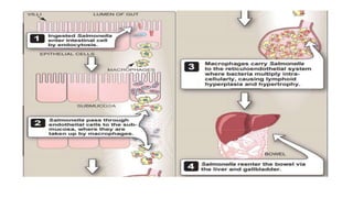
• The organisms enter, multiply in the mononuclear phagocytes of Peyer’s
patches, and then spread to the phagocytes of the liver, gallbladder, and spleen.
• This leads to bacteremia, which is associated with the onset of fever and other
symptoms, probably caused by endotoxin.
• Survival and growth of the organism within phagosomes in phagocytic cells are
a striking feature of this disease, as is the predilection for invasion of the
gallbladder, which can result in establishment of the carrier state and excretion
of the bacteria in the feces for long periods.
Typhoid fever remains a global health problem. In the United States, however,
typhoid fever has become less prevalent, and is now primarily a disease of
travelers and immigrants.
3. Other sites of Salmonella infection
• Sustained bacteremia (Septicemia) is often associated with vascular
Salmonella infections that occur when bacteria seed atherosclerotic
plaque.
• Salmonella can also cause abdominal infections (often of the
hepatobiliary tract and spleen), osteomyelitis, septic arthritis, and, rarely,
infections of other tissues or organs. Chronic carriage may, rarely,
develop.
Laboratory diagnosis
prevention
• Don’t kiss cats, dogs, chickens, turtles, lizards, or other pets or
animals.
• Don’t put your hands in your mouth after petting or playing with
animals.
• Take your pet to the veterinarian regularly.
• Wash your hands.
• Clean your pet’s bed, cage.
III. Shigella: General characteristics
• Non–lactose-fermenting.
• Non motile
• That can be distinguished from salmonellae by three criteria:
they produce no gas from the fermentation of glucose, they do not
produce H2S, and they are nonmotile.
• All shigellae have O antigens (polysaccharide) in their cell
walls, and these antigens are used to divide the genus into
four groups: A, B, C, and D.
• Spread from person to person, with contaminated stools serving as a
major source of organisms. Flies and contaminated food or water can
also transmit the disease
• Fewer than 200 viable organisms are sufficient to cause disease.
Classification : According Antigen
Their serologic specificity depends on the polysaccharide. There are
more than 40 serotypes.
The classification of shigellae relies on biochemical and antigenic
characteristics. The pathogenic species are
1. S. sonnei
2. S. flexneri
3. S. dysenteriae
4. S. boydii
Pathogenesis and clinical significance
• Shigella invade and destroy the mucosa of the large intestine.
Infection rarely penetrates to deeper layers of the intestine.
• does not lead to shigella bacteremia.
• Exotoxin (Shiga toxin) with enterotoxic and cytotoxic properties has
been isolated from these organisms, and its toxicity may play a
secondary role in development of intestinal lesions.
• Shigellae cause classic bacillary dysentery, characterized by diarrhea
with blood, mucus, and painful abdominal cramping
• The disease is generally most severe in the very young and elderly, and
among malnourished individuals, in whom shigellosis may lead to
severe dehydration and sometimes death.
Toxins
1. Endotoxin Upon autolysis: all shigellae release their toxic
lipopolysaccharide. This endotoxin probably contributes to the
irritation of the bowel wall.
2. Shigella Dysenteriae Exotoxin: S. dysenteriae type 1 (Shiga bacillus)
produces a heat-labile exotoxin that affects both the gut and the
central nervous system.
the exotoxin also inhibits sugar and amino acid absorption in the small
intestine
IV. OTHER ENTEROBACTERIACEAE
1. Klebsiella
2. Enterobacter
3. Proteus
4. Serratia
5. Citrobacter
6. Providecia
7. Morganella
8. Hafnia and Erwinia
9. Raoutella, Cronobacter, Pantoea
which can be found as normal inhabitants of the large intestine, include
organisms that are primarily opportunistic and often nosocomial pathogens.
Widespread antibiotic resistance among these organisms necessitates
sensitivity testing to determine the appropriate antibiotic treatment.
Distribution of gram-
negative pathogens.
1.Klebsiella
• Klebsiellae are large
• nonmotile
• bacilli that possess a luxurious capsule
• They are Lac+
• K. pneumoniae and K. oxytoca cause necrotizing lobar pneumonia in
individuals compromised by alcoholism, diabetes, or chronic
obstructive pulmonary disease.
• K. pneumoniae also causes urinary tract infections and bacteremia,
particularly in hospitalized patients.
2. Enterobacter
• motile and Lac+.
• They rarely cause primary disease in humans, but frequently colonize
hospitalized patients, especially in association with antibiotic
treatment, indwelling catheters, or invasive procedures.
• These organisms may infect burns, wounds, and the respiratory
(causing pneumonia), or urinary tracts.
• E. cloacae and E. aerogenes are two most important Enterobacter
species responsible for a variety of nosocomial infections.
3. Serratia
• Serratia are motile and ferment lactose slowly, if at all.
The species of Serratia that most frequently causes human infection is
S.marcescens.
• Serratia can cause extraintestinal infections such as
those of the lower respiratory and urinary tracts, especially among
hospitalized patients.
Proteus, Providencia, and Morganella
• Are normal intestinal microbiota, and are recognized as opportunistic
pathogens.
• Members of these genera are agents of urinary tract and other
extraintestinal infections.
• Proteus species are relatively common causes of uncomplicated as
well as nosocomial UTIs. Other extraintestinal infections, such as
wound infections, pneumonias, and septicemias, are associated with
compromised patients.
• Proteus organisms produce urease, which catalyzes the hydrolysis of
ureato ammonia. The resulting alkaline environment promotes the
precipitation of struvite stones containing insoluble phosphates of
magnesium and phosphate.
Erwinia and Hafnia
Erwinia
organisms are usually found in soils and they cause
infection in plants. Erwinia herbicola is the only species that
has occasionally been isolated from respiratory and urinary
infections in chronic debilitated(weaken) patients and in hospitalized
patients.
Hafnia
Hafnia alvei is the only species of the genus Hafnia. It is found
in human and animal feces, sewage, soil, and water. H. alvei is
motile. It does not ferment lactose, The bacteria have been isolated
from abscesses, wounds
Diagnosis of Enterobacteriaceae
Sources
1.Lippincot – chapter 13. third ed.
2. Jawetz-chapter 15. 28th ed.
3. Levinson chapter-18. 14th ed.

More Related Content

What's hot (20)

Mechanism of bacterial pathogenesis
Mechanism of bacterial pathogenesisMechanism of bacterial pathogenesis
Mechanism of bacterial pathogenesis
 
Enterobacteriaceae
EnterobacteriaceaeEnterobacteriaceae
Enterobacteriaceae
 
Campylobacter & Helicobacter.ppt
Campylobacter & Helicobacter.pptCampylobacter & Helicobacter.ppt
Campylobacter & Helicobacter.ppt
 
Shigella.ppt
Shigella.pptShigella.ppt
Shigella.ppt
 
SALMONELLA
SALMONELLASALMONELLA
SALMONELLA
 
Neisseria ppt mahadi
Neisseria ppt mahadiNeisseria ppt mahadi
Neisseria ppt mahadi
 
Clostridium
ClostridiumClostridium
Clostridium
 
Bacillus anthracis
Bacillus anthracisBacillus anthracis
Bacillus anthracis
 
E coli, klebsiella, enterobacter lecture notes
E coli, klebsiella, enterobacter lecture notesE coli, klebsiella, enterobacter lecture notes
E coli, klebsiella, enterobacter lecture notes
 
Bacterial pathogenesis
Bacterial pathogenesisBacterial pathogenesis
Bacterial pathogenesis
 
Virus-cell and virus-host interaction (virology 7)
Virus-cell and virus-host interaction (virology 7)Virus-cell and virus-host interaction (virology 7)
Virus-cell and virus-host interaction (virology 7)
 
Shigella
ShigellaShigella
Shigella
 
Escherichia coli
Escherichia coliEscherichia coli
Escherichia coli
 
Microbial pathogenicity
Microbial pathogenicityMicrobial pathogenicity
Microbial pathogenicity
 
Bacillus anthracis
Bacillus anthracisBacillus anthracis
Bacillus anthracis
 
Shigella
ShigellaShigella
Shigella
 
Bacillus cereus.pptx
Bacillus cereus.pptxBacillus cereus.pptx
Bacillus cereus.pptx
 
E coli - Microbiology
E coli - Microbiology E coli - Microbiology
E coli - Microbiology
 
Enterococci ppt mahadi
Enterococci  ppt mahadiEnterococci  ppt mahadi
Enterococci ppt mahadi
 
Clostridium species
Clostridium species Clostridium species
Clostridium species
 

Similar to Gram-Negative Rods of the Family Enterobacteriaceae

E.coli and others dr .ihsan alsaimary
E.coli and others dr .ihsan alsaimaryE.coli and others dr .ihsan alsaimary
E.coli and others dr .ihsan alsaimarydr.Ihsan alsaimary
 
Enteric infection and food poisoning
Enteric infection and food poisoningEnteric infection and food poisoning
Enteric infection and food poisoningdr.Ihsan alsaimary
 
Enteric infection and food poisoning
Enteric infection and food poisoningEnteric infection and food poisoning
Enteric infection and food poisoningdr.Ihsan alsaimary
 
Salmonella and E.coli infection
Salmonella and E.coli infectionSalmonella and E.coli infection
Salmonella and E.coli infectionAnik Banik
 
Gram negative bacilli (Enterobacteriaceae)
Gram negative bacilli (Enterobacteriaceae)Gram negative bacilli (Enterobacteriaceae)
Gram negative bacilli (Enterobacteriaceae)Sijo A
 
Bacterial diarrhea 2013 4th year unza medical,by DR MWANSA
Bacterial diarrhea 2013 4th year unza medical,by DR MWANSABacterial diarrhea 2013 4th year unza medical,by DR MWANSA
Bacterial diarrhea 2013 4th year unza medical,by DR MWANSAJonathan Bwalya
 
LINDA TORKUMA POWER POINT SEMINAR PRESENTATION
LINDA TORKUMA POWER POINT SEMINAR PRESENTATIONLINDA TORKUMA POWER POINT SEMINAR PRESENTATION
LINDA TORKUMA POWER POINT SEMINAR PRESENTATIONamee terdue
 
Food borne diseases.ppt
Food borne diseases.pptFood borne diseases.ppt
Food borne diseases.pptAnjaliPn2
 
Listeriosis and Colibacillosis
Listeriosis and ColibacillosisListeriosis and Colibacillosis
Listeriosis and ColibacillosisAsfar Zaman
 
10 Microbiology of the Gastrointestinal Tract final .pptx
10 Microbiology of the Gastrointestinal Tract final .pptx10 Microbiology of the Gastrointestinal Tract final .pptx
10 Microbiology of the Gastrointestinal Tract final .pptxBogaleDegefa1
 
Food born disease
Food born disease Food born disease
Food born disease China
 

Similar to Gram-Negative Rods of the Family Enterobacteriaceae (20)

E.coli and others dr .ihsan alsaimary
E.coli and others dr .ihsan alsaimaryE.coli and others dr .ihsan alsaimary
E.coli and others dr .ihsan alsaimary
 
Microlect13enterobacteriaceae
Microlect13enterobacteriaceaeMicrolect13enterobacteriaceae
Microlect13enterobacteriaceae
 
Enteric infection and food poisoning
Enteric infection and food poisoningEnteric infection and food poisoning
Enteric infection and food poisoning
 
Enteric infection and food poisoning
Enteric infection and food poisoningEnteric infection and food poisoning
Enteric infection and food poisoning
 
E. coli
E. coli  E. coli
E. coli
 
Microb e.coli
Microb e.coliMicrob e.coli
Microb e.coli
 
Genus Escherichia coli
Genus Escherichia coliGenus Escherichia coli
Genus Escherichia coli
 
Salmonella and E.coli infection
Salmonella and E.coli infectionSalmonella and E.coli infection
Salmonella and E.coli infection
 
E.coli Dr. Mahadi
E.coli Dr. Mahadi E.coli Dr. Mahadi
E.coli Dr. Mahadi
 
Enterobactaraecae.pptx
Enterobactaraecae.pptxEnterobactaraecae.pptx
Enterobactaraecae.pptx
 
Gram negative bacilli (Enterobacteriaceae)
Gram negative bacilli (Enterobacteriaceae)Gram negative bacilli (Enterobacteriaceae)
Gram negative bacilli (Enterobacteriaceae)
 
Amoebiasis.pptx
Amoebiasis.pptxAmoebiasis.pptx
Amoebiasis.pptx
 
Bacterial diarrhea 2013 4th year unza medical,by DR MWANSA
Bacterial diarrhea 2013 4th year unza medical,by DR MWANSABacterial diarrhea 2013 4th year unza medical,by DR MWANSA
Bacterial diarrhea 2013 4th year unza medical,by DR MWANSA
 
LINDA TORKUMA POWER POINT SEMINAR PRESENTATION
LINDA TORKUMA POWER POINT SEMINAR PRESENTATIONLINDA TORKUMA POWER POINT SEMINAR PRESENTATION
LINDA TORKUMA POWER POINT SEMINAR PRESENTATION
 
Food borne diseases.ppt
Food borne diseases.pptFood borne diseases.ppt
Food borne diseases.ppt
 
Lecture no 1.pptx
Lecture no 1.pptxLecture no 1.pptx
Lecture no 1.pptx
 
Listeriosis and Colibacillosis
Listeriosis and ColibacillosisListeriosis and Colibacillosis
Listeriosis and Colibacillosis
 
10 Microbiology of the Gastrointestinal Tract final .pptx
10 Microbiology of the Gastrointestinal Tract final .pptx10 Microbiology of the Gastrointestinal Tract final .pptx
10 Microbiology of the Gastrointestinal Tract final .pptx
 
Escherichia coli
Escherichia coliEscherichia coli
Escherichia coli
 
Food born disease
Food born disease Food born disease
Food born disease
 

More from MusFa1

The next pandemic: AMR
The next pandemic: AMRThe next pandemic: AMR
The next pandemic: AMRMusFa1
 
Humanistic learning theory.pptx
Humanistic learning theory.pptxHumanistic learning theory.pptx
Humanistic learning theory.pptxMusFa1
 
phage therapy.pptx
phage therapy.pptxphage therapy.pptx
phage therapy.pptxMusFa1
 
Giant viruses.pptx
Giant viruses.pptxGiant viruses.pptx
Giant viruses.pptxMusFa1
 
CNS infections.pptx
CNS infections.pptxCNS infections.pptx
CNS infections.pptxMusFa1
 
Pseudomonas.pptx
Pseudomonas.pptxPseudomonas.pptx
Pseudomonas.pptxMusFa1
 
Proteus spp.
Proteus spp.Proteus spp.
Proteus spp.MusFa1
 
Protozoa and Entamoeba histolytica
Protozoa and Entamoeba histolyticaProtozoa and Entamoeba histolytica
Protozoa and Entamoeba histolyticaMusFa1
 
Alternatives for conventional Antibiotics
Alternatives for conventional AntibioticsAlternatives for conventional Antibiotics
Alternatives for conventional AntibioticsMusFa1
 

More from MusFa1 (9)

The next pandemic: AMR
The next pandemic: AMRThe next pandemic: AMR
The next pandemic: AMR
 
Humanistic learning theory.pptx
Humanistic learning theory.pptxHumanistic learning theory.pptx
Humanistic learning theory.pptx
 
phage therapy.pptx
phage therapy.pptxphage therapy.pptx
phage therapy.pptx
 
Giant viruses.pptx
Giant viruses.pptxGiant viruses.pptx
Giant viruses.pptx
 
CNS infections.pptx
CNS infections.pptxCNS infections.pptx
CNS infections.pptx
 
Pseudomonas.pptx
Pseudomonas.pptxPseudomonas.pptx
Pseudomonas.pptx
 
Proteus spp.
Proteus spp.Proteus spp.
Proteus spp.
 
Protozoa and Entamoeba histolytica
Protozoa and Entamoeba histolyticaProtozoa and Entamoeba histolytica
Protozoa and Entamoeba histolytica
 
Alternatives for conventional Antibiotics
Alternatives for conventional AntibioticsAlternatives for conventional Antibiotics
Alternatives for conventional Antibiotics
 

Recently uploaded

Recombination DNA Technology (Nucleic Acid Hybridization )
Recombination DNA Technology (Nucleic Acid Hybridization )Recombination DNA Technology (Nucleic Acid Hybridization )
Recombination DNA Technology (Nucleic Acid Hybridization )aarthirajkumar25
 
Call Us ≽ 9953322196 ≼ Call Girls In Mukherjee Nagar(Delhi) |
Call Us ≽ 9953322196 ≼ Call Girls In Mukherjee Nagar(Delhi) |Call Us ≽ 9953322196 ≼ Call Girls In Mukherjee Nagar(Delhi) |
Call Us ≽ 9953322196 ≼ Call Girls In Mukherjee Nagar(Delhi) |aasikanpl
 
Physiochemical properties of nanomaterials and its nanotoxicity.pptx
Physiochemical properties of nanomaterials and its nanotoxicity.pptxPhysiochemical properties of nanomaterials and its nanotoxicity.pptx
Physiochemical properties of nanomaterials and its nanotoxicity.pptxAArockiyaNisha
 
Scheme-of-Work-Science-Stage-4 cambridge science.docx
Scheme-of-Work-Science-Stage-4 cambridge science.docxScheme-of-Work-Science-Stage-4 cambridge science.docx
Scheme-of-Work-Science-Stage-4 cambridge science.docxyaramohamed343013
 
STERILITY TESTING OF PHARMACEUTICALS ppt by DR.C.P.PRINCE
STERILITY TESTING OF PHARMACEUTICALS ppt by DR.C.P.PRINCESTERILITY TESTING OF PHARMACEUTICALS ppt by DR.C.P.PRINCE
STERILITY TESTING OF PHARMACEUTICALS ppt by DR.C.P.PRINCEPRINCE C P
 
Hubble Asteroid Hunter III. Physical properties of newly found asteroids
Hubble Asteroid Hunter III. Physical properties of newly found asteroidsHubble Asteroid Hunter III. Physical properties of newly found asteroids
Hubble Asteroid Hunter III. Physical properties of newly found asteroidsSérgio Sacani
 
G9 Science Q4- Week 1-2 Projectile Motion.ppt
G9 Science Q4- Week 1-2 Projectile Motion.pptG9 Science Q4- Week 1-2 Projectile Motion.ppt
G9 Science Q4- Week 1-2 Projectile Motion.pptMAESTRELLAMesa2
 
Labelling Requirements and Label Claims for Dietary Supplements and Recommend...
Labelling Requirements and Label Claims for Dietary Supplements and Recommend...Labelling Requirements and Label Claims for Dietary Supplements and Recommend...
Labelling Requirements and Label Claims for Dietary Supplements and Recommend...Lokesh Kothari
 
Isotopic evidence of long-lived volcanism on Io
Isotopic evidence of long-lived volcanism on IoIsotopic evidence of long-lived volcanism on Io
Isotopic evidence of long-lived volcanism on IoSérgio Sacani
 
Traditional Agroforestry System in India- Shifting Cultivation, Taungya, Home...
Traditional Agroforestry System in India- Shifting Cultivation, Taungya, Home...Traditional Agroforestry System in India- Shifting Cultivation, Taungya, Home...
Traditional Agroforestry System in India- Shifting Cultivation, Taungya, Home...jana861314
 
Call Girls in Munirka Delhi 💯Call Us 🔝8264348440🔝
Call Girls in Munirka Delhi 💯Call Us 🔝8264348440🔝Call Girls in Munirka Delhi 💯Call Us 🔝8264348440🔝
Call Girls in Munirka Delhi 💯Call Us 🔝8264348440🔝soniya singh
 
Nightside clouds and disequilibrium chemistry on the hot Jupiter WASP-43b
Nightside clouds and disequilibrium chemistry on the hot Jupiter WASP-43bNightside clouds and disequilibrium chemistry on the hot Jupiter WASP-43b
Nightside clouds and disequilibrium chemistry on the hot Jupiter WASP-43bSérgio Sacani
 
Unlocking the Potential: Deep dive into ocean of Ceramic Magnets.pptx
Unlocking  the Potential: Deep dive into ocean of Ceramic Magnets.pptxUnlocking  the Potential: Deep dive into ocean of Ceramic Magnets.pptx
Unlocking the Potential: Deep dive into ocean of Ceramic Magnets.pptxanandsmhk
 
Is RISC-V ready for HPC workload? Maybe?
Is RISC-V ready for HPC workload? Maybe?Is RISC-V ready for HPC workload? Maybe?
Is RISC-V ready for HPC workload? Maybe?Patrick Diehl
 
PossibleEoarcheanRecordsoftheGeomagneticFieldPreservedintheIsuaSupracrustalBe...
PossibleEoarcheanRecordsoftheGeomagneticFieldPreservedintheIsuaSupracrustalBe...PossibleEoarcheanRecordsoftheGeomagneticFieldPreservedintheIsuaSupracrustalBe...
PossibleEoarcheanRecordsoftheGeomagneticFieldPreservedintheIsuaSupracrustalBe...Sérgio Sacani
 
Orientation, design and principles of polyhouse
Orientation, design and principles of polyhouseOrientation, design and principles of polyhouse
Orientation, design and principles of polyhousejana861314
 
Animal Communication- Auditory and Visual.pptx
Animal Communication- Auditory and Visual.pptxAnimal Communication- Auditory and Visual.pptx
Animal Communication- Auditory and Visual.pptxUmerFayaz5
 
NAVSEA PEO USC - Unmanned & Small Combatants 26Oct23.pdf
NAVSEA PEO USC - Unmanned & Small Combatants 26Oct23.pdfNAVSEA PEO USC - Unmanned & Small Combatants 26Oct23.pdf
NAVSEA PEO USC - Unmanned & Small Combatants 26Oct23.pdfWadeK3
 
Boyles law module in the grade 10 science
Boyles law module in the grade 10 scienceBoyles law module in the grade 10 science
Boyles law module in the grade 10 sciencefloriejanemacaya1
 

Recently uploaded (20)

Recombination DNA Technology (Nucleic Acid Hybridization )
Recombination DNA Technology (Nucleic Acid Hybridization )Recombination DNA Technology (Nucleic Acid Hybridization )
Recombination DNA Technology (Nucleic Acid Hybridization )
 
Call Us ≽ 9953322196 ≼ Call Girls In Mukherjee Nagar(Delhi) |
Call Us ≽ 9953322196 ≼ Call Girls In Mukherjee Nagar(Delhi) |Call Us ≽ 9953322196 ≼ Call Girls In Mukherjee Nagar(Delhi) |
Call Us ≽ 9953322196 ≼ Call Girls In Mukherjee Nagar(Delhi) |
 
Physiochemical properties of nanomaterials and its nanotoxicity.pptx
Physiochemical properties of nanomaterials and its nanotoxicity.pptxPhysiochemical properties of nanomaterials and its nanotoxicity.pptx
Physiochemical properties of nanomaterials and its nanotoxicity.pptx
 
Scheme-of-Work-Science-Stage-4 cambridge science.docx
Scheme-of-Work-Science-Stage-4 cambridge science.docxScheme-of-Work-Science-Stage-4 cambridge science.docx
Scheme-of-Work-Science-Stage-4 cambridge science.docx
 
The Philosophy of Science
The Philosophy of ScienceThe Philosophy of Science
The Philosophy of Science
 
STERILITY TESTING OF PHARMACEUTICALS ppt by DR.C.P.PRINCE
STERILITY TESTING OF PHARMACEUTICALS ppt by DR.C.P.PRINCESTERILITY TESTING OF PHARMACEUTICALS ppt by DR.C.P.PRINCE
STERILITY TESTING OF PHARMACEUTICALS ppt by DR.C.P.PRINCE
 
Hubble Asteroid Hunter III. Physical properties of newly found asteroids
Hubble Asteroid Hunter III. Physical properties of newly found asteroidsHubble Asteroid Hunter III. Physical properties of newly found asteroids
Hubble Asteroid Hunter III. Physical properties of newly found asteroids
 
G9 Science Q4- Week 1-2 Projectile Motion.ppt
G9 Science Q4- Week 1-2 Projectile Motion.pptG9 Science Q4- Week 1-2 Projectile Motion.ppt
G9 Science Q4- Week 1-2 Projectile Motion.ppt
 
Labelling Requirements and Label Claims for Dietary Supplements and Recommend...
Labelling Requirements and Label Claims for Dietary Supplements and Recommend...Labelling Requirements and Label Claims for Dietary Supplements and Recommend...
Labelling Requirements and Label Claims for Dietary Supplements and Recommend...
 
Isotopic evidence of long-lived volcanism on Io
Isotopic evidence of long-lived volcanism on IoIsotopic evidence of long-lived volcanism on Io
Isotopic evidence of long-lived volcanism on Io
 
Traditional Agroforestry System in India- Shifting Cultivation, Taungya, Home...
Traditional Agroforestry System in India- Shifting Cultivation, Taungya, Home...Traditional Agroforestry System in India- Shifting Cultivation, Taungya, Home...
Traditional Agroforestry System in India- Shifting Cultivation, Taungya, Home...
 
Call Girls in Munirka Delhi 💯Call Us 🔝8264348440🔝
Call Girls in Munirka Delhi 💯Call Us 🔝8264348440🔝Call Girls in Munirka Delhi 💯Call Us 🔝8264348440🔝
Call Girls in Munirka Delhi 💯Call Us 🔝8264348440🔝
 
Nightside clouds and disequilibrium chemistry on the hot Jupiter WASP-43b
Nightside clouds and disequilibrium chemistry on the hot Jupiter WASP-43bNightside clouds and disequilibrium chemistry on the hot Jupiter WASP-43b
Nightside clouds and disequilibrium chemistry on the hot Jupiter WASP-43b
 
Unlocking the Potential: Deep dive into ocean of Ceramic Magnets.pptx
Unlocking  the Potential: Deep dive into ocean of Ceramic Magnets.pptxUnlocking  the Potential: Deep dive into ocean of Ceramic Magnets.pptx
Unlocking the Potential: Deep dive into ocean of Ceramic Magnets.pptx
 
Is RISC-V ready for HPC workload? Maybe?
Is RISC-V ready for HPC workload? Maybe?Is RISC-V ready for HPC workload? Maybe?
Is RISC-V ready for HPC workload? Maybe?
 
PossibleEoarcheanRecordsoftheGeomagneticFieldPreservedintheIsuaSupracrustalBe...
PossibleEoarcheanRecordsoftheGeomagneticFieldPreservedintheIsuaSupracrustalBe...PossibleEoarcheanRecordsoftheGeomagneticFieldPreservedintheIsuaSupracrustalBe...
PossibleEoarcheanRecordsoftheGeomagneticFieldPreservedintheIsuaSupracrustalBe...
 
Orientation, design and principles of polyhouse
Orientation, design and principles of polyhouseOrientation, design and principles of polyhouse
Orientation, design and principles of polyhouse
 
Animal Communication- Auditory and Visual.pptx
Animal Communication- Auditory and Visual.pptxAnimal Communication- Auditory and Visual.pptx
Animal Communication- Auditory and Visual.pptx
 
NAVSEA PEO USC - Unmanned & Small Combatants 26Oct23.pdf
NAVSEA PEO USC - Unmanned & Small Combatants 26Oct23.pdfNAVSEA PEO USC - Unmanned & Small Combatants 26Oct23.pdf
NAVSEA PEO USC - Unmanned & Small Combatants 26Oct23.pdf
 
Boyles law module in the grade 10 science
Boyles law module in the grade 10 scienceBoyles law module in the grade 10 science
Boyles law module in the grade 10 science
 

Gram-Negative Rods of the Family Enterobacteriaceae

  • 2. Outlines • Enterobacteriaceae : Overview • I. Escherichia coli : General characteristics • II. Salmonella : General characteristics • III. Shigella : General characteristics • IV. OTHER ENTEROBACTERIACEAE • General Diagnosis
  • 3. Enterobacteriaceae : Overview 1. Are a large heterogeneous group of Gram-negative rods. 2. Natural habitat is the human or animal intestinal. 3. Many also have alternative habitats in soil or water. 4. The family includes many genera. 5. Are facultative anaerobes or aerobes. 6. Ferment a wide range of carbohydrates. 7. possess a complex antigenic Structure, and produce a variety of toxins.
  • 4. Enterobacteriaceae : Overview 8. Many as part of the normal flora. 9. Grow well on MacConkey agar. 10. Catalase positive. 11. Oxidase negative (except for Plesiomonas). 12. Reduce nitrate to nitrite. 13. Non spore forming. 14. Motile And Non-Motile. 15. Some has capsule. 16. Some are nosocomial infections.
  • 6. Enterobacteriaceae: Classification Gram-negative rods related to the enteric tract include a large number of genera. These genera have therefore been divided into three groups depending on the major anatomic location of disease, namely, 1. pathogens both within and outside the enteric tract. 2. pathogens primarily within the enteric tract. 3. pathogens outside the enteric tract. Source of Site of Infection Enteric tract Genus 1. Both within and outside Escherichia, Salmonella 2. Primarily within Shigella, Vibrio, Campylobacter, Helicobacter 3. Outside only Klebsiella–Enterobacter–Serratia group, Proteus- Pseudomonas, Bacteroides, Prevotella, Fusobacterium Providencia–Morganella group CHAPTER 18-levinson
  • 7. Classification according biochemical characteristics • Ewing grouped bacterial species with similar biochemical characteristics. Within the tribes.
  • 9. Lactose fermented rapidly Escherichia coli: metallic sheen on differential media; motile; flat, nonviscous colonies Enterobacter aerogenes: raised colonies, no metallic sheen; often motile; more viscous growth Enterobacter cloacae: similar to Enterobacter aerogenes Klebsiella pneumoniae: very viscous, mucoid growth; nonmotile Lactose fermented slowly Edwardsiella, Serratia, Citrobacter, Salmonella Arizona subgroup, Erwinia, some strains of Shigella sonnei (on extended incubation) Lactose not fermented Shigella species: nonmotile; no gas from dextrose Salmonella species: motile; acid and usually gas from dextrose Proteus species: “swarming” on agar; urea rapidly hydrolyzed (smell of ammonia) Providencia species, other than P. stuartii Morganella species. Yersinia species Rapid, Presumptive Identification of Gram-Negative Enteric Bacteria-jawtez
  • 10. I. Escherichia coli : General characteristics • Escherichia is the most important genus in Enterobacteriaceae and has many species, Escherichia coli (E.coli) is the most medical important species. • Most used in applied biotechnology. • Motile by peritrichous flagella. • Habitat GI tract, is part of the normal flora. • Facultative anaerobes. • Has fimbriae or pili. • Opportunistic pathogenic. • Some species: E. albertii, E. fergusonii, E. hermannii E. marmotae, E. vulneris.
  • 11. Functions of E.coli as normal flora • Protects the intestinal tract from bactreial infection • Produce our main source of vitamins B12 and K. • Lives symbiotically. • Aids in digestion.
  • 12. Antigenic structure and Virulence factor 1.The O antigens: (somatic or cell wall antigens) are found on the polysaccharide portion of the LPS. These antigens are heat-stable, and may be shared among different Enterobacteriaceae genera. 2. The H antigens: are associated with flagella; therefore, only flagellated (motile)Enterobacteriaceae such as E. coli have H antigen. 3. The K antigen: are most often associated with the capsule or, less commonly, with the fimbriae. The antigenic classification of Enterobacteriaceae often indicates the presence of each specific antigen; for example, the antigenic formula of an E. coli may be O55:K5:H21
  • 13. Enterobacteriaceae have a complex antigenic They are classified by more than 150 different heat- stable somatic O (lipopolysaccharide) antigens, more than 100 heat-labile K (capsular) antigens, more than 50 H (flagellar) antigens Antigenic structure Although each genus of Enterobacteriaceae is associated with specific O groups, a single organism may carry several O antigens. Thus, most shigellae share one or more O antigens with E. coli
  • 14. Clinical significance: E. coli-associated diarrheal diseases • Transmission of intestinal disease is commonly by the fecal/oral route. • At least five types of intestinal infections that differ in pathogenic mechanisms have been identified .
  • 15. 1.Enterotoxigenic Escherichia coli. ETEC • In the United States and other Western industrialized nations, ETEC diarrhea is the most common cause of a diarrheal disease sometimes referred to as traveler’s diarrhea. • ETEC spread commonly via consumption of contaminated food or water. Poor hygiene. • A high infective dose (10*610*10 organisms) is necessary to initiate disease. • They produce a heat-labile toxin (LT), which is similar in action and amino acid sequence to cholera toxin from Vibrio cholerae
  • 16. During infection, the A portion activates cellular adenylate Cyclase causing an increase in the conversion of adenosine triphosphate to cyclic adenosine monophosphate (cAMP). The consequence of accumulation of cAMP is hypersecretion of both electrolytes and fluids into the intestinal lumen, resulting in watery diarrhea similar to cholera.
  • 17. 2.Enterohemorrhagic Escherichia coli EHEC • The EHEC strain serotype O157:H7 has since been associated with hemorrhagic diarrhea, colitis, and hemolytic uremic syndrome (HUS). HUS is characterized by low platelet count, hemolytic anemia, and kidney failure. • E. coli O157:H7 produces two cytotoxins: verotoxin I and verotoxin II. (Verotoxin I is a phage-encoded cytotoxin identical to the Shiga toxin (Stx) produced by Shigella dysenteriae). • In EHEC the stool contains no leukocytes, which distinguishes it from dysentery caused by Shigella spp. or EIEC infections.
  • 18. 3.Enteroinvasive Escherichia coli. EIEC • EIEC differs greatly from EPEC and ETEC strains, less commonly in developing countries than others two. • EIEC strains produce dysentery with direct penetration, invasion, and destruction of the intestinal mucosa. • This diarrheal illness is very similar to that produced by Shigella spp. • The organisms might be easily misidentified because of their similarity to shigellae. • EIEC strains can be nonmotile and generally do not ferment lactose • the infective dose of EIEC necessary to produce disease is much higher (106) than that of shigellae (about 100 bacterial cells).
  • 19. Clinical significance: extraintestinal disease • The source of infection for extraintestinal disease is frequently the patient's own flora, in which the individual’s own E. coli is non pathogenic in the intestine. However, it causes disease in that individual when the organism is found, for example, in the bladder or bloodstream . 1. Urinary tract infections (UTI): E. coli is the most common cause of UTI, including cystitis and pyelonephritis. Women are particularly at risk for infection. Uncomplicated cystitis (the most commonly encountered UTI). The E. coli strains that cause UTIs usually originate in the large intestine as resident biota.
  • 20. 2. Neonatal meningitis: E. coli is a major cause of this disease occurring within the first month of life. The K1 (capsular) antigen is particularly associated with such infections. 3. Sepsis: When normal host defenses are inadequate, E. coli may reach the bloodstream and cause sepsis. Newborns may be highly susceptible to E. coli sepsis because they lack IgM antibodies. 4. Hemolytic uremic syndrome (HUS): is a condition that can occur when the small blood vessels in your kidneys become damaged and inflamed. This damage can cause clots to form in the vessels. The clots clog the filtering system in the kidneys and lead to kidney failure, which could be life-threatening. 5. pneumonia.
  • 22. Prevention • Practice proper hygiene, especially good handwashing. 1. Wash your hands thoroughly after using the bathroom. 2. ash your hands thoroughly after contact with animals or their environments 3. Wash your hands thoroughly before and after preparing or eating food. • Avoid raw milk, unpasteurized dairy products, and unpasteurized juices (such as fresh apple cider). • Cook meats thoroughly.
  • 23.
  • 24. II. SALMONELLA General characteristics • Most strains of Salmonella are Lac– • produce acid and gas during fermentation of glucose. • They also produce H2S from sulfur-containing amino acids. • Salmonellae possess multiple types of pili. • Salmonellae are both commensal as well as pathogenic. • cause of enteritis and systemic infection. • transmitted via contaminated water or food from animals and animal products to humans. • Salmonella are widely distributed in nature.
  • 25. Classification : according species & sup species • The taxonomic classification of salmonellae is complex. • The names (eg, Salmonella Typhi and Salmonella typhimurium) were originally written as if they were genus and species; this form of the nomenclature remains in widespread but incorrect use. • Currently, the genus Salmonella is divided into two species each with multiple subspecies and serotypes. The two species are Salmonella enterica and Salmonella bongori. Based on the phenotypic profiles, S. enterica is further subdivided into six subspecies, which are subspecies enterica (subspecies I)(subspecies II), (subspecies IIIa), (subspecies IIIb),(subspecies IV)(subspecies VI). • Most human illness is caused by S. enterica subspecies I strains.
  • 26. Classification: According Antigenic • Furthermore, salmonellae can be classified by their serotype; serotypes are assigned according to antigenically diverse surface structures: somatic O antigens and flagellar H antigens. • The 2 species of Salmonella and their respective subspecies consist of more than 2500 serotypes (serovars), including more than 1400 in DNA hybridization group I. Some spp. Has (Vi polysaccharide) is part of the bacterial capsule. Vi capsular polysaccharide vaccine. present
  • 27. Classification: Salmonella Typhimurium. The widely accepted nomenclature for classification at the present time is as follows: for example, S. enterica subspecies enterica serotype Typhimurium. which can be shortened to Salmonella Typhimurium. with the genus name in italics and the serotype name in roman type.
  • 28. Classification: typhoidal and nontyphoidal. Based on their serotype, Salmonella species (specifically S. enterica) are further classified as “typhoidal” and “nontyphoidal”. 1. Typhoidal Salmonella: refers to those specific serotypes that cause typhoid (“enteric”) fever,and include the serotypes Typhi, Paratyphi A, Paratyphi B, and Paratyphoid C. 2.Non-typhoidal Salmonella: refers to all other serotypes.
  • 29.
  • 30.
  • 31. Virulence Factors • The role of fimbriae in adherence in initiating intestinal infection has been cited. • Their ability to traverse(cut off) intestinal mucosa. • Enterotoxin produced by certain Salmonella strains that cause gastroenteritis has been implicated as a significant virulence factor.
  • 32. Clinical significance 1. Gastroenteritis: This localized disease (also called salmonellosis) • is caused primarily by serotypes enteriditis and typhimurium (nontyphoidal salmonella). • It is characterized by nausea, vomiting, and diarrhea (usually nonbloody) which develop generally within 48 hours of ingesting contaminated food or water. • Fever and abdominal cramping are common. • In uncompromised patients, disease is generally self-limiting (48 to 72 hours), although convalescent carriage of organisms may persist for a month or more.
  • 33. 2. Enteric (typhoid) fever: • infection begins in the small intestine, but few gastrointestinal symptoms occur. • The organisms enter, multiply in the mononuclear phagocytes of Peyer’s patches, and then spread to the phagocytes of the liver, gallbladder, and spleen. • This leads to bacteremia, which is associated with the onset of fever and other symptoms, probably caused by endotoxin. • Survival and growth of the organism within phagosomes in phagocytic cells are a striking feature of this disease, as is the predilection for invasion of the gallbladder, which can result in establishment of the carrier state and excretion of the bacteria in the feces for long periods. Typhoid fever remains a global health problem. In the United States, however, typhoid fever has become less prevalent, and is now primarily a disease of travelers and immigrants.
  • 34.
  • 35. 3. Other sites of Salmonella infection • Sustained bacteremia (Septicemia) is often associated with vascular Salmonella infections that occur when bacteria seed atherosclerotic plaque. • Salmonella can also cause abdominal infections (often of the hepatobiliary tract and spleen), osteomyelitis, septic arthritis, and, rarely, infections of other tissues or organs. Chronic carriage may, rarely, develop.
  • 37. prevention • Don’t kiss cats, dogs, chickens, turtles, lizards, or other pets or animals. • Don’t put your hands in your mouth after petting or playing with animals. • Take your pet to the veterinarian regularly. • Wash your hands. • Clean your pet’s bed, cage.
  • 38.
  • 39. III. Shigella: General characteristics • Non–lactose-fermenting. • Non motile • That can be distinguished from salmonellae by three criteria: they produce no gas from the fermentation of glucose, they do not produce H2S, and they are nonmotile. • All shigellae have O antigens (polysaccharide) in their cell walls, and these antigens are used to divide the genus into four groups: A, B, C, and D. • Spread from person to person, with contaminated stools serving as a major source of organisms. Flies and contaminated food or water can also transmit the disease • Fewer than 200 viable organisms are sufficient to cause disease.
  • 40. Classification : According Antigen Their serologic specificity depends on the polysaccharide. There are more than 40 serotypes. The classification of shigellae relies on biochemical and antigenic characteristics. The pathogenic species are 1. S. sonnei 2. S. flexneri 3. S. dysenteriae 4. S. boydii
  • 41.
  • 42. Pathogenesis and clinical significance • Shigella invade and destroy the mucosa of the large intestine. Infection rarely penetrates to deeper layers of the intestine. • does not lead to shigella bacteremia. • Exotoxin (Shiga toxin) with enterotoxic and cytotoxic properties has been isolated from these organisms, and its toxicity may play a secondary role in development of intestinal lesions. • Shigellae cause classic bacillary dysentery, characterized by diarrhea with blood, mucus, and painful abdominal cramping • The disease is generally most severe in the very young and elderly, and among malnourished individuals, in whom shigellosis may lead to severe dehydration and sometimes death.
  • 43. Toxins 1. Endotoxin Upon autolysis: all shigellae release their toxic lipopolysaccharide. This endotoxin probably contributes to the irritation of the bowel wall. 2. Shigella Dysenteriae Exotoxin: S. dysenteriae type 1 (Shiga bacillus) produces a heat-labile exotoxin that affects both the gut and the central nervous system. the exotoxin also inhibits sugar and amino acid absorption in the small intestine
  • 44.
  • 45. IV. OTHER ENTEROBACTERIACEAE 1. Klebsiella 2. Enterobacter 3. Proteus 4. Serratia 5. Citrobacter 6. Providecia 7. Morganella 8. Hafnia and Erwinia 9. Raoutella, Cronobacter, Pantoea which can be found as normal inhabitants of the large intestine, include organisms that are primarily opportunistic and often nosocomial pathogens. Widespread antibiotic resistance among these organisms necessitates sensitivity testing to determine the appropriate antibiotic treatment. Distribution of gram- negative pathogens.
  • 46. 1.Klebsiella • Klebsiellae are large • nonmotile • bacilli that possess a luxurious capsule • They are Lac+ • K. pneumoniae and K. oxytoca cause necrotizing lobar pneumonia in individuals compromised by alcoholism, diabetes, or chronic obstructive pulmonary disease. • K. pneumoniae also causes urinary tract infections and bacteremia, particularly in hospitalized patients.
  • 47. 2. Enterobacter • motile and Lac+. • They rarely cause primary disease in humans, but frequently colonize hospitalized patients, especially in association with antibiotic treatment, indwelling catheters, or invasive procedures. • These organisms may infect burns, wounds, and the respiratory (causing pneumonia), or urinary tracts. • E. cloacae and E. aerogenes are two most important Enterobacter species responsible for a variety of nosocomial infections.
  • 48. 3. Serratia • Serratia are motile and ferment lactose slowly, if at all. The species of Serratia that most frequently causes human infection is S.marcescens. • Serratia can cause extraintestinal infections such as those of the lower respiratory and urinary tracts, especially among hospitalized patients.
  • 49. Proteus, Providencia, and Morganella • Are normal intestinal microbiota, and are recognized as opportunistic pathogens. • Members of these genera are agents of urinary tract and other extraintestinal infections. • Proteus species are relatively common causes of uncomplicated as well as nosocomial UTIs. Other extraintestinal infections, such as wound infections, pneumonias, and septicemias, are associated with compromised patients. • Proteus organisms produce urease, which catalyzes the hydrolysis of ureato ammonia. The resulting alkaline environment promotes the precipitation of struvite stones containing insoluble phosphates of magnesium and phosphate.
  • 50. Erwinia and Hafnia Erwinia organisms are usually found in soils and they cause infection in plants. Erwinia herbicola is the only species that has occasionally been isolated from respiratory and urinary infections in chronic debilitated(weaken) patients and in hospitalized patients. Hafnia Hafnia alvei is the only species of the genus Hafnia. It is found in human and animal feces, sewage, soil, and water. H. alvei is motile. It does not ferment lactose, The bacteria have been isolated from abscesses, wounds
  • 52.
  • 53.
  • 54. Sources 1.Lippincot – chapter 13. third ed. 2. Jawetz-chapter 15. 28th ed. 3. Levinson chapter-18. 14th ed.

Editor's Notes

  1. Characteristics of intestinal infections caused by E. coli. Fluoroquinolones are commonly used in adults for travelerʼs diarrhea, but are not recommended for children. Rehydration and correction of electrolyte abnormalities are essential for all diarrhea illnesses. Rifaximin is approved for the treatment of diarrhea caused by noninvasive strains of Escherichia coli in patients 12 years of age and older. Rifaximin is a nonabsorable, gastrointestinal-selective, oral antibiotic.
  2. Hemolytic uremic syndrome (HUS) is a condition that can occur when the small blood vessels in your kidneys become damaged and inflamed. This damage can cause clots to form in the vessels. The clots clog the filtering system in the kidneys and lead to kidney failure, which could be life-threatening.