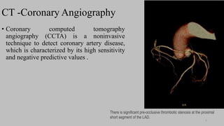
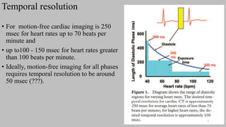
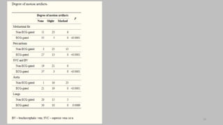
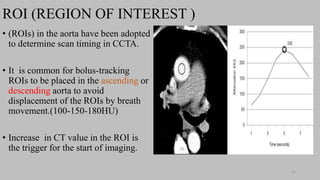
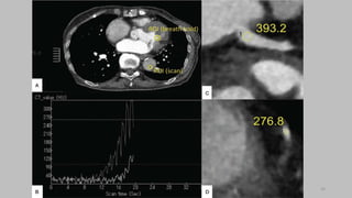
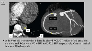
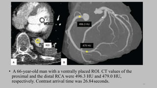
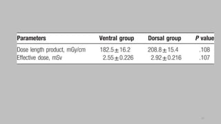
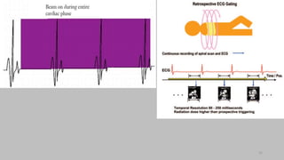
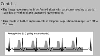
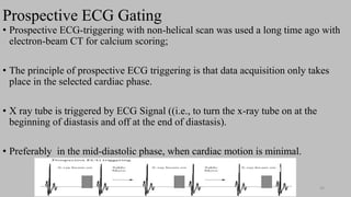
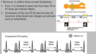
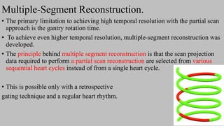
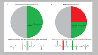
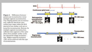
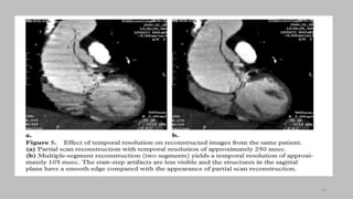
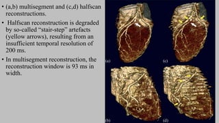
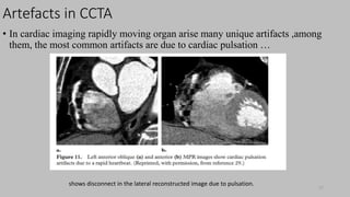
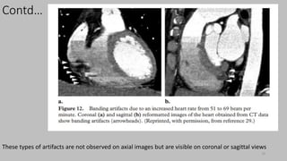
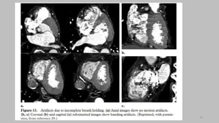
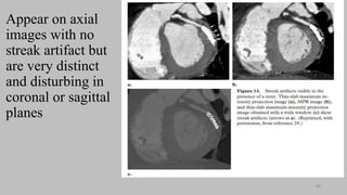
Ct   coronary  angiography edited 1st

Editor's Notes

  • #5 Sensitivity -test correctly generates a positive result for people who have the condition Specificity - ability to correctly generate a negative result for people who don’t have the condition NPV - represents the probability that a person does not have a disease or condition,
  • #8 MDCT most important advancement similar spatial resolution in all directions, also expressed as isotropic spatial resolution
  • #10 Obtuse – greater thana 90 but less than 180
  • #13 Oxygen saturation of left atrium is about 93-90% why ?????? Coz of coronary drainage and bronchopulmonary drainage in to the LF atrium Foramen ovale and ductus arterious , fetal circulation
  • #17  Peripheral pulse gating cannot be used for heart imaging due to the long and unpredictable delay between cardiac contraction and arrival of the pulse wave to the finger.
  • #21 The axial scanning method in cardiac CT is always prospectively ECG triggered. 
  • #24  Using 4D-MRI, Miyazaki et al. have shown a tendency for a faster flow velocity at the dorsal side of the middle thoracic descending aorta This technique has shown more consistent aortic and coronary enhancement with reduced interpatient variability.
  • #25 . When the left ventricle reached 100 HU, patients were instructed to breathe in and hold; when the descending aorta reached 260 HU  A circular ROI was placed in the proximal (C) and distal (D) 
  • #30 Parse out = through out The minimum amount of projection data required to construct a complete CT image is 180° plus the fan angle of the CT detectors in the axial plane. ????/
  • #32 They do however retain their clinical relevance in patients with arrhythmia and for volumetry and functional analysis.
  • #39 This can be overcome with the latest 320-slice CT scanner, which enables coverage of the cardiac volume in a single heartbeat.  ECG-triggered acquisition targets only a specific phase of the cardiac cycle; thus, functional information about cardiac valve or ventricular wall motion is not available 
  • #41 if one chooses to select half of the data set required for partial scan reconstruction from one heart cycle and the rest from another heart cycle, this results in temporal resolution that is about one-fourth of the gantry rotation time.
  • #47 enabling acquisition of complete data in a single cardiac cycle with excellent image quality at a consistent dose lower than 1.0 mSv  In high-pitch spiral acquisition, the heart rate should even be below 60 bpm. 
  • #50 Atrial fibrillation is an irregular and often rapid heart rate that occurs when the two upper chambers of your heart experience chaotic electrical signals.
  • #55 Equivocal - uncertain or questionable in nature.
  • #56 Discordant – unusual , unfavorable
  • #57 Note that dual source scanners, owing to the higher temporal resolution, can permit the scanning of patients with irregular rhythms and reduce the need for b-blocker administration.
  • #58 Median basilic vein (superior arm vein )
  • #59 Positioning of the electrocardiogram (ECG) leads. The ECG electrodes are usually positioned around the left and right shoulder region at locations that are not affected by disturbing activity from the musculature underneath. The third lead is attached to the lower abdomen. may need to be given 1 hour before the scan if the patient is not naturally bradycardic.
  • #61 which is defined as a heart rate less than 60 bpm; systolic blood pressure of less than 100 mm Hg; may need to be given 1 hour before the scan if the patient is not naturally bradycardic. Patients intolerant of b blockers can be given a calcium channel blocker.
  • #62 left arm might result in high-attenuation “streak” artifacts in the left subclavian vein and internal mammary artery. An injection rate of at least 5 mL/s is preferable, with increased rates at higher cardiac outputs (8). An increase in injection rate results in an earlier and higher arterial enhancement 
  • #69 Indicator of CAD Stenosis = the abnormal narrowing of a passage in the body.
  • #71 When doing a CT calcium score , stents should be recognized so that they are not included in the images measured, as they will artificially increase the score
  • #73 iodinated contrast agents are biologically inert and diffuse passively into the increased extracellular matrix of infarcted myocardium with a distribution half-life of approximately 20 min. . In the setting of acute myocardial infarction, delayed enhancement is explained by myocyte necrosis, sarcomere membrane rupture, and passive diffusion of contrast into the intracellular space.
  • #78 These types of artifacts are minimized by multiple-segment reconstruction or by scanning at even higher temporal resolution on the order of 50 msec.
  • #81 These types of artifacts are to some extent handled by special artifact reduction software developed by manufacturers