Downloaded 464 times


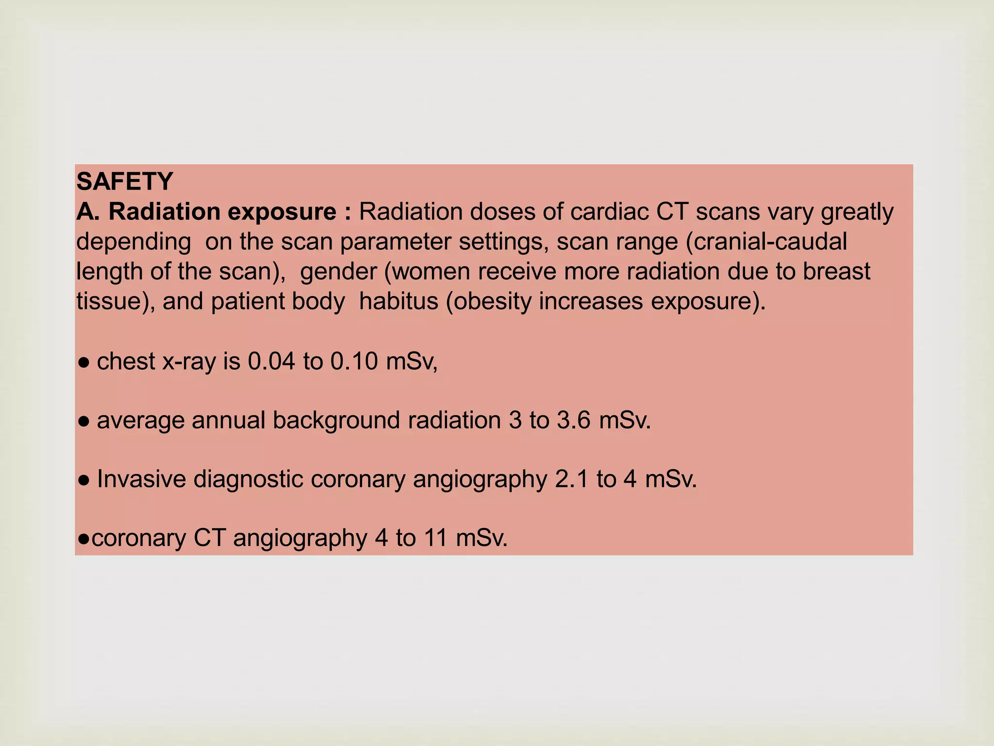


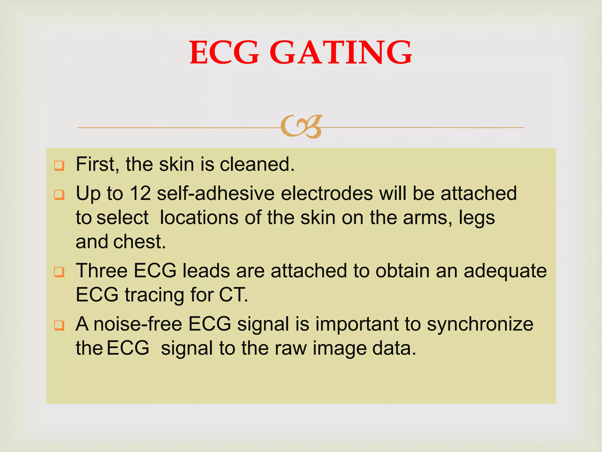


Coronary CT angiography is a noninvasive imaging modality used to evaluate coronary artery disease. It has a high sensitivity of 87-99% and specificity of 93-96% for detecting coronary artery stenosis. Coronary CT angiography is most useful in low- to intermediate-risk patients with chest pain to rule out coronary artery disease given its high negative predictive value of 93-100%. Coronary CT angiography involves acquiring images using ionizing radiation as the patient holds their breath and synchronizing the images with the patient's ECG signal.


![CARDIOVASCULAR SYSTEM new [Autosaved].pptx](https://cdn.slidesharecdn.com/ss_thumbnails/cardiovascularsystemnewautosaved-240711150130-2b5a8369-thumbnail.jpg?width=640&height=640&fit=bounds)


