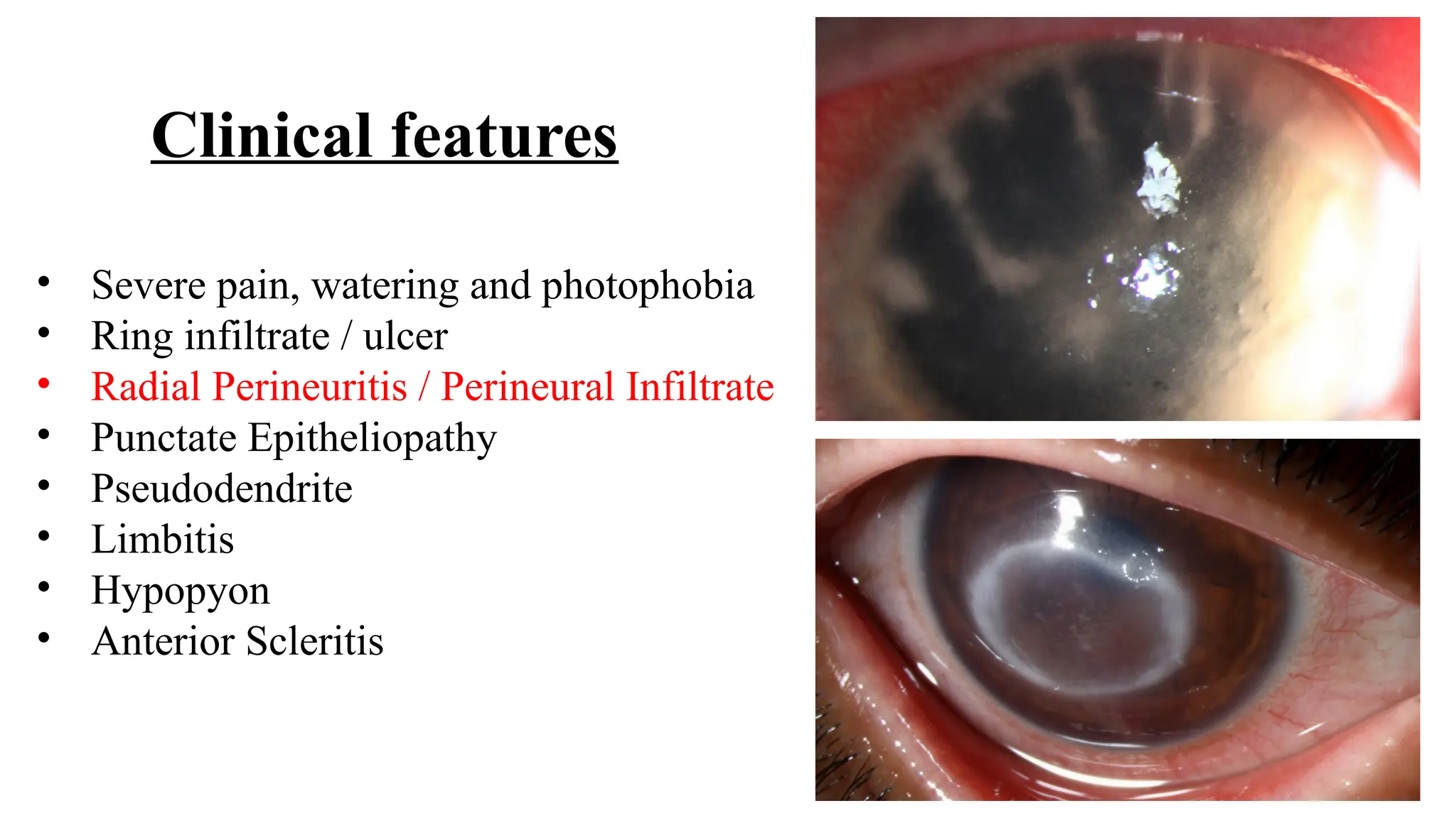
CORNEAL ULCER
Moderator : Dr Rajesh R Nayak
Presenter : Dr Sriraj Alapati
ETIOLOGY
SOURCE
• Exogenous
• Endogenous : immunological
Phlyctenular - TB
IK – Syphilis
• Contiguous:
Conjunctiva – trachoma, VKH
Sclera – sclerosing scleritis
Uveal – HSV endothelitis
STAGES
1. Progressive : infiltrates
2. Active : ulceration
3. Regressive : resolution of infiltrates
4. Cicatrizing : scarring
LOCATION
• Superficial
• Deep
• Impending perforation
• Perforation
ETIOLOGY
Infection, immune, degenerative,
neoplastic, traumatic
PATHOLOGY
• Suppurative (purulent) – bacteria,
fungi
• Non Suppurative (non purulent) –
virus, parasitic
Rapidly Progressive Suppurative Infiltrate
• Bacteria:
S. aureus
S. pneumoniae
Pseudomonas
N. Gonorrhoea
• Viral
• Mixed Infection
Slowly Progressive Localized Infiltrate
• Bacteria:
S. epidermidis
Alpha hemolytic Streptococci
Actinomycetales
Moraxella
ATM
• Fungi
• Protozoa
Clinical Evaluation
BACTERIAL ULCER
Penetrate intact epithelium:
o Neisseria
o Corynebacterium diptheria
o H. influenzae
o Shigella
o Listeria
Etiology
Gram positive cocci:
• Staphylococci (MC)
• Streptococci
Gram negative cocci:
• Neisseria
Gram positive bacilli:
• Actinomyces
• Corynebacterium
• Listeria
Gram negative bacilli:
• Pseudomonas
• E.coli
•Trauma
•Contact lens (PSEUDOMONAS)
•Loose sutures
•Steroids
•Dry eye
•Misdirected cilia
•Lagophthalmos
•Dacrocystitis
•Recurrent epithelial erosions
Predisposing Factors
Disruption of integrity of
corneal epithelium
Symptoms:
• Pain
• Redness
• Photophobia
• Blurred vision
• Mucopurulent discharge
Signs:
• Eyelid swelling
• Chemosis
• Epithelial defect with infiltrate
• Circumcorneal injection
• Stromal oedema
• DM folds
• Hypopyon (STERILE & MOBILE)
• Posterior synechiae
Clinical features
Organism specific features
Gram positive cocci:
(staph, strepococci)
Well defined margins
Localized, deep penetration
Gram negative bacilli:
(pseudomonas)
Ill defined margins due to more
tissue destruction (Exotoxins)
ULCUS SERPANS
 S. Pneumococcus
 One edge - infiltration , other –
cicatrization
 Healing at leading edge,
progress at advancing edge
• Needle-like, crystalline
opacities, arborizing without
cellular infiltrate or ocular
inflammation.
• Corneal grafts, contact lens
wear, topical anesthetic,
steroids abuse.
• Streptococci viridans
Infectious crystalline keratopathy (ICK)
Gram negative cocci:
(N. goncocci)
Rapidly progressive
Corneal melting <24 hrs
STD
Systemic treatment
Moraxella:
Steroids
Mild reaction
Well defind lesion
Nocardia:
Beaded, branching,
slender, AFB,
filamentous bacteria.
Multiple calcareous
elevations
Wreath ulcer (ring)/
cracked wind shield app.
Rx : AG’s
NTM:
(M. chelonae)
Aerobic, non motile, AFB,
non - sporing bacteria.
Sx, trauma, keratoplasty
Cracked wind shield app
Persistent infiltrates
On & off S/S
Rx : Macrolides / AG’s
Corneal ulcer scrapings to be taken before starting treatment
Margins (More bacteria) & base of ulcer with 15 blade or kimura spatula
• Sac syringing
• RBS
Investigations
PCR
FUNGI
GRAM staining
Antibiotic Susceptibility Testing
Treatment
Topical antibiotics:
• Eye drops
• Eye ointment ( To compensate night lag period )
Atropine (mydriatic – break PS & cycloplegic – reduce pain)
NSAIDS
AGM
T. VIT C 500 MG ( Improves wound healing )
Collagen cross linking
System antibiotics (no role) are indicated only in case of:
• Endophthalmitis
• Perforated corneal ulcer
• Ulcer extended beyond limbus
• Gonococci
Gram Positive-
• Cephalosporins : Cefazolin 50 mg/ml – 5%
Ceftazidime 50 mg/ml– 5%
• Vancomycin 50 mg/ml– 5%
• Chloramphenicol
• 2nd
gen fluroquinolones: Gatifloxacin / Moxifloxacin
Gram Negative-
• Aminoglycosides : Tobramicin 14 mg/ml – 1.4%
Getamicin 14 mg/ml– 1.4%
Amikacin 50 mg/ml – 5%
• 1st gen fluroquinolones: Ciprofloxacin / ofloxacin
Aminoglycosides for nocordia & NTM
MOXI : G+>G-
GATI : G->G+
Topical : no vitreous penetration
Oral GATI : vitreous penetration
OFLOX : CORNEAL PERFORATION
CIPLOX : CORNEAL DEPOSITS
LEVO, NOR, BESIFLOXACIN 0.6%
GENTA – nephro, oto, retinotoxic
Sofra, neo, tobra, amikacin
Empirical Therapy:
{Fluoroquinolone monotherapy / fortified vancomycin + ceftazidime}
• Initial treatment
• Small non-severe ulcers
• Close follow up
• Perform microbiology if worsens
All severe (> 3 mm) cases must be treated based on microbiology
G+ COCCI G- RODS
Q1H for 48 hours
Q2H in morning, Q4H in night
Q6H (healing)
Ceftazidime: 50mg/ml(5%)
Method: Reconstitute parenteral Ceftazidime 500mg with 2ml sterile
water/BSS available with the injection and add to 8ml of artificial tears.
Storage: Refrigerate in 4 degrees C.
Shelf Life: week under refrigeration at 4 degrees C and 3 days in room
temperature.
Vancomycin: 50mg/ml(5%)
Method: Reconstitute 500mg of vancomycin powder for injection with 2 ml
sterile water/BSS. Add to 8ml of artificial tears.
Storage: Refrigerate at 4 Degrees C.
Shelf Life: 28 days at 4 Degrees C.
Preparation of Fortified antibiotic eye drops
FUNGAL ULCER
Classification of Fungi:
• Yeasts (Candida)
• Yeast like fungi
• Filamentous
• Dimorphic (Blastomyces)
Etiology
Candida
• Immunocompromised hosts
(ICU, DM)
• Alcoholics
• Vitamin deficiency
• Protracted epithelial ulceration
Predisposing factors
Filamentous fungi
• Immunocompetent hosts
• Trauma with vegetative matter
• Agricultural & warehouse workers
• June – September ( humid )
Pathogenesis
ADHERANCE PENETRATION HOST RESPONSE
Disruption of
integrity of corneal
epithelium
• Parallel growth of hyphae to
stroma
• Release of :
Mycotoxins
Proteolytic enzymes
Soluble fungal antigen
Release of lysosomal
substances by PMN
Destruction of corneal stroma
Clinical features
SIGNS > SYMPTOMS
( large organisms, Inflammation process takes time )
•Dry, greyish, elevated, irregular margins
•Feathery edges ( HALLMARK)
•Satellite lesions ( multiple small ulcers )
•Hyphae
•Hypopyon ( non sterile, non mobile )
•Non specific infiltrate
•Pseudodendrite ( knobs absent )
•Endothelial plaques
•Immune ring of wessely
•Pigmentation
Flower pot - pseudodendrite on FS Feathery edges
Satellite lesions ( multiple small ulcers ) Endothelial plaques
Immune ring Pigmentation ( Aspergillus. Niger )
Investigations
Corneal ulcer scrapings feels gritty
Helps in debulking & improves drug penetration
Staining Methods-
A. Light Microscopy: 10% KOH ( dissolves the tissue
so fungi are visualized ), Gram’s, Giemsa, Grocott-
Gomori Methenamine Silver (GMS) Stains, PAS
B. Fluorescent Microscopy:
Calcofluor White (CCF) staining
GMS
CCF
10% KOH Gram’s
Culture : SDA,PDA, BHI,
cooked meat broth
Confocal microscopy
Only fungi & acanthamoeba are visualized
Treatment
Filamentous fungi – NATAMYCIN
Yeast - NYSTATIN + AMP.B
Intrastromal / intracameral injections:
• Voriconazole : 50 microgram in 0.1ml
• Amphotericin B: 5-10 microgram in 0.1ml
Oral antifungal therapy:
• Indication: Scleritis, Endophthalmitis
• Drugs : Ketoconazole 200-600mg
Voriconazole 200-400mg
Fluconozole 200-400mg
Itraconazole 200mg
• Duration of treatment : 14-21 days.
• LFT & Hallucinations
TST Protocol
( Topical, Systemic, Targeted therapy )
• First line of Rx: Topical Natamycin 5%.
• Addition of Oral Ketoconazole / Voriconazole in severe ulcers.
• Second Line of Rx: Addition of topical Voriconazole 1%.
• Third line of Rx: Intrastromal + Intracameral antifungals.
• Fourth Line of Rx: TPK.
BACTERIAL FUNGAL
Diffusion of bacterial toxins Invasion of fungal hyphae
Sterile Non sterile
Fluid Fibrinous network
Mobile (gravity, head position) Non mobile
Easily absorbed Not absorbed
HYPOPYON (PMN’S)
• Virulence of organism
• Resistance of tissues
ACANTHAMOEBIC
KERATITIS
Free living amoeba
2 forms : trophozoite ( active )
cyst ( dormant )
Risk factors:
• Contact lens users ( pseudomonas > acanthamoeba )
• Swimming pool
• Trauma with soiled matter
Clinical features
• Severe pain, watering and photophobia
• Ring infiltrate / ulcer
• Radial Perineuritis / Perineural Infiltrate
• Punctate Epitheliopathy
• Pseudodendrite
• Limbitis
• Hypopyon
• Anterior Scleritis
Investigations
Giemsa, GMS, CCF, PAS Stains
Non nutrient agar with E.COLI
Confocal microscopy
• Bright spots (89 %)
• Double walled cysts (83%)
• Signet rings (17%)
Bacteria cant be visualized
Biguanides:
•Poly Hexa Methylene Biguanide (PHMB) 0.02% - 0.06 %
•Chlorhexidine 0.02% - 0.2%
Diamidines:
•Propamidine Isethionate 0.1%
•Hexamidine 0.1%
MOA : Highly charged cationic molecules bind to negatively charged
phospholipid membrane of amoeba causing denaturation of proteins.
Aminoglycosides:
•Neomycin
MOA : inhibition of protein synthesis
Treatment
Duration : 6- 9 months
MICROSPORIDIA
• Epithelial keratitis
• Typical stuck on
appearance of SPK
• Treatment : 0.3%
fluconazole
Complications
A. Small perforation
B. Large perforation
Pseudocornea Anterior staphyloma
Adherent leucoma
• Keratectasia
• Keratocoele / descemetocoele (DM herniates through ulcer)
• Perforation
Coughing, sneezing, straining, spasm of orbicularis
Rise in BP
Rise in IOP
Perforation
Prolapse of iris
<2mm : tissue adhesive (cyanoacrylate)
2-4mm : patch graft/tenoplasty
>4mm : tectonic keratoplasty
• Secondary glaucoma (iris & cicatricial tissue are too weak to
support the restored IOP)
• Anterior staphyloma
• ASCC (contact with ulcer)
• Corneal fistula
• Pseudocornea
• Spontaneous expulsion of lens and vitreous
• Expulsive hemorrhage (sudden reduction in IOP)
• Endophthalmitis
• Panophthalmitis
• Scarring (irregular astigmatism)
Non healing ulcer always check for:
 Diabetes mellitus (immunocompromised)
 Chronic dacryocystitis
 Contamination of medications
 Drug resistance
 Atypical microorganisms
 Incomplete treatment
 Improper diagnosis
Non infectious causes:
• Chronic epithelial defect
• Autoimmune disease
• Rheumatoid arthritis
• Mooren's ulcer
• Terrien's marginal degeneration
• Staphylococcal marginal disease
• Phlyctenulosis
• Contact lens-related infiltration
• Vernal keratoconjunctivitis (shield ulcer)
• Smokable drug-induced
• Anesthetic abuse
• Xerophthalmia, keratomalacia
Confocal
microscopy
COLOUR
CODING OF
CORNEAL
ULCER
THANK YOU

Corneal Ulcer (bacterial, fungal, acanthamoeba).pptx

  • 1.
    CORNEAL ULCER Moderator :Dr Rajesh R Nayak Presenter : Dr Sriraj Alapati
  • 3.
  • 4.
    SOURCE • Exogenous • Endogenous: immunological Phlyctenular - TB IK – Syphilis • Contiguous: Conjunctiva – trachoma, VKH Sclera – sclerosing scleritis Uveal – HSV endothelitis STAGES 1. Progressive : infiltrates 2. Active : ulceration 3. Regressive : resolution of infiltrates 4. Cicatrizing : scarring LOCATION • Superficial • Deep • Impending perforation • Perforation ETIOLOGY Infection, immune, degenerative, neoplastic, traumatic PATHOLOGY • Suppurative (purulent) – bacteria, fungi • Non Suppurative (non purulent) – virus, parasitic
  • 5.
    Rapidly Progressive SuppurativeInfiltrate • Bacteria: S. aureus S. pneumoniae Pseudomonas N. Gonorrhoea • Viral • Mixed Infection Slowly Progressive Localized Infiltrate • Bacteria: S. epidermidis Alpha hemolytic Streptococci Actinomycetales Moraxella ATM • Fungi • Protozoa Clinical Evaluation
  • 6.
  • 7.
    Penetrate intact epithelium: oNeisseria o Corynebacterium diptheria o H. influenzae o Shigella o Listeria Etiology Gram positive cocci: • Staphylococci (MC) • Streptococci Gram negative cocci: • Neisseria Gram positive bacilli: • Actinomyces • Corynebacterium • Listeria Gram negative bacilli: • Pseudomonas • E.coli
  • 8.
    •Trauma •Contact lens (PSEUDOMONAS) •Loosesutures •Steroids •Dry eye •Misdirected cilia •Lagophthalmos •Dacrocystitis •Recurrent epithelial erosions Predisposing Factors Disruption of integrity of corneal epithelium
  • 9.
    Symptoms: • Pain • Redness •Photophobia • Blurred vision • Mucopurulent discharge Signs: • Eyelid swelling • Chemosis • Epithelial defect with infiltrate • Circumcorneal injection • Stromal oedema • DM folds • Hypopyon (STERILE & MOBILE) • Posterior synechiae Clinical features
  • 10.
    Organism specific features Grampositive cocci: (staph, strepococci) Well defined margins Localized, deep penetration Gram negative bacilli: (pseudomonas) Ill defined margins due to more tissue destruction (Exotoxins)
  • 11.
    ULCUS SERPANS  S.Pneumococcus  One edge - infiltration , other – cicatrization  Healing at leading edge, progress at advancing edge
  • 12.
    • Needle-like, crystalline opacities,arborizing without cellular infiltrate or ocular inflammation. • Corneal grafts, contact lens wear, topical anesthetic, steroids abuse. • Streptococci viridans Infectious crystalline keratopathy (ICK)
  • 13.
    Gram negative cocci: (N.goncocci) Rapidly progressive Corneal melting <24 hrs STD Systemic treatment Moraxella: Steroids Mild reaction Well defind lesion
  • 14.
    Nocardia: Beaded, branching, slender, AFB, filamentousbacteria. Multiple calcareous elevations Wreath ulcer (ring)/ cracked wind shield app. Rx : AG’s
  • 15.
    NTM: (M. chelonae) Aerobic, nonmotile, AFB, non - sporing bacteria. Sx, trauma, keratoplasty Cracked wind shield app Persistent infiltrates On & off S/S Rx : Macrolides / AG’s
  • 16.
    Corneal ulcer scrapingsto be taken before starting treatment Margins (More bacteria) & base of ulcer with 15 blade or kimura spatula • Sac syringing • RBS Investigations PCR FUNGI
  • 18.
  • 20.
  • 21.
    Treatment Topical antibiotics: • Eyedrops • Eye ointment ( To compensate night lag period ) Atropine (mydriatic – break PS & cycloplegic – reduce pain) NSAIDS AGM T. VIT C 500 MG ( Improves wound healing ) Collagen cross linking System antibiotics (no role) are indicated only in case of: • Endophthalmitis • Perforated corneal ulcer • Ulcer extended beyond limbus • Gonococci
  • 22.
    Gram Positive- • Cephalosporins: Cefazolin 50 mg/ml – 5% Ceftazidime 50 mg/ml– 5% • Vancomycin 50 mg/ml– 5% • Chloramphenicol • 2nd gen fluroquinolones: Gatifloxacin / Moxifloxacin Gram Negative- • Aminoglycosides : Tobramicin 14 mg/ml – 1.4% Getamicin 14 mg/ml– 1.4% Amikacin 50 mg/ml – 5% • 1st gen fluroquinolones: Ciprofloxacin / ofloxacin Aminoglycosides for nocordia & NTM
  • 23.
    MOXI : G+>G- GATI: G->G+ Topical : no vitreous penetration Oral GATI : vitreous penetration OFLOX : CORNEAL PERFORATION CIPLOX : CORNEAL DEPOSITS LEVO, NOR, BESIFLOXACIN 0.6% GENTA – nephro, oto, retinotoxic Sofra, neo, tobra, amikacin
  • 24.
    Empirical Therapy: {Fluoroquinolone monotherapy/ fortified vancomycin + ceftazidime} • Initial treatment • Small non-severe ulcers • Close follow up • Perform microbiology if worsens All severe (> 3 mm) cases must be treated based on microbiology G+ COCCI G- RODS Q1H for 48 hours Q2H in morning, Q4H in night Q6H (healing)
  • 25.
    Ceftazidime: 50mg/ml(5%) Method: Reconstituteparenteral Ceftazidime 500mg with 2ml sterile water/BSS available with the injection and add to 8ml of artificial tears. Storage: Refrigerate in 4 degrees C. Shelf Life: week under refrigeration at 4 degrees C and 3 days in room temperature. Vancomycin: 50mg/ml(5%) Method: Reconstitute 500mg of vancomycin powder for injection with 2 ml sterile water/BSS. Add to 8ml of artificial tears. Storage: Refrigerate at 4 Degrees C. Shelf Life: 28 days at 4 Degrees C. Preparation of Fortified antibiotic eye drops
  • 26.
  • 27.
    Classification of Fungi: •Yeasts (Candida) • Yeast like fungi • Filamentous • Dimorphic (Blastomyces) Etiology
  • 29.
    Candida • Immunocompromised hosts (ICU,DM) • Alcoholics • Vitamin deficiency • Protracted epithelial ulceration Predisposing factors Filamentous fungi • Immunocompetent hosts • Trauma with vegetative matter • Agricultural & warehouse workers • June – September ( humid )
  • 30.
    Pathogenesis ADHERANCE PENETRATION HOSTRESPONSE Disruption of integrity of corneal epithelium • Parallel growth of hyphae to stroma • Release of : Mycotoxins Proteolytic enzymes Soluble fungal antigen Release of lysosomal substances by PMN Destruction of corneal stroma
  • 31.
    Clinical features SIGNS >SYMPTOMS ( large organisms, Inflammation process takes time ) •Dry, greyish, elevated, irregular margins •Feathery edges ( HALLMARK) •Satellite lesions ( multiple small ulcers ) •Hyphae •Hypopyon ( non sterile, non mobile ) •Non specific infiltrate •Pseudodendrite ( knobs absent ) •Endothelial plaques •Immune ring of wessely •Pigmentation
  • 32.
    Flower pot -pseudodendrite on FS Feathery edges
  • 33.
    Satellite lesions (multiple small ulcers ) Endothelial plaques
  • 34.
    Immune ring Pigmentation( Aspergillus. Niger )
  • 35.
    Investigations Corneal ulcer scrapingsfeels gritty Helps in debulking & improves drug penetration Staining Methods- A. Light Microscopy: 10% KOH ( dissolves the tissue so fungi are visualized ), Gram’s, Giemsa, Grocott- Gomori Methenamine Silver (GMS) Stains, PAS B. Fluorescent Microscopy: Calcofluor White (CCF) staining
  • 36.
  • 37.
    Culture : SDA,PDA,BHI, cooked meat broth
  • 38.
    Confocal microscopy Only fungi& acanthamoeba are visualized
  • 39.
    Treatment Filamentous fungi –NATAMYCIN Yeast - NYSTATIN + AMP.B
  • 40.
    Intrastromal / intracameralinjections: • Voriconazole : 50 microgram in 0.1ml • Amphotericin B: 5-10 microgram in 0.1ml Oral antifungal therapy: • Indication: Scleritis, Endophthalmitis • Drugs : Ketoconazole 200-600mg Voriconazole 200-400mg Fluconozole 200-400mg Itraconazole 200mg • Duration of treatment : 14-21 days. • LFT & Hallucinations
  • 41.
    TST Protocol ( Topical,Systemic, Targeted therapy ) • First line of Rx: Topical Natamycin 5%. • Addition of Oral Ketoconazole / Voriconazole in severe ulcers. • Second Line of Rx: Addition of topical Voriconazole 1%. • Third line of Rx: Intrastromal + Intracameral antifungals. • Fourth Line of Rx: TPK.
  • 42.
    BACTERIAL FUNGAL Diffusion ofbacterial toxins Invasion of fungal hyphae Sterile Non sterile Fluid Fibrinous network Mobile (gravity, head position) Non mobile Easily absorbed Not absorbed HYPOPYON (PMN’S) • Virulence of organism • Resistance of tissues
  • 43.
  • 44.
    Free living amoeba 2forms : trophozoite ( active ) cyst ( dormant ) Risk factors: • Contact lens users ( pseudomonas > acanthamoeba ) • Swimming pool • Trauma with soiled matter
  • 45.
    Clinical features • Severepain, watering and photophobia • Ring infiltrate / ulcer • Radial Perineuritis / Perineural Infiltrate • Punctate Epitheliopathy • Pseudodendrite • Limbitis • Hypopyon • Anterior Scleritis
  • 46.
    Investigations Giemsa, GMS, CCF,PAS Stains Non nutrient agar with E.COLI
  • 47.
    Confocal microscopy • Brightspots (89 %) • Double walled cysts (83%) • Signet rings (17%) Bacteria cant be visualized
  • 48.
    Biguanides: •Poly Hexa MethyleneBiguanide (PHMB) 0.02% - 0.06 % •Chlorhexidine 0.02% - 0.2% Diamidines: •Propamidine Isethionate 0.1% •Hexamidine 0.1% MOA : Highly charged cationic molecules bind to negatively charged phospholipid membrane of amoeba causing denaturation of proteins. Aminoglycosides: •Neomycin MOA : inhibition of protein synthesis Treatment Duration : 6- 9 months
  • 49.
    MICROSPORIDIA • Epithelial keratitis •Typical stuck on appearance of SPK • Treatment : 0.3% fluconazole
  • 50.
    Complications A. Small perforation B.Large perforation Pseudocornea Anterior staphyloma Adherent leucoma
  • 51.
    • Keratectasia • Keratocoele/ descemetocoele (DM herniates through ulcer) • Perforation Coughing, sneezing, straining, spasm of orbicularis Rise in BP Rise in IOP Perforation Prolapse of iris <2mm : tissue adhesive (cyanoacrylate) 2-4mm : patch graft/tenoplasty >4mm : tectonic keratoplasty
  • 52.
    • Secondary glaucoma(iris & cicatricial tissue are too weak to support the restored IOP) • Anterior staphyloma • ASCC (contact with ulcer) • Corneal fistula • Pseudocornea • Spontaneous expulsion of lens and vitreous • Expulsive hemorrhage (sudden reduction in IOP) • Endophthalmitis • Panophthalmitis • Scarring (irregular astigmatism)
  • 53.
    Non healing ulceralways check for:  Diabetes mellitus (immunocompromised)  Chronic dacryocystitis  Contamination of medications  Drug resistance  Atypical microorganisms  Incomplete treatment  Improper diagnosis
  • 54.
    Non infectious causes: •Chronic epithelial defect • Autoimmune disease • Rheumatoid arthritis • Mooren's ulcer • Terrien's marginal degeneration • Staphylococcal marginal disease • Phlyctenulosis • Contact lens-related infiltration • Vernal keratoconjunctivitis (shield ulcer) • Smokable drug-induced • Anesthetic abuse • Xerophthalmia, keratomalacia
  • 56.
  • 58.
  • 59.