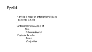
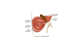
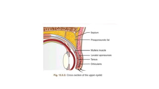
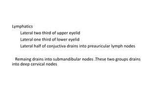
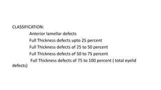
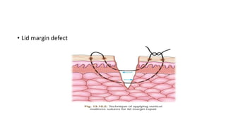
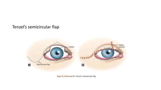
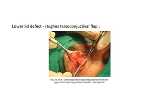
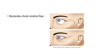
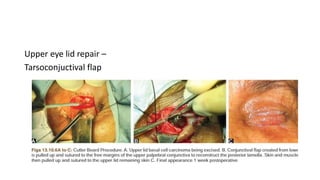
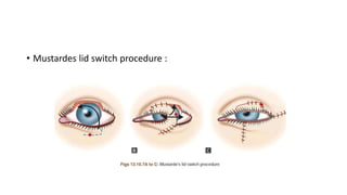
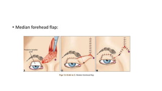
EYELID RECONSTRUCTION
-By B.Lakshmi preethi
Eyelid
• Eyelid is made of anterior lamella and
posterior lamella
Anterior lamella consist of
Skin
Orbicularis oculi
Posterior lamella
Tarsus
Conjuctiva
• Arterial supply
Arterial supply of lid comes from anastomosis
between internal and external carotid artery
Internal carotid artery Supplies lid through
terminal branches of ophthalmic artery via
dorsal nasal artery , supratrochlear artery , supraorbital
Artery ,lacrimal artery
• External carotid artery supplies via facia artery ,
Internal maxillary artery , superficial temporal artery .
• Venous drainage
Lymphatics
Lateral two third of upper eyelid
Lateral one third of lower eyelid
Lateral half of conjuctiva drains into preauricular lymph nodes
Remaing drains into submandibular nodes .These two groups drains
into deep cervical nodes
• Nerve supply
sensory nerve supply by Ophthalmic and maxillary division
of trigeminal nerve and motor nerve supply by facial nerve
Eyelid reconstruction
GOAL :
To restore the normal anatomy and function of eyelid .
AIM :
To restore the layer by layer alignment of the lid tissues.
CLASSIFICATION:
Anterior lamellar defects
Full Thickness defects upto 25 percent
Full Thickness defects of 25 to 50 percent
Full Thickness defects of 50 to 75 percent
Full Thickness defects of 75 to 100 percent ( total eyelid
defects)
• ANTERIOR LAMELLAR DEFECTS
There is Several options to repair skin and muscle
defects in the periocular region :
Healing by granulation
Primary closure
Free skin grafts
Myocutaneous advancement flap
Healing by Granulation ( laissez faire technique) :
This technique generally reserved for small , isolated
anterior lamellar defects in the pretarsal and medial canthal areas
away from important landmarks .
Skin graft and flap to medial canthus can often have
poor esthetic outcome.
Healing is prolonged affair taking a few weeks to months
and may be associated with scar contracture .
primary closure is generally better option
Primary Closure :
It is performed if enough reduntant skin is availabe
adjacent to defect.
Patients with wrinkled and sagging skin usually have
enough reduntant skin to allow primary closure of small defects .
It requires undermining of adjacent tissues ( level at
which undermining carried out is important , and it should done
cautiously to avoid damage to blood vessels and nerve ) .
Care needs to be taken when undermining any tissues in the
path of seventh nerve extending from tragus of ear to tail of eyebrow
While closing the wound , direction should be such as to avoid
any tissue distortion and maximize scar camouflage .
Free Skin Grafts :
Useful in large anterior lamellar defects that can’t be covered by
a myocutaneous flap
Full thickness skin grafts (FTSG) consist of entire thickness of
epidermis and dermis .It heals with less shrinkage compared to split
thickness skin grafts making the full thickness technique more suited
for eyelid reconstruction .
However, donor site should be matched in terms of colour,
thickness and texture.
Upper eyelid , retroauricular skin , preauricular skin ,
supraclavicular or upper inner arm skin are thus , the preffered sites for
harvesting FTSGs.
In upper and lower eyelids, underlying orbicularis provides an
excellent vascular bed for the graft .If it is absent , muscle should be
moved in from around the defect .
common complications are graft contraction and its sequele
like ectropian and lid retraction , graft hypertrophy , partial graft failure
image
Advantages of split skin graft is that a large area of skin may be
harvested because no skin closure required at donor site and can
survive on bare bone .
Inspite of these advantages split skin graft is rarely used because
of its poor skin colour match and shrinkage
Myocutaneous flaps
Advancement flaps fashioned from skin and orbicularis from the
periocular area offer the best possible match in terms of graft color,
thickness and vascularity.
It is useful to remember while closing any lower eyelid defect
that the tension on the lid at the end of the procedure should be
horizontal rather tha vertical to avoid any complication like ectropian or
lid retraction .
• Full thickness defects upto 25 percent
lid margin repair
lateral canthal defect less than 25 percent
medial canthal defect less than 25 percent
• Lid margin defect
A 6 0 silk vertical matress suture is
Passed through meibomian gland orifices to align the
lid margin and should be tightened to ensure slight
eversion .
This is followed by placement of anterior lid margin
Suture keeping the lashes aligned and another posterior marginal
Suture.
• Lateral canthal defect less than 25 percent :
lateral canthal strip – A strip is created from free available
edge of tarsus and sutured to periorbita over inner part of lateral
orbital wall
Medial canthal defect upto 25 percent
Canalicular laceration should repaired earliest after
trauma with monocanalicular or bicanalicular stents
Free medial edge of tarsus sutured with periorbita over posterior
lacrimal crest or medial edge of frontal process of maxilla
• Full thickness lid defect from 25 to 50 percent :
Tenzel’s semicircular flap
V-Y flap
Tenzel’s semicircular flap
Edges of defect is made perpendicular to lid margin .
An arching semicircular line is drawn from lateral canthus .
A lateral canthotomy is done and musculocutaneous flap is
undermined along the line drawn .
The flap is then rotated inward and edges of the lid defect can be
closed .
with the tissue mobilized adequately,primary lid defect is closed.
Lateral edge of the incision can be then closed directly with interupted
sutures .
V-y flap :
It is an alternative myocutaneous flap where the
Upper margin of flap is placed in line with lid crease .
The lateral flap is then mobilized medially on a
pedicle of Orbicularis
• Full thickness lid defect from 50 to 75 percent
lower eyelid
Hughes tarsoconjuctival flap
Mustardes cheek rotation flap
Upper eyelid
Tarsoconjuctival flap
Mustardes lid switch procedure
Median forehead flap
Lower lid defect - Hughes tarsoconjuctival flap :
The defect in lower eyelid is measure in its horizontal extent . A
corresponding flap of tarsus and conjuctiva is fashioned from upper
eyelid .
The tarso conjuctival flapis then brought down to bridge the gap in
the lower eyelid . Tarsus is anchored to residual Tarsus of lower eyelid
Anterior lamella is formed by mucocutaneous advancement flap .
• Mustardes cheek rotation flap :
This procedure involves making an arched incision from lateral
canthus ,drawn towards the hairline and then extending inferiorly
anterior to tragus .
The akin is undermined , a flap created and rotated to fill the
anterior lamella defect .
Upper eye lid repair –
Tarsoconjuctival flap
• Mustardes lid switch procedure :
It is creating a full thickness transposition flap from the lower lid
which is atleast 4mm wide ,which is rotated medially or laterally to
cover the defect in the upper lid
Advantages are
It provides more stable
Presence of eye lashes
• Median forehead flap:
• A vertically oriented skin flap is elevt from forehead area and rotated
into the lid defect .
• Used in massive tissue loss of upperlid .
• Full thickness eyelid defects of 75 to 100 percent
Full loss of thickness of upper and lower
eyelid defect
For large defect , mobilize conjuctival flap from remaining
Forniceal conjuctiva and securing over cornea
Anterior lamella formed midlineforehead flap
Division of new eyelid formed several weeks later.
Reconstructed eyelid have poor function in the setting of total
Upper eyelid and lower eyelid loss and multiple surgeries required to
improve the outcome.
EYELID RECONSTRUCTION.pptx

EYELID RECONSTRUCTION.pptx

  • 1.
  • 2.
    Eyelid • Eyelid ismade of anterior lamella and posterior lamella Anterior lamella consist of Skin Orbicularis oculi Posterior lamella Tarsus Conjuctiva
  • 7.
    • Arterial supply Arterialsupply of lid comes from anastomosis between internal and external carotid artery Internal carotid artery Supplies lid through terminal branches of ophthalmic artery via dorsal nasal artery , supratrochlear artery , supraorbital Artery ,lacrimal artery
  • 8.
    • External carotidartery supplies via facia artery , Internal maxillary artery , superficial temporal artery .
  • 9.
  • 10.
    Lymphatics Lateral two thirdof upper eyelid Lateral one third of lower eyelid Lateral half of conjuctiva drains into preauricular lymph nodes Remaing drains into submandibular nodes .These two groups drains into deep cervical nodes
  • 11.
    • Nerve supply sensorynerve supply by Ophthalmic and maxillary division of trigeminal nerve and motor nerve supply by facial nerve
  • 12.
    Eyelid reconstruction GOAL : Torestore the normal anatomy and function of eyelid . AIM : To restore the layer by layer alignment of the lid tissues.
  • 13.
    CLASSIFICATION: Anterior lamellar defects FullThickness defects upto 25 percent Full Thickness defects of 25 to 50 percent Full Thickness defects of 50 to 75 percent Full Thickness defects of 75 to 100 percent ( total eyelid defects)
  • 14.
    • ANTERIOR LAMELLARDEFECTS There is Several options to repair skin and muscle defects in the periocular region : Healing by granulation Primary closure Free skin grafts Myocutaneous advancement flap
  • 15.
    Healing by Granulation( laissez faire technique) : This technique generally reserved for small , isolated anterior lamellar defects in the pretarsal and medial canthal areas away from important landmarks . Skin graft and flap to medial canthus can often have poor esthetic outcome. Healing is prolonged affair taking a few weeks to months and may be associated with scar contracture . primary closure is generally better option
  • 16.
    Primary Closure : Itis performed if enough reduntant skin is availabe adjacent to defect. Patients with wrinkled and sagging skin usually have enough reduntant skin to allow primary closure of small defects . It requires undermining of adjacent tissues ( level at which undermining carried out is important , and it should done cautiously to avoid damage to blood vessels and nerve ) .
  • 17.
    Care needs tobe taken when undermining any tissues in the path of seventh nerve extending from tragus of ear to tail of eyebrow While closing the wound , direction should be such as to avoid any tissue distortion and maximize scar camouflage .
  • 18.
    Free Skin Grafts: Useful in large anterior lamellar defects that can’t be covered by a myocutaneous flap Full thickness skin grafts (FTSG) consist of entire thickness of epidermis and dermis .It heals with less shrinkage compared to split thickness skin grafts making the full thickness technique more suited for eyelid reconstruction . However, donor site should be matched in terms of colour, thickness and texture.
  • 19.
    Upper eyelid ,retroauricular skin , preauricular skin , supraclavicular or upper inner arm skin are thus , the preffered sites for harvesting FTSGs. In upper and lower eyelids, underlying orbicularis provides an excellent vascular bed for the graft .If it is absent , muscle should be moved in from around the defect . common complications are graft contraction and its sequele like ectropian and lid retraction , graft hypertrophy , partial graft failure image
  • 20.
    Advantages of splitskin graft is that a large area of skin may be harvested because no skin closure required at donor site and can survive on bare bone . Inspite of these advantages split skin graft is rarely used because of its poor skin colour match and shrinkage
  • 22.
    Myocutaneous flaps Advancement flapsfashioned from skin and orbicularis from the periocular area offer the best possible match in terms of graft color, thickness and vascularity. It is useful to remember while closing any lower eyelid defect that the tension on the lid at the end of the procedure should be horizontal rather tha vertical to avoid any complication like ectropian or lid retraction .
  • 23.
    • Full thicknessdefects upto 25 percent lid margin repair lateral canthal defect less than 25 percent medial canthal defect less than 25 percent
  • 24.
  • 25.
    A 6 0silk vertical matress suture is Passed through meibomian gland orifices to align the lid margin and should be tightened to ensure slight eversion . This is followed by placement of anterior lid margin Suture keeping the lashes aligned and another posterior marginal Suture.
  • 26.
    • Lateral canthaldefect less than 25 percent : lateral canthal strip – A strip is created from free available edge of tarsus and sutured to periorbita over inner part of lateral orbital wall
  • 27.
    Medial canthal defectupto 25 percent Canalicular laceration should repaired earliest after trauma with monocanalicular or bicanalicular stents Free medial edge of tarsus sutured with periorbita over posterior lacrimal crest or medial edge of frontal process of maxilla
  • 28.
    • Full thicknesslid defect from 25 to 50 percent : Tenzel’s semicircular flap V-Y flap
  • 29.
  • 30.
    Edges of defectis made perpendicular to lid margin . An arching semicircular line is drawn from lateral canthus . A lateral canthotomy is done and musculocutaneous flap is undermined along the line drawn . The flap is then rotated inward and edges of the lid defect can be closed . with the tissue mobilized adequately,primary lid defect is closed. Lateral edge of the incision can be then closed directly with interupted sutures .
  • 31.
    V-y flap : Itis an alternative myocutaneous flap where the Upper margin of flap is placed in line with lid crease . The lateral flap is then mobilized medially on a pedicle of Orbicularis
  • 32.
    • Full thicknesslid defect from 50 to 75 percent lower eyelid Hughes tarsoconjuctival flap Mustardes cheek rotation flap Upper eyelid Tarsoconjuctival flap Mustardes lid switch procedure Median forehead flap
  • 33.
    Lower lid defect- Hughes tarsoconjuctival flap :
  • 34.
    The defect inlower eyelid is measure in its horizontal extent . A corresponding flap of tarsus and conjuctiva is fashioned from upper eyelid . The tarso conjuctival flapis then brought down to bridge the gap in the lower eyelid . Tarsus is anchored to residual Tarsus of lower eyelid Anterior lamella is formed by mucocutaneous advancement flap .
  • 35.
    • Mustardes cheekrotation flap :
  • 36.
    This procedure involvesmaking an arched incision from lateral canthus ,drawn towards the hairline and then extending inferiorly anterior to tragus . The akin is undermined , a flap created and rotated to fill the anterior lamella defect .
  • 37.
    Upper eye lidrepair – Tarsoconjuctival flap
  • 38.
    • Mustardes lidswitch procedure :
  • 39.
    It is creatinga full thickness transposition flap from the lower lid which is atleast 4mm wide ,which is rotated medially or laterally to cover the defect in the upper lid Advantages are It provides more stable Presence of eye lashes
  • 41.
  • 42.
    • A verticallyoriented skin flap is elevt from forehead area and rotated into the lid defect . • Used in massive tissue loss of upperlid .
  • 43.
    • Full thicknesseyelid defects of 75 to 100 percent Full loss of thickness of upper and lower eyelid defect For large defect , mobilize conjuctival flap from remaining Forniceal conjuctiva and securing over cornea Anterior lamella formed midlineforehead flap Division of new eyelid formed several weeks later. Reconstructed eyelid have poor function in the setting of total Upper eyelid and lower eyelid loss and multiple surgeries required to improve the outcome.