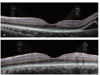
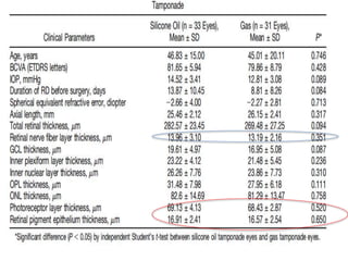
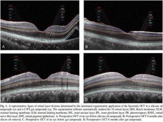
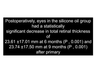
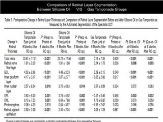
RETINAL LAYER
SEGMENTATION AFTER
SILICONE OIL
OR
GAS TAMPONADE
FOR MACULA-ON RETINAL
DETACHMENT USING OCT
SUNG HOON LEE, MD,* JONG WOOK HAN, MD,* SUK HO BYEON, MD,* SUNG SOO KIM, MD,†
HYOUNG JUN KOH, MD,† SUNG CHUL LEE, MD,† MIN KIM, MD*
RETINA 0:1–10, 2017
To evaluate and compare the effect
of silicone oil and gas on the
thickness of all retinal layers in
eyes with macula-on retinal
detachment.
• Silicone Tamponade = 367
• Gas Tamponade = 310
• Rheg RD NOT involving Macula +
underwent Vitrectomy with R.Error
+/- 5D SE & +/-2D Astig.
RETROSPECTIVE COMPARITIVE INTERVENTIONAL CASE SERIES .
GANGMAN SEVERANCE HOSPITAL
(2011-2015)
• Thickness of all retinal layers & BCVA at
6 months and 9 months after primary
RD surgery with Spectralis OC
• Silicone Oil used of same brand i.e
Oxane 1300 (Bausch & Lomb, Inc, New
York, NY) ,removed after 101+/- 42
days
• Gas C3F8
• Xxxxxxxxxxxxxxxxxxxxxxxxxxxxxxx
DR with ME , Uveitis with ME
Mac.Hole, ERM,Glaucoma & ONH
abnormalities, PFCL usage in Sx, who
required 2nd sx
STUDY DESIGN
• 33 eyes that underwent 1,000 cs silicone oil injection &
• 31 eyes with gas tamponade were finally enrolled in the
study
• Preoperative SD-OCT measurements were performed the day before surgery,
and postoperative measurements were performed 6 months after silicone oil
removal or 6 months after gas tamponade
• The new technique for automated retinal segmentation of SD-OCT retinal images
was applied to distinguish each retinal layer and quantify its thickness
(Segmentation Technology; Heidelberg Engineering, Inc). The inbuilt Spectralis
software, Heidelberg Eye Explorer (version 6.0.9.0), was used for segmentation.
• The Spectralis software measures the total retinal thickness as the vertical
distance between the internal limiting membrane and Bruch membrane.
• The degree of change in thickness of each retinal layer within 1 mm ETDRS
subfields was analyzed.
Postoperatively, eyes in the silicone oil group
had a statistically
significant decrease in total retinal thickness
of
23.61 ±17.01 mm at 6 months (P , 0.001) and
23.74 ±17.50 mm at 9 months (P , 0.001)
after primary
RD surgery
Comparison of Retinal Layer Segmentation
Between Silicone Oil V/S Gas Tamponade Groups
The change in thickness of each retinal layer within a 1 mm ETDRS subfield in the
silicone oil group was compared with that in the gas group.
• Postoperatively, there was a significant drop in BCVA in the silicone oil group
(66.57 ETDRS letters, P = 0.004 at 6 months after RD surgery, 66.35 ETDRS
letters, P = 0.003 at 9 months after RD surgery), but no significant change in the
gas group (79.81 ETDRS letters, P = 0.976).
• The postoperative BCVA in the silicone oil group was significantly worse than
that in the gas group (6 months after RD surgery: P = 0.018, 9 months after RD
surgery: P = 0.016)
• The mean duration of the silicone oil tamponade was 101 days (±42 days). After
adjusting for age, sex, and axial length, silicone oil tamponade duration was found to
have no statistically significant correlation with either postoperative final visual acuity
or the degree of retinal thinning (all P . 0.05).
DISCUSSION
• Silicone oil tamponade had a significant effect on the postoperative
decrease in thickness of all retinal layers, except for the photoreceptor
layer.
• Decreased thickness was noted not only in the inner retinal layer but in
the outer retinal layer as well.
• No significant reduction of retinal thickness in any of the retinal layers
after gas tamponade.
• Decrease of GCL, OPL, and ONL thickness showed the strongest
correlation with changes in visual acuity in the silicone oil group.
SILICONE OIL DAMAGE ACTION
• Possible causes include mechanical stress to the fovea
induced by silicone oil, and subsequent early loss of
ONL cell bodies.
• Emulsified silicone oil may induce an internal limiting
membrane defect and may be toxic to the retina by
entering theM intraretinal space and causing retinal
thinning
Removal of silicone oil, performed as
early as possible, may minimize
complications, such as secondary
glaucoma, cataract, and vision loss,
and may help avoid potential toxic
effects.
• Thank you everyone for patiently listening to this journal by DR.PUSHKAR DHIR
• For feedbacks & brickbats plz mail at
• ykush@yahoo.co.in./drdhir2014@gmail.com
Questions?
• 1. what was FAX? During suregeries
• 2.Any hypothesis why photoreceptor layer
didn’t get affected?
• 3. Any Cryo procedure was done along
• 4. Suregeon same or different?
5.

Silicon vs Gas HRA Segmentation.pptx

  • 1.
    RETINAL LAYER SEGMENTATION AFTER SILICONEOIL OR GAS TAMPONADE FOR MACULA-ON RETINAL DETACHMENT USING OCT SUNG HOON LEE, MD,* JONG WOOK HAN, MD,* SUK HO BYEON, MD,* SUNG SOO KIM, MD,† HYOUNG JUN KOH, MD,† SUNG CHUL LEE, MD,† MIN KIM, MD* RETINA 0:1–10, 2017
  • 2.
    To evaluate andcompare the effect of silicone oil and gas on the thickness of all retinal layers in eyes with macula-on retinal detachment.
  • 3.
    • Silicone Tamponade= 367 • Gas Tamponade = 310 • Rheg RD NOT involving Macula + underwent Vitrectomy with R.Error +/- 5D SE & +/-2D Astig. RETROSPECTIVE COMPARITIVE INTERVENTIONAL CASE SERIES . GANGMAN SEVERANCE HOSPITAL (2011-2015) • Thickness of all retinal layers & BCVA at 6 months and 9 months after primary RD surgery with Spectralis OC • Silicone Oil used of same brand i.e Oxane 1300 (Bausch & Lomb, Inc, New York, NY) ,removed after 101+/- 42 days • Gas C3F8 • Xxxxxxxxxxxxxxxxxxxxxxxxxxxxxxx DR with ME , Uveitis with ME Mac.Hole, ERM,Glaucoma & ONH abnormalities, PFCL usage in Sx, who required 2nd sx STUDY DESIGN • 33 eyes that underwent 1,000 cs silicone oil injection & • 31 eyes with gas tamponade were finally enrolled in the study
  • 4.
    • Preoperative SD-OCTmeasurements were performed the day before surgery, and postoperative measurements were performed 6 months after silicone oil removal or 6 months after gas tamponade • The new technique for automated retinal segmentation of SD-OCT retinal images was applied to distinguish each retinal layer and quantify its thickness (Segmentation Technology; Heidelberg Engineering, Inc). The inbuilt Spectralis software, Heidelberg Eye Explorer (version 6.0.9.0), was used for segmentation. • The Spectralis software measures the total retinal thickness as the vertical distance between the internal limiting membrane and Bruch membrane. • The degree of change in thickness of each retinal layer within 1 mm ETDRS subfields was analyzed.
  • 8.
    Postoperatively, eyes inthe silicone oil group had a statistically significant decrease in total retinal thickness of 23.61 ±17.01 mm at 6 months (P , 0.001) and 23.74 ±17.50 mm at 9 months (P , 0.001) after primary RD surgery
  • 9.
    Comparison of RetinalLayer Segmentation Between Silicone Oil V/S Gas Tamponade Groups
  • 10.
    The change inthickness of each retinal layer within a 1 mm ETDRS subfield in the silicone oil group was compared with that in the gas group. • Postoperatively, there was a significant drop in BCVA in the silicone oil group (66.57 ETDRS letters, P = 0.004 at 6 months after RD surgery, 66.35 ETDRS letters, P = 0.003 at 9 months after RD surgery), but no significant change in the gas group (79.81 ETDRS letters, P = 0.976). • The postoperative BCVA in the silicone oil group was significantly worse than that in the gas group (6 months after RD surgery: P = 0.018, 9 months after RD surgery: P = 0.016) • The mean duration of the silicone oil tamponade was 101 days (±42 days). After adjusting for age, sex, and axial length, silicone oil tamponade duration was found to have no statistically significant correlation with either postoperative final visual acuity or the degree of retinal thinning (all P . 0.05).
  • 11.
    DISCUSSION • Silicone oiltamponade had a significant effect on the postoperative decrease in thickness of all retinal layers, except for the photoreceptor layer. • Decreased thickness was noted not only in the inner retinal layer but in the outer retinal layer as well. • No significant reduction of retinal thickness in any of the retinal layers after gas tamponade. • Decrease of GCL, OPL, and ONL thickness showed the strongest correlation with changes in visual acuity in the silicone oil group.
  • 12.
    SILICONE OIL DAMAGEACTION • Possible causes include mechanical stress to the fovea induced by silicone oil, and subsequent early loss of ONL cell bodies. • Emulsified silicone oil may induce an internal limiting membrane defect and may be toxic to the retina by entering theM intraretinal space and causing retinal thinning
  • 13.
    Removal of siliconeoil, performed as early as possible, may minimize complications, such as secondary glaucoma, cataract, and vision loss, and may help avoid potential toxic effects.
  • 14.
    • Thank youeveryone for patiently listening to this journal by DR.PUSHKAR DHIR • For feedbacks & brickbats plz mail at • ykush@yahoo.co.in./drdhir2014@gmail.com
  • 16.
    Questions? • 1. whatwas FAX? During suregeries • 2.Any hypothesis why photoreceptor layer didn’t get affected? • 3. Any Cryo procedure was done along • 4. Suregeon same or different? 5.