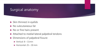
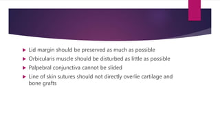
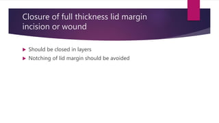
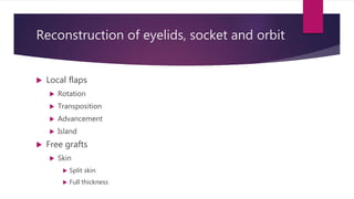
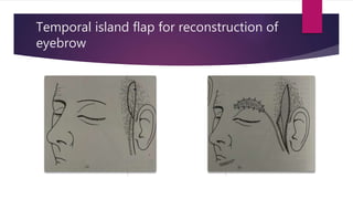
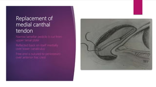
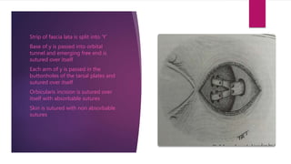
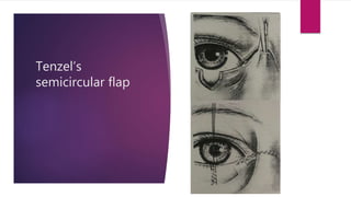
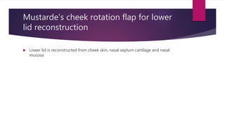
Lid reconstruction
PRESENTED BY:
DR. VANDANA SHARMA
Surgical anatomy
 Skin thinnest in eyelids
 No subcutaneous fat
 No or fine hairs present
 Attached to medial lateral palpebral tendons
 Dimensions of palpebral fissure:
 Vertical: 8 – 11mm
 Horizontal: 25 – 30 mm
 Lateral canthus lies 1-3 mm above medial canthus
 Highest point of palpebral fissure is at junction of medial and
central thirds
 Structures of lid margin:
 Cilia
 Grey line
 Meibomian ducts
 Intermarginal strip has sharp posterior border
 Tarsal plate is firmly attached to connective tissue and skin at
lid margin
 Muscles of the lids:
 Orbicularis oculi
 Levator palpebre superioris
 Corrugator supercilii
 Muller’s muscle
 Orbital septum
 Unites tarsal plate to supra and infra orbital margins
 Not uniform in thickness
 Pierced by vessels and nerves
 Medial palpebral tendon:
 Connects both tarsal plates with medial wall of orbit on lacrimal crest
 Gives origin to orbicularis oculi
 Lateral palpebral tendon
 Closely connected to tarsal plates, lateral raphe of orbicularis oculi
and lateral expansion of LPS
 Attaches on a tubercle about 3 mm posterior to orbital margin
General considerations before surgery
 Final shape and outline of features should be comparable on
both sides
 Patient’s general state of health should be known
 Local condition of the wound should be noted
 Skin defects in the lids should be covered by sliding adjacent
skin or using flap from redundant skin
 Lid margin should be preserved as much as possible
 Orbicularis muscle should be disturbed as little as possible
 Palpebral conjunctiva cannot be slided
 Line of skin sutures should not directly overlie cartilage and
bone grafts
Technique of suturing
 All blood clots and debris should be removed
 Sutures should be applied with 6 – 0 braided silk with spatulated curved
eyeless needle
 Slight eversion of cut edges should be done when sutures are tied
 Colour of skin edge should remain pink
 Sutures should be placed about 6 mm apart
 Skin wound of 3 – 4 mm along skin crease needs no suturing
 Wound of orbicularis at right angles to the muscle should be sutured with
6- 0 absorbable suture
Closure of full thickness lid margin
incision or wound
 Should be closed in layers
 Notching of lid margin should be avoided
Absorbable 6 – 0 suture is placed as
close to lid margin as possible
Single knot is tied and approximation
is assessed
2 more absorbable sutures are passed
through tarsal plate
Silk 6 – 0 suture is passed through
grey line and left loose
1st absorbable suture is then tied
Orbicularis is sutured
Grey line suture is tied and left long
Lash line is sutured
Skin is sutured and long ends of
previous sutures are caught in them
Deep subcutaneous knots
Burying a
subcutaneous knot Figure of 8 stitch
Reconstruction of eyelids, socket and orbit
 Local flaps
 Rotation
 Transposition
 Advancement
 Island
 Free grafts
 Skin
 Split skin
 Full thickness
 Hair bearing
 Mucous membrane
 Other like fat, bone, cartilage etc
 Pedicle grafts
 Expanded grafts
 Muscle grafts
 Free flaps
Local flaps
 Main sources are
 Forehead
 Glabella
 Cheek
 Transposition flaps such as Z plasty may be done
 Advancement flap such as V Y operation may be used
 In island flap, skin is raised in subcutaneous pedicle along
with blood supply from the pedicle and advanced over short
distances
Free grafts
 Skin grafts:
 Split skin grafts:
 Preparation of donor site
 Usually medial aspect of upper arm or thigh
 General anaesthesia is used
 Harvesting the graft
 Donor site lubricated with sterile liquid paraffin or petroleum jelly
 Donor skin stretched with skin graft boards
 Graft taken with skin graft knife
 Dressing donor site
 Dressed with tulle gras and melolin
 Light gauze bandage is applied
 Left undisturbed for 10 days
 Hemostasis achieved at recipient site
 Fixation of graft
 Graft stitched to edges of defect with fine silk
 Dressing with tulle gras and proflavine wool is sutured with the graft
 Post op care
 Inspection of dressing at 24 and 48 hrs
 Removal of sutures after 10 days
 Full thickness grafts
 May be taken from
 Redundant skin in upper eyelid
 post auricular skin
 Pattern of recipient area is taken
 Full thickness graft is taken and donor site is closed
 Skin flap is defatted
 Edges of graft are sutured to edges of the defect
 Proflavine bolster sutured
 Anchoring sutures may be applied
 Daily inspection of graft to ensure no seroma or hematoma is formed
 Stitches removed after 7 – 14 days
 Hair bearing grafts:
 Full thickness free grafts containing hair follicles
 Local scalp flaps containing hair
Temporal island flap for reconstruction of
eyebrow
 Mucous membrane:
 Conjunctiva
 Free conjunctival flaps
 Local transposition of conjunctival flap
 Buccal mucosa
 To cover defects in palpebral and bulbar conjunctiva after injury, burns
excision of neoplasms
 May be taken from inner aspect of lower lip or cheek
 Fat:
 Free fat grafts shrink to almost half of their size
 So not preferred for contour reconstruction
 Used to repair depressed scars
 Cartilage:
 Thin cartilage grafts needed to reconstruct tarsus taken from auricle
or nasal septum
 Cartilage grafts for reconstruction of orbital margins taken from rib
 Bone:
 Reconstruction of orbital floor is done with bone graft from iliac crest
 Periosteal layer should be retained with the grafts
 Fascia:
 Used in ptosis surgery
 Taken from fascia lata or extensor tendon of little toe
Pedicle grafts
 Consists of skin, and subcutaneous tissue along with its own
blood supply
 Skin of temporofrontal region is commonly used in eyelids
 Good color match
 May be hair bearing
 Have to be advanced in stages
Expanded flaps
 To cover large defects, flaps are expanded with tissue
expanders
 More complications are seen with these
Muscle grafts
 Microvascular transplantation of muscle will ensure its
survival at transplanted site
 Function can be retained by preserving nerve supply
Free flaps
 Transfer of skin flaps with their own arterial and venous
supply
 Direct anastomosis of arteries and veins is done under
operating microscope
 Large areas of skin, muscle and cartilage can be transferred
Lateral canthotomy
 Indications:
 To decompress orbit in cases with orbital cellulitis
 Correction of partial ankyloblepharon or blepharophimosis
 In cataract surgery for better exposure of globe as in deep socket
 Procedure:
 Skin at lateral canthus is made taut and lifted up
 Hemostasis is achieved by crushing with mosquito forcep
 One blade of a blunt ended scissors is passed into lateral fornix
 Scissors is closed thereby cutting the tissue
 Closure of canthotomy:
 One interrupted silk suture passed through the two cut ends is
sufficient
Cantholysis
 Indications:
 Reconstruction of full thickness defect for quarter of length of lid or
more
 Facilitate forward movement of lids in tarsorraphy
 Procedure:
 Dicision of one or both bands extending from tarsal plate to lateral
canthal tendon
 Division of orbital septum from orbital margin may also be done
Canthoplasty
 Lateral canthotomy is done
 Bulbar and froniceal conjunctiva is undermined
 Sliding flap of conjunctiva is carried through the skin 4 mm
lateral to extent of canthotomy
 Mattress sutures are passed between skin and conjunctival
flaps to cover the raw surfaces
 If conjunctiva is insufficient, free flaps may be taken
Canthal tendon operations
 Division and fixation of medial canthal tendon:
 Medial canthal tendon may get damaged due to injury or DCR
 May lead to downward displacement of medial canthus
 Repair:
Tendon is exposed
Anchored into a pre bored tunnel in
frontal process of maxilla
If the tendon is short, a non
absorbable suture passed at end of
the tendon is passed into the
tunnel, back into the tendon and
sutured
Replacement of
medial canthal
tendon
Narrow lamellar pedicle is cut from
upper tarsal plate
Reflected back on itself medially
over lower canaliculus
Free end is sutured to periosteum
over anterior iliac crest
 Division of lateral palpebral tendon:
 Done in rapidly progressive exophthalmos
 Procedure:
 Lateral canthotomy is done
 Lateral margin of orbit is identified
 Blunt ended scissors is spread posteriorly upto orbital tubercle
 Scissors is turned so that the blades open in vertical direction
 Tendon is engaged and cut
 Reconstruction of lateral palpebral tendon
 Fascia lata may be used
 Procedure:
 1.5 cm long incision is made at lateral canthus along skin crease
 Orbicularis is retracted
 Lateral ends of upper and lower tarsal plates are identified
 Vertical incisions are made into each tarsus 4 mm long, 3 mm from its
ends
 Tunnel of diameter 4 mm is drilled into orbital wall after retracting orbital
contents medially
Strip of fascia lata is split into ‘Y’
Base of y is passed into orbital
tunnel and emerging free end is
sutured over itself
Each arm of y is passed in the
buttonholes of the tarsal plates and
sutured over itself
Orbicularis incision is sutured over
itself with absorbable sutures
Skin is sutured with non absorbable
sutures
Closure of lid defects
 Small defects:
 Closed by advancement of adjacent skin or V – Y plasty
 Full thickness defect for quarter of length or less:
 Modified Wheeler’s operation is done
 Full thickness defect for more then quarter of length:
 Mustarde’s operation
 Upper lid rotation from lower lid
 Loss of more than half of eyelid
 Cheek rotation flap for reconstruction of lowerlid
 Lower lid rotation and cheek rotation flap for upper lid reconstruction
Modified wheeler’s
operation for lid
defects
Mustarde’s
operation for large
defects
Lower lid reconstruction
Tenzel’s
semicircular flap
Mustarde’s cheek rotation flap for lower
lid reconstruction
 Lower lid is reconstructed from cheek skin, nasal septum cartilage and nasal
mucosa
Mustarde’s cheek
rotation flap
Mobilisation of cheek flap
Nasal mucous membrane and septal
cartilage graft
• size of mucous membrane graft: 2.5
cm long and 1.5 cm wide
Fixation of cheek flap
Hughe’s
tarsoconjunctival
advancement graft
Reconstruction of upper lid
 Severe damage to upper lid is rare
 Immediately jeopardizes safety of the eye
 Points to be considered while reconstructing upper lid
 Should be lined with conjunctiva or buccal mucosa
 Should contain a supporting plate
 Should have a muscle action if possible in form of orbicularis or LPS attachment
Mustarde’s
rotation flap from
lower lid
 LPS may be torn in trauma
 Retraction is prevented by lateral attachments of the muscle
 Repair:
 Local anaesthetic is injected
 Site of torn aponeurosis is revealed by asking the patient to look up
 Aponeurosis is held in a clamp and sutured to anterior surface of tarsus with 4 – 0
absorbable sutures
 If primary repair has not been done timely, 6 – 12 months should be allowed to elapse
before attempting repair
Mustarde’s operation for reconstruction of
upper lid in 2 stages
 Broad based full thickness flap of lower lid is made
 Rotated into the defect as far as possible and sutured
 2 weeks later base of flap is cut and remaining flap sutured to remaining defect
 Lower lid is reconstructed with cheek flap and nasal septal cartilage and mucous
membrane
 Principles for creating lower lid flaps:
 If remnant of upper lid is present: hinge made on same side
 If defect is total or central: hinge is placed laterally
 Lacrimal punctum is not included in the flap
Mustarde’s
operation for
reconstruction of
upper lid in 2
stages
Cutler beard
technique
Neoplasm excision
 Proliferative type of BCC does not invade deeper tissues
 Lesions over lid margins also do not tend to involve tarsal plate
 2 different sets of instruments should be used for excision of tumor and plastic
repair
 Excision of neoplasm:
 Proliferative lesion 3 mm from lid margin: lid margin structures should be preserved
 Deeply infiltrative lesions: full thickness resection has to be done
Excision of neoplasms
Collar stud excision
with partial
tarsectomy
Done when main mass of lesion lies
away from lid margin with possible
involvement of tarsus at lid margin
Tarsal coloboma is closed with
interrupted absorbable sutures
Cantholysis may be used
Tarsal sutures are tied
Orbicularis bridge flap is sutuered
Full thickness skin flap is placed
over the defect
 Medial canthus reconstruction:
 When plane of excision is not upto periosteum: full thickness skin grafts are used
 When plane of excision is upto periosteum:
 V Y advancement of frontoglabellar flap for defect upto 10 mm
 Midline frontal pedicle for defect more than 10 mm
Frontoglabellar flap
Inverted V incision is given
Flap is mobilized
Defect is closed
Flap is sutures in Y shape
Free conjunctival or buccal mucosal
flap is attached to undersurface
Medial end is sutured to periosteum
Flap is cut in centre to create upper
and lower lid
Midline frontal pedicle flap
Incision is given on glabella
and flap is raised
Anterior end of flap is divided
so as to fit upper and lower
lid defects
Anterior end is lined by free
conjunctival or buccal
mucosal flap
Anterior end of flap is
sutured to the defects
Vertical incisions on forehead
are undermined,
approximated and sutured
Medial limit of graft is
marked
Tulle is passed beneath the
pedicle bridge
After 18 days, pedicle is cut at
marked site
Fibrous tissue from
undersurface is excised and it
is sutured back to frontal
region
Medial edge of graft is
sutured
Total loss of lower lid and partial loss of
upper lid
 Lower lid is reconstructed using cheek flap
 If medial half of upper lid is lost, midline frontoglabellar pedicle flap is used
 If lateral half of upper lid is lost, temporofrontal flap is used
Total loss of upper lid and partial loss of
lower lid
 If lateral part of lower lid is conserved it is rotated to medial side of upper lid
 Lateral part of upper lid is reconstructed with temporofrontal flap
 Lower part of lid is reconstructed with cheek flap
 When medial part of lower lid is conserved
 Lateral defect closed with cheek flap
 Upper lid reconstructed with supraorbital flap
Total loss of both eyelids
 Four stage repair is done
 Stage 1:
 Remnants of conjunctiva mobilized and sutured with absorbable suture to cover the
cornea
 Thin shaving of auricular cartilage may be placed to replace tarsus
 Bridge pedicles of orbicularis are placed over the eye
 Split skin graft is placed
 Stage 2:
 After 1 month, split skin graft is replaced with full thickness skin graft
 Stage 3:
 Eyebrow bearing transplants may be used for creating lash line
 1 – 2 mm skin is kept between the two lash lines
 Stage 4:
 After 3 – 4 months after 1st operation, tarsorraphy is divided
Bibliography
 Stallard’s eye surgery;7th edition
 Lids, orbits, extraocular muscles, volume 1; Von noorden
Lid reconstruction

Lid reconstruction

  • 1.
  • 2.
    Surgical anatomy  Skinthinnest in eyelids  No subcutaneous fat  No or fine hairs present  Attached to medial lateral palpebral tendons  Dimensions of palpebral fissure:  Vertical: 8 – 11mm  Horizontal: 25 – 30 mm
  • 3.
     Lateral canthuslies 1-3 mm above medial canthus  Highest point of palpebral fissure is at junction of medial and central thirds  Structures of lid margin:  Cilia  Grey line  Meibomian ducts
  • 4.
     Intermarginal striphas sharp posterior border  Tarsal plate is firmly attached to connective tissue and skin at lid margin  Muscles of the lids:  Orbicularis oculi  Levator palpebre superioris  Corrugator supercilii  Muller’s muscle
  • 5.
     Orbital septum Unites tarsal plate to supra and infra orbital margins  Not uniform in thickness  Pierced by vessels and nerves  Medial palpebral tendon:  Connects both tarsal plates with medial wall of orbit on lacrimal crest  Gives origin to orbicularis oculi
  • 6.
     Lateral palpebraltendon  Closely connected to tarsal plates, lateral raphe of orbicularis oculi and lateral expansion of LPS  Attaches on a tubercle about 3 mm posterior to orbital margin
  • 7.
    General considerations beforesurgery  Final shape and outline of features should be comparable on both sides  Patient’s general state of health should be known  Local condition of the wound should be noted  Skin defects in the lids should be covered by sliding adjacent skin or using flap from redundant skin
  • 8.
     Lid marginshould be preserved as much as possible  Orbicularis muscle should be disturbed as little as possible  Palpebral conjunctiva cannot be slided  Line of skin sutures should not directly overlie cartilage and bone grafts
  • 9.
    Technique of suturing All blood clots and debris should be removed  Sutures should be applied with 6 – 0 braided silk with spatulated curved eyeless needle  Slight eversion of cut edges should be done when sutures are tied  Colour of skin edge should remain pink  Sutures should be placed about 6 mm apart  Skin wound of 3 – 4 mm along skin crease needs no suturing  Wound of orbicularis at right angles to the muscle should be sutured with 6- 0 absorbable suture
  • 10.
    Closure of fullthickness lid margin incision or wound  Should be closed in layers  Notching of lid margin should be avoided
  • 12.
    Absorbable 6 –0 suture is placed as close to lid margin as possible Single knot is tied and approximation is assessed 2 more absorbable sutures are passed through tarsal plate Silk 6 – 0 suture is passed through grey line and left loose 1st absorbable suture is then tied Orbicularis is sutured Grey line suture is tied and left long Lash line is sutured Skin is sutured and long ends of previous sutures are caught in them
  • 13.
    Deep subcutaneous knots Buryinga subcutaneous knot Figure of 8 stitch
  • 14.
    Reconstruction of eyelids,socket and orbit  Local flaps  Rotation  Transposition  Advancement  Island  Free grafts  Skin  Split skin  Full thickness
  • 15.
     Hair bearing Mucous membrane  Other like fat, bone, cartilage etc  Pedicle grafts  Expanded grafts  Muscle grafts  Free flaps
  • 16.
    Local flaps  Mainsources are  Forehead  Glabella  Cheek  Transposition flaps such as Z plasty may be done  Advancement flap such as V Y operation may be used  In island flap, skin is raised in subcutaneous pedicle along with blood supply from the pedicle and advanced over short distances
  • 17.
    Free grafts  Skingrafts:  Split skin grafts:  Preparation of donor site  Usually medial aspect of upper arm or thigh  General anaesthesia is used  Harvesting the graft  Donor site lubricated with sterile liquid paraffin or petroleum jelly  Donor skin stretched with skin graft boards  Graft taken with skin graft knife
  • 18.
     Dressing donorsite  Dressed with tulle gras and melolin  Light gauze bandage is applied  Left undisturbed for 10 days  Hemostasis achieved at recipient site  Fixation of graft  Graft stitched to edges of defect with fine silk  Dressing with tulle gras and proflavine wool is sutured with the graft
  • 19.
     Post opcare  Inspection of dressing at 24 and 48 hrs  Removal of sutures after 10 days
  • 20.
     Full thicknessgrafts  May be taken from  Redundant skin in upper eyelid  post auricular skin  Pattern of recipient area is taken  Full thickness graft is taken and donor site is closed  Skin flap is defatted  Edges of graft are sutured to edges of the defect  Proflavine bolster sutured
  • 21.
     Anchoring suturesmay be applied  Daily inspection of graft to ensure no seroma or hematoma is formed  Stitches removed after 7 – 14 days
  • 22.
     Hair bearinggrafts:  Full thickness free grafts containing hair follicles  Local scalp flaps containing hair
  • 23.
    Temporal island flapfor reconstruction of eyebrow
  • 24.
     Mucous membrane: Conjunctiva  Free conjunctival flaps  Local transposition of conjunctival flap  Buccal mucosa  To cover defects in palpebral and bulbar conjunctiva after injury, burns excision of neoplasms  May be taken from inner aspect of lower lip or cheek
  • 25.
     Fat:  Freefat grafts shrink to almost half of their size  So not preferred for contour reconstruction  Used to repair depressed scars  Cartilage:  Thin cartilage grafts needed to reconstruct tarsus taken from auricle or nasal septum  Cartilage grafts for reconstruction of orbital margins taken from rib
  • 26.
     Bone:  Reconstructionof orbital floor is done with bone graft from iliac crest  Periosteal layer should be retained with the grafts  Fascia:  Used in ptosis surgery  Taken from fascia lata or extensor tendon of little toe
  • 27.
    Pedicle grafts  Consistsof skin, and subcutaneous tissue along with its own blood supply  Skin of temporofrontal region is commonly used in eyelids  Good color match  May be hair bearing  Have to be advanced in stages
  • 28.
    Expanded flaps  Tocover large defects, flaps are expanded with tissue expanders  More complications are seen with these
  • 29.
    Muscle grafts  Microvasculartransplantation of muscle will ensure its survival at transplanted site  Function can be retained by preserving nerve supply
  • 30.
    Free flaps  Transferof skin flaps with their own arterial and venous supply  Direct anastomosis of arteries and veins is done under operating microscope  Large areas of skin, muscle and cartilage can be transferred
  • 31.
    Lateral canthotomy  Indications: To decompress orbit in cases with orbital cellulitis  Correction of partial ankyloblepharon or blepharophimosis  In cataract surgery for better exposure of globe as in deep socket  Procedure:  Skin at lateral canthus is made taut and lifted up  Hemostasis is achieved by crushing with mosquito forcep
  • 32.
     One bladeof a blunt ended scissors is passed into lateral fornix  Scissors is closed thereby cutting the tissue  Closure of canthotomy:  One interrupted silk suture passed through the two cut ends is sufficient
  • 33.
    Cantholysis  Indications:  Reconstructionof full thickness defect for quarter of length of lid or more  Facilitate forward movement of lids in tarsorraphy  Procedure:  Dicision of one or both bands extending from tarsal plate to lateral canthal tendon  Division of orbital septum from orbital margin may also be done
  • 34.
    Canthoplasty  Lateral canthotomyis done  Bulbar and froniceal conjunctiva is undermined  Sliding flap of conjunctiva is carried through the skin 4 mm lateral to extent of canthotomy  Mattress sutures are passed between skin and conjunctival flaps to cover the raw surfaces  If conjunctiva is insufficient, free flaps may be taken
  • 35.
    Canthal tendon operations Division and fixation of medial canthal tendon:  Medial canthal tendon may get damaged due to injury or DCR  May lead to downward displacement of medial canthus  Repair:
  • 36.
    Tendon is exposed Anchoredinto a pre bored tunnel in frontal process of maxilla If the tendon is short, a non absorbable suture passed at end of the tendon is passed into the tunnel, back into the tendon and sutured
  • 37.
    Replacement of medial canthal tendon Narrowlamellar pedicle is cut from upper tarsal plate Reflected back on itself medially over lower canaliculus Free end is sutured to periosteum over anterior iliac crest
  • 38.
     Division oflateral palpebral tendon:  Done in rapidly progressive exophthalmos  Procedure:  Lateral canthotomy is done  Lateral margin of orbit is identified  Blunt ended scissors is spread posteriorly upto orbital tubercle  Scissors is turned so that the blades open in vertical direction  Tendon is engaged and cut
  • 39.
     Reconstruction oflateral palpebral tendon  Fascia lata may be used  Procedure:  1.5 cm long incision is made at lateral canthus along skin crease  Orbicularis is retracted  Lateral ends of upper and lower tarsal plates are identified  Vertical incisions are made into each tarsus 4 mm long, 3 mm from its ends  Tunnel of diameter 4 mm is drilled into orbital wall after retracting orbital contents medially
  • 40.
    Strip of fascialata is split into ‘Y’ Base of y is passed into orbital tunnel and emerging free end is sutured over itself Each arm of y is passed in the buttonholes of the tarsal plates and sutured over itself Orbicularis incision is sutured over itself with absorbable sutures Skin is sutured with non absorbable sutures
  • 41.
    Closure of liddefects  Small defects:  Closed by advancement of adjacent skin or V – Y plasty  Full thickness defect for quarter of length or less:  Modified Wheeler’s operation is done  Full thickness defect for more then quarter of length:  Mustarde’s operation  Upper lid rotation from lower lid  Loss of more than half of eyelid  Cheek rotation flap for reconstruction of lowerlid  Lower lid rotation and cheek rotation flap for upper lid reconstruction
  • 42.
  • 43.
  • 44.
  • 45.
  • 46.
    Mustarde’s cheek rotationflap for lower lid reconstruction  Lower lid is reconstructed from cheek skin, nasal septum cartilage and nasal mucosa
  • 47.
    Mustarde’s cheek rotation flap Mobilisationof cheek flap Nasal mucous membrane and septal cartilage graft • size of mucous membrane graft: 2.5 cm long and 1.5 cm wide Fixation of cheek flap
  • 48.
  • 50.
    Reconstruction of upperlid  Severe damage to upper lid is rare  Immediately jeopardizes safety of the eye  Points to be considered while reconstructing upper lid  Should be lined with conjunctiva or buccal mucosa  Should contain a supporting plate  Should have a muscle action if possible in form of orbicularis or LPS attachment
  • 51.
  • 52.
     LPS maybe torn in trauma  Retraction is prevented by lateral attachments of the muscle  Repair:  Local anaesthetic is injected  Site of torn aponeurosis is revealed by asking the patient to look up  Aponeurosis is held in a clamp and sutured to anterior surface of tarsus with 4 – 0 absorbable sutures  If primary repair has not been done timely, 6 – 12 months should be allowed to elapse before attempting repair
  • 53.
    Mustarde’s operation forreconstruction of upper lid in 2 stages  Broad based full thickness flap of lower lid is made  Rotated into the defect as far as possible and sutured  2 weeks later base of flap is cut and remaining flap sutured to remaining defect  Lower lid is reconstructed with cheek flap and nasal septal cartilage and mucous membrane  Principles for creating lower lid flaps:  If remnant of upper lid is present: hinge made on same side  If defect is total or central: hinge is placed laterally  Lacrimal punctum is not included in the flap
  • 54.
  • 55.
  • 57.
    Neoplasm excision  Proliferativetype of BCC does not invade deeper tissues  Lesions over lid margins also do not tend to involve tarsal plate  2 different sets of instruments should be used for excision of tumor and plastic repair  Excision of neoplasm:  Proliferative lesion 3 mm from lid margin: lid margin structures should be preserved  Deeply infiltrative lesions: full thickness resection has to be done
  • 58.
  • 59.
    Collar stud excision withpartial tarsectomy Done when main mass of lesion lies away from lid margin with possible involvement of tarsus at lid margin Tarsal coloboma is closed with interrupted absorbable sutures Cantholysis may be used
  • 60.
    Tarsal sutures aretied Orbicularis bridge flap is sutuered Full thickness skin flap is placed over the defect
  • 61.
     Medial canthusreconstruction:  When plane of excision is not upto periosteum: full thickness skin grafts are used  When plane of excision is upto periosteum:  V Y advancement of frontoglabellar flap for defect upto 10 mm  Midline frontal pedicle for defect more than 10 mm
  • 62.
    Frontoglabellar flap Inverted Vincision is given Flap is mobilized Defect is closed Flap is sutures in Y shape Free conjunctival or buccal mucosal flap is attached to undersurface Medial end is sutured to periosteum Flap is cut in centre to create upper and lower lid
  • 63.
    Midline frontal pedicleflap Incision is given on glabella and flap is raised Anterior end of flap is divided so as to fit upper and lower lid defects Anterior end is lined by free conjunctival or buccal mucosal flap Anterior end of flap is sutured to the defects Vertical incisions on forehead are undermined, approximated and sutured Medial limit of graft is marked Tulle is passed beneath the pedicle bridge After 18 days, pedicle is cut at marked site Fibrous tissue from undersurface is excised and it is sutured back to frontal region Medial edge of graft is sutured
  • 64.
    Total loss oflower lid and partial loss of upper lid  Lower lid is reconstructed using cheek flap  If medial half of upper lid is lost, midline frontoglabellar pedicle flap is used  If lateral half of upper lid is lost, temporofrontal flap is used
  • 65.
    Total loss ofupper lid and partial loss of lower lid  If lateral part of lower lid is conserved it is rotated to medial side of upper lid  Lateral part of upper lid is reconstructed with temporofrontal flap  Lower part of lid is reconstructed with cheek flap  When medial part of lower lid is conserved  Lateral defect closed with cheek flap  Upper lid reconstructed with supraorbital flap
  • 66.
    Total loss ofboth eyelids  Four stage repair is done  Stage 1:  Remnants of conjunctiva mobilized and sutured with absorbable suture to cover the cornea  Thin shaving of auricular cartilage may be placed to replace tarsus  Bridge pedicles of orbicularis are placed over the eye  Split skin graft is placed
  • 67.
     Stage 2: After 1 month, split skin graft is replaced with full thickness skin graft  Stage 3:  Eyebrow bearing transplants may be used for creating lash line  1 – 2 mm skin is kept between the two lash lines  Stage 4:  After 3 – 4 months after 1st operation, tarsorraphy is divided
  • 68.
    Bibliography  Stallard’s eyesurgery;7th edition  Lids, orbits, extraocular muscles, volume 1; Von noorden