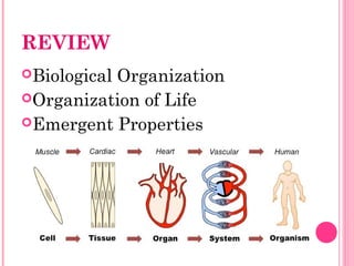
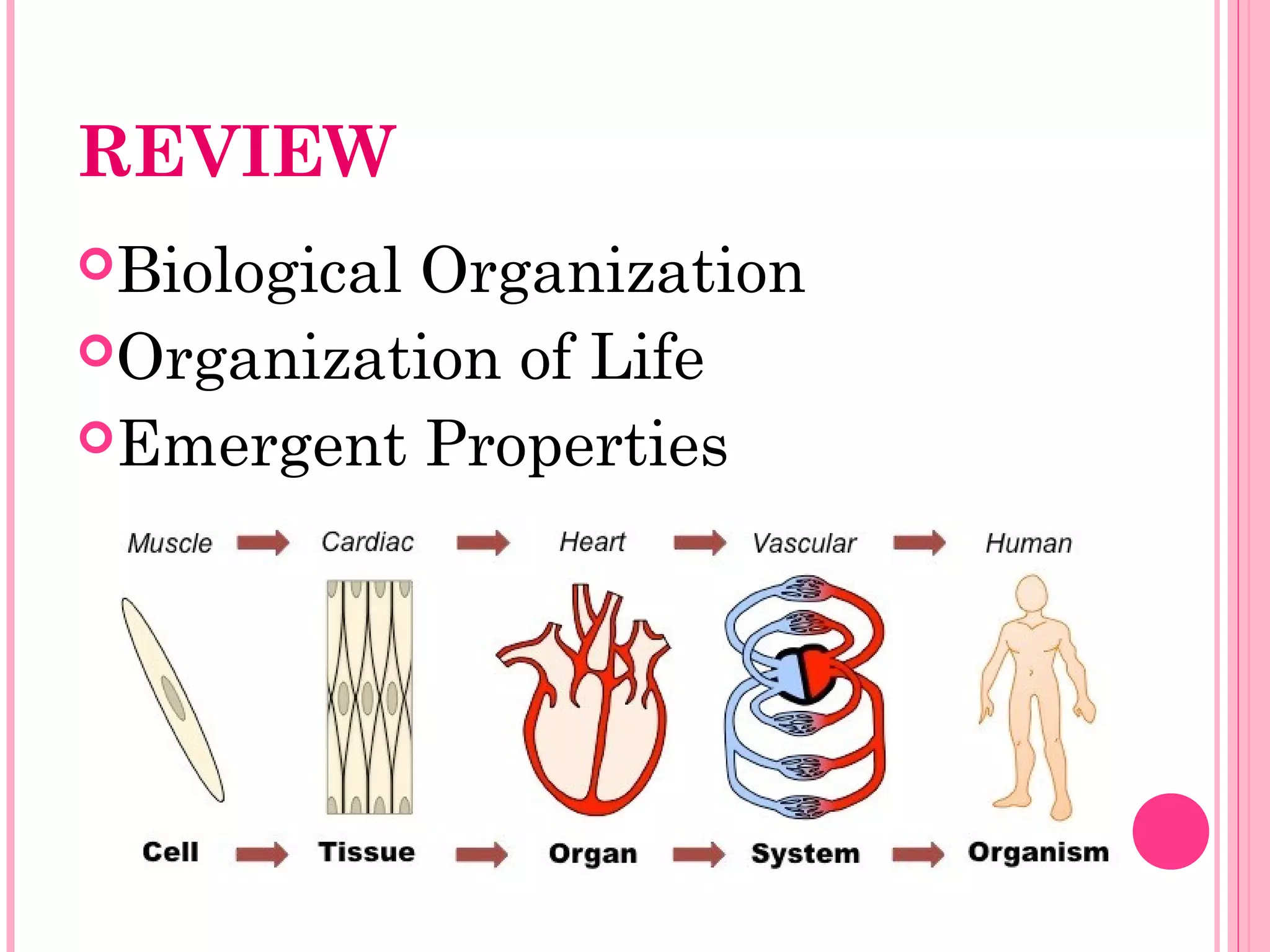
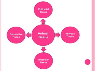
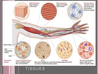
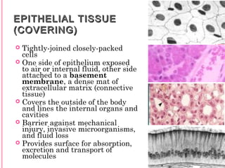
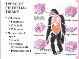
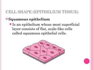
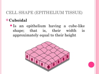
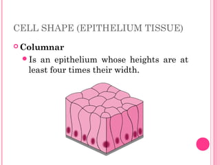
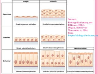
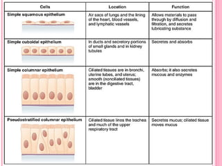
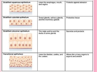
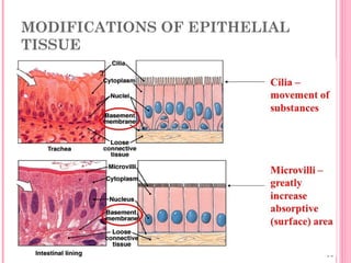
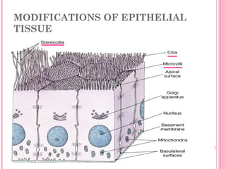
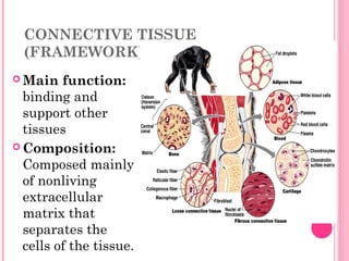
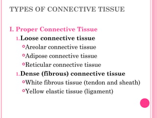
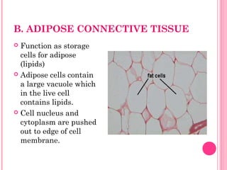
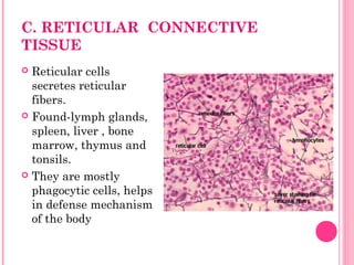
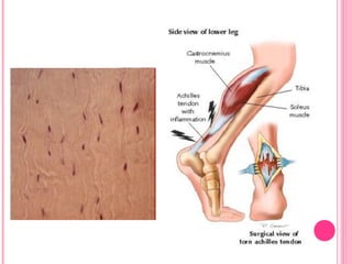
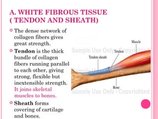
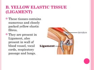
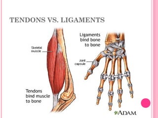
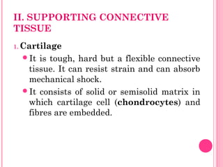
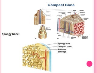
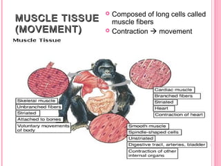
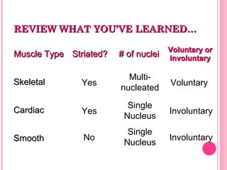
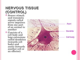
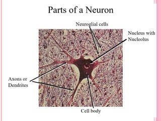
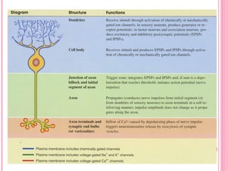
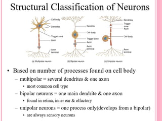
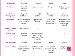
REVIEW
Biological Organization
Organization of Life
Emergent Properties
STRUCTURE AND
FUNCTIONS OF
ANIMAL AND PLANT
TISSUES
Prepared by:
MS. LOUIE JANE T. ELECCION
Animal
Tissue
Epithelial
Tissue
Nervous
Tissue
Muscular
Tissue
Connective
Tissue
FOUR TYPES OF ANIMALFOUR TYPES OF ANIMAL
TISSUESTISSUES
EPITHELIAL TISSUEEPITHELIAL TISSUE
(COVERING)(COVERING)
 Tightly-joined closely-packed
cells
 One side of epithelium exposed
to air or internal fluid, other side
attached to a basement
membrane, a dense mat of
extracellular matrix (connective
tissue)
 Covers the outside of the body
and lines the internal organs and
cavities
 Barrier against mechanical
injury, invasive microorganisms,
and fluid loss
 Provides surface for absorption,
excretion and transport of
molecules
TYPES OFTYPES OF
EPITHELIALEPITHELIAL
TISSUETISSUE
 Cell shape
Squamous
Cuboidal
Columnar
 Number of cell
layers
Simple
Stratified
(Transitional)
Pseudostratified
CELL SHAPE (EPITHELIUM TISSUE)
 Squamous epithelium
Is an epithelium whose most superficial
layer consists of flat, scale-like cells
called squamous epithelial cells.
CELL SHAPE (EPITHELIUM TISSUE)
 Cuboidal
Is an epithelium having a cube-like
shape; that is, their width is
approximately equal to their height
CELL SHAPE (EPITHELIUM TISSUE)
 Columnar
Is an epithelium whose heights are at
least four times their width.
Source:
Biologydictionary.net
Editors. (2014).
Organ. Retrieved
November 4, 2014,
from
https://biologydictionary.ne
MODIFICATIONS OF EPITHELIAL
TISSUE
MODIFICATIONS OF EPITHELIAL
TISSUE
CHECK YOUR UNDERSTANDING
1. Which of these epithelia can stretch
to increase the volume of the organ
based on internal water pressure?
A. Simple squamous epithelia
B. Simple columnar epithelia
C. Transitional epithelia
D. Stratified columnar epithelia
CHECK YOUR UNDERSTANDING
2. How is a pseudostratified epithelium
different from a truly stratified tissue?
A. The nuclei are at the same level in
pseudostratified epithelia.
B. The apical and basal surfaces are aligned.
C. There are multiple cell layers in
pseudostratified epithelia.
D. All the cells in a pseudostratified epithelium
interact with the basement membrane.
CHECK YOUR UNDERSTANDING
3. Which of these is an important function of
epithelial cells?
A. Protection from chemical abrasion
B. Secretion of hormones and enzymes
C. Absorption of nutrients
D. All of the above
CONNECTIVE TISSUE
(FRAMEWORK)
 Main function:
binding and
support other
tissues
 Composition:
Composed mainly
of nonliving
extracellular
matrix that
separates the
cells of the tissue.
FUNCTIONS OF CONNECTIVE
TISSUE
 It binds various tissue together like skin
with the muscles and muscles with bones
 It forms intercellular substance between
cells of different types of tissue, so that it
will help in friction less movement of the
body organ
 It forms sheaths around the body organs
and make a kind of packaging tissue
TYPES OF CONNECTIVE TISSUE
I. Proper Connective Tissue
1.Loose connective tissue
Areolar connective tissue
Adipose connective tissue
Reticular connective tissue
1.Dense (fibrous) connective tissue
White fibrous tissue (tendon and sheath)
Yellow elastic tissue (ligament)
TYPES OF CONNECTIVE TISSUE
II. Supporting Connective Tissue
1.Cartilage
 Hyaline cartilage, Elastic cartilage,
Fibrous cartilage
1.Bone
 Spongy bone, Compact bone
III. Fluid or Liquid Connective Tissue
1. Blood
2. Lymph
I. PROPER CONNECTIVE TISSUE
1. Loose Connective Tissue
This tissue fills the space between organs
and serves as packing materials
surrounding the elements of other tissues
This binds muscle cells together and binds
skin to underlying tissues.
a. Areolar CT
b. Adipose CT
c. Reticular CT
A. AREOLAR CONNECTIVE TISSUE
 Gel like ground with both
elastic and non-elastic fibers
running though the ground
in many directions.
 Fibers are loosely arranged
and there is space between
fibers, areolae, which
derives its name.
 Wraps and cushions organs
under the skin.
B. ADIPOSE CONNECTIVE TISSUE
 Function as storage
cells for adipose
(lipids)
 Adipose cells contain
a large vacuole which
in the live cell
contains lipids.
 Cell nucleus and
cytoplasm are pushed
out to edge of cell
membrane.
C. RETICULAR CONNECTIVE
TISSUE
 Reticular cells
secretes reticular
fibers.
 Found-lymph glands,
spleen, liver , bone
marrow, thymus and
tonsils.
 They are mostly
phagocytic cells, helps
in defense mechanism
of the body
II. PROPER CONNECTIVE TISSUE
2. Dense (fibrous) Connective Tissue
Made up of thick collagen fibers and dark,
compressed cells between the fiber
bundles.
Functions: (1) for flexibility and support,
(2) shock absorption and (3) reduction of
friction.
Ex. Tendon, ligament, urinary tract
and collagen
A. WHITE FIBROUS TISSUE
( TENDON AND SHEATH)
 The dense network of
collagen fibers gives
great strength.
 Tendon is the thick
bundle of collagen
fibers running parallel
to each other, giving
strong, flexible but
inextensible strength.
It joins skeletal
muscles to bones.
 Sheath forms
covering of cartilage
and bones.
B. YELLOW ELASTIC TISSUE
(LIGAMENT)
 These tissues contains
numerous and closely
packed yellow elastic
fibres.
 They are present in
Ligament, also
present in wall of
blood vessel, vocal
cords, respiratory
passage and lungs.
TENDONS VS. LIGAMENTS
II. SUPPORTING CONNECTIVE
TISSUE
1. Cartilage
It is tough, hard but a flexible connective
tissue. It can resist strain and can absorb
mechanical shock.
It consists of solid or semisolid matrix in
which cartilage cell (chondrocytes) and
fibres are embedded.
II. SUPPORTING CONNECTIVE
TISSUE
1. Cartilage
Structure:
cartilage is enclosed in a sheath of white fibrous
connective tissue called Below the perichondrium,
there is layer chondroblast cell, which form
chondrocytes.
Chondrocytes are dispersed in the matrix and
occur in the fluid filled space called lacunae.
The matrix lacks the blood vessels.
II. SUPPORTING CONNECTIVE
TISSUE
1. Cartilage
3 Types:
Hyaline cartilage
The matrix is glass like semi transparent,
homogenous and has fine collagen fibres
It is slightly elastic and compressible.
Found in- nose, larynx, trachea, ends of bone. It
forms skeleton of cartilage fish
Elastic cartilage
Fibrous cartilage
II. SUPPORTING CONNECTIVE
TISSUE
1. Cartilage
3 Types:
Hyaline cartilage
Elastic cartilage
The matrix is semi opaque and has network of
yellow elastic fibres
It is highly elastic and flexible. The tissue
recover the shape quickly
Found in-external ear, epiglottis, and pharynx
Fibrous cartilage
II. SUPPORTING CONNECTIVE
TISSUE
1. Cartilage
3 Types:
Hyaline cartilage
Elastic cartilage
Fibrous cartilage
The matrix has bundles of densely packed
white collagen fibres
It provides great strength and little degree of
flexibility
It acts as a shock absorber by giving a
cushioning effect
Found in- intervertebral discs and at pubis
symphysis
II. SUPPORTING CONNECTIVE
TISSUE
2. Bone
• It is supportive and protective tissue.
• The matrix is solid and calcified.
• Bone cell (Osteocyte) and collagen fibre are
embedded in solid matrix
II. SUPPORTING CONNECTIVE
TISSUE
2. Bone
Structure:
Each bone is enclosed in a layer of white fibrous
connective tissue, called periosteum.
Matrix is arranged in concentric circles called
lamellae.
Number of osteoblast cell and osteocytes are arranged
in between the lamellae, in the fluid filed space called
Lacunae.
Each lacunae has fine cytoplasmic processes called
canaliculi, which connect with other lacunae.
Osteoblast are active bone cell, while osteocytes are
inactive osteroblast.
II. SUPPORTING CONNECTIVE
TISSUE
2. Bone
Types:
Compact Bone
 In the centre of compact bone, bone marrow cavity
is present, filled with yellow bone marrow.
Spongy Bone
The matrix is web like rather than solid with
number of space in between.
Also known as trabeculae
Found in epiphysis of long bone
It contains red bone marrow, which forms RBC and
WBC.
III. FLUID OR LIQUID CONNECTIVE
TISSUE
MUSCLE TISSUEMUSCLE TISSUE
(MOVEMENT)(MOVEMENT)
 Composed of long cells called
muscle fibers
 Contraction  movement
CLASSIFICATION OF MUSCLE
CELLS
 Striated vs. Nonstriated
striated have a banded appearance (stripes)
 Single nucleus or multinucleated cells
 Muscle cells can be controlled
voluntarily (consciously)
or
involuntarily (automatically)
Skeletal Muscle CellsSkeletal Muscle Cells

Striated, voluntary, and multinucleatedStriated, voluntary, and multinucleated

are long, thin and cylindrical; they areare long, thin and cylindrical; they are
attached to bones and move our skeletonattached to bones and move our skeleton

are usually called “are usually called “muscle fibersmuscle fibers””

do not divide to create new cellsdo not divide to create new cells

new fibers are produced by stem cellsnew fibers are produced by stem cells

Striations -- cross stripes (bands) runStriations -- cross stripes (bands) run
perpendicular to the cellsperpendicular to the cells
SKELETAL MUSCLE TISSUE
CARDIAC MUSCLE CELLSCARDIAC MUSCLE CELLS
are called “cardiocytescardiocytes” and found in
heart walls
branching cells connect at intercalatedintercalated
disksdisks which allow contractions to occur
faster
are regulated by pacemaker cells which
control contraction of the heart muscles
striated, involuntary, and singlestriated, involuntary, and single
nucleusnucleus
CARDIAC MUSCLE TISSUE
SMOOTH MUSCLE CELLSSMOOTH MUSCLE CELLS
are small and pointed at their ends
can divide and regenerate new cells
non-striated, involuntary, and singlenon-striated, involuntary, and single
nucleusnucleus
found in hollow organs like the intestine,
bladder, lungs, and blood vessels
move substances through hollow opening
by contracting slowly; they squeeze things
through like a tube of toothpaste
SMOOTH MUSCLE TISSUE
REVIEW WHAT YOU’VE LEARNED…REVIEW WHAT YOU’VE LEARNED…
Muscle TypeMuscle Type Striated?Striated? # of nuclei# of nuclei Voluntary orVoluntary or
InvoluntaryInvoluntary
SkeletalSkeletal
CardiacCardiac
SmoothSmooth
Yes
Yes
No
Single
Nucleus
Single
Nucleus
Multi-
nucleated
Voluntary
Involuntary
Involuntary
NERVOUS TISSUENERVOUS TISSUE
(CONTROL)(CONTROL)
 Senses stimuli
and transmits
signals called
nerve impulses
from one part
of an animal to
another
 Consists of a
cell body and
long extensions
called dendrites
(towards cell
body) and
axons (towards
another cell or
an effector)
Axon
Dendrite
Cell body
Tissue Type Epithelial Connective Muscle Nerve
Cell Shape Flattened,
cuboidal,
columnar
Irregular or round Elongated Cell appendages
branched
Cell
Arrangement
Single 
multilayered
Scattered in
matrix
In sheets or
bundles
Isolated or
networked
Location Body covering or
lining organs or
cavities
Supports other
organs
Lining internal
organs, make
skeletal muscles
Concentrated in
brain and spinal
cord + all over
the body
Surface Feature
of Cells
Cilia, microvilli - - -
Matrix Type Basement
membrane
Varied – protein
fibers + liquid,
gelatinous, firm
to calcified
- -
Matrix Amount Minimal Extensive Absent Absent
Unique Feature No direct blood
supply, except for
glands
Cartilage has no
blood supply
Can generate
electrical signals,
force and
movement
Can generate
electrical signal
REFERENCES
 Biologydictionary.net Editors. (2014). Organ.
Retrieved November 4, 2014, from
https://biologydictionary.net/organ/
 http://www.onlinebiologynotes.com/connective-tissue-charac

Animal Tissues

  • 1.
  • 6.
    STRUCTURE AND FUNCTIONS OF ANIMALAND PLANT TISSUES Prepared by: MS. LOUIE JANE T. ELECCION
  • 7.
  • 8.
    FOUR TYPES OFANIMALFOUR TYPES OF ANIMAL TISSUESTISSUES
  • 9.
    EPITHELIAL TISSUEEPITHELIAL TISSUE (COVERING)(COVERING) Tightly-joined closely-packed cells  One side of epithelium exposed to air or internal fluid, other side attached to a basement membrane, a dense mat of extracellular matrix (connective tissue)  Covers the outside of the body and lines the internal organs and cavities  Barrier against mechanical injury, invasive microorganisms, and fluid loss  Provides surface for absorption, excretion and transport of molecules
  • 10.
    TYPES OFTYPES OF EPITHELIALEPITHELIAL TISSUETISSUE Cell shape Squamous Cuboidal Columnar  Number of cell layers Simple Stratified (Transitional) Pseudostratified
  • 11.
    CELL SHAPE (EPITHELIUMTISSUE)  Squamous epithelium Is an epithelium whose most superficial layer consists of flat, scale-like cells called squamous epithelial cells.
  • 12.
    CELL SHAPE (EPITHELIUMTISSUE)  Cuboidal Is an epithelium having a cube-like shape; that is, their width is approximately equal to their height
  • 13.
    CELL SHAPE (EPITHELIUMTISSUE)  Columnar Is an epithelium whose heights are at least four times their width.
  • 14.
  • 17.
  • 18.
  • 19.
    CHECK YOUR UNDERSTANDING 1.Which of these epithelia can stretch to increase the volume of the organ based on internal water pressure? A. Simple squamous epithelia B. Simple columnar epithelia C. Transitional epithelia D. Stratified columnar epithelia
  • 20.
    CHECK YOUR UNDERSTANDING 2.How is a pseudostratified epithelium different from a truly stratified tissue? A. The nuclei are at the same level in pseudostratified epithelia. B. The apical and basal surfaces are aligned. C. There are multiple cell layers in pseudostratified epithelia. D. All the cells in a pseudostratified epithelium interact with the basement membrane.
  • 21.
    CHECK YOUR UNDERSTANDING 3.Which of these is an important function of epithelial cells? A. Protection from chemical abrasion B. Secretion of hormones and enzymes C. Absorption of nutrients D. All of the above
  • 22.
    CONNECTIVE TISSUE (FRAMEWORK)  Mainfunction: binding and support other tissues  Composition: Composed mainly of nonliving extracellular matrix that separates the cells of the tissue.
  • 23.
    FUNCTIONS OF CONNECTIVE TISSUE It binds various tissue together like skin with the muscles and muscles with bones  It forms intercellular substance between cells of different types of tissue, so that it will help in friction less movement of the body organ  It forms sheaths around the body organs and make a kind of packaging tissue
  • 24.
    TYPES OF CONNECTIVETISSUE I. Proper Connective Tissue 1.Loose connective tissue Areolar connective tissue Adipose connective tissue Reticular connective tissue 1.Dense (fibrous) connective tissue White fibrous tissue (tendon and sheath) Yellow elastic tissue (ligament)
  • 25.
    TYPES OF CONNECTIVETISSUE II. Supporting Connective Tissue 1.Cartilage  Hyaline cartilage, Elastic cartilage, Fibrous cartilage 1.Bone  Spongy bone, Compact bone III. Fluid or Liquid Connective Tissue 1. Blood 2. Lymph
  • 27.
    I. PROPER CONNECTIVETISSUE 1. Loose Connective Tissue This tissue fills the space between organs and serves as packing materials surrounding the elements of other tissues This binds muscle cells together and binds skin to underlying tissues. a. Areolar CT b. Adipose CT c. Reticular CT
  • 28.
    A. AREOLAR CONNECTIVETISSUE  Gel like ground with both elastic and non-elastic fibers running though the ground in many directions.  Fibers are loosely arranged and there is space between fibers, areolae, which derives its name.  Wraps and cushions organs under the skin.
  • 29.
    B. ADIPOSE CONNECTIVETISSUE  Function as storage cells for adipose (lipids)  Adipose cells contain a large vacuole which in the live cell contains lipids.  Cell nucleus and cytoplasm are pushed out to edge of cell membrane.
  • 30.
    C. RETICULAR CONNECTIVE TISSUE Reticular cells secretes reticular fibers.  Found-lymph glands, spleen, liver , bone marrow, thymus and tonsils.  They are mostly phagocytic cells, helps in defense mechanism of the body
  • 31.
    II. PROPER CONNECTIVETISSUE 2. Dense (fibrous) Connective Tissue Made up of thick collagen fibers and dark, compressed cells between the fiber bundles. Functions: (1) for flexibility and support, (2) shock absorption and (3) reduction of friction. Ex. Tendon, ligament, urinary tract and collagen
  • 33.
    A. WHITE FIBROUSTISSUE ( TENDON AND SHEATH)  The dense network of collagen fibers gives great strength.  Tendon is the thick bundle of collagen fibers running parallel to each other, giving strong, flexible but inextensible strength. It joins skeletal muscles to bones.  Sheath forms covering of cartilage and bones.
  • 34.
    B. YELLOW ELASTICTISSUE (LIGAMENT)  These tissues contains numerous and closely packed yellow elastic fibres.  They are present in Ligament, also present in wall of blood vessel, vocal cords, respiratory passage and lungs.
  • 35.
  • 36.
    II. SUPPORTING CONNECTIVE TISSUE 1.Cartilage It is tough, hard but a flexible connective tissue. It can resist strain and can absorb mechanical shock. It consists of solid or semisolid matrix in which cartilage cell (chondrocytes) and fibres are embedded.
  • 37.
    II. SUPPORTING CONNECTIVE TISSUE 1.Cartilage Structure: cartilage is enclosed in a sheath of white fibrous connective tissue called Below the perichondrium, there is layer chondroblast cell, which form chondrocytes. Chondrocytes are dispersed in the matrix and occur in the fluid filled space called lacunae. The matrix lacks the blood vessels.
  • 38.
    II. SUPPORTING CONNECTIVE TISSUE 1.Cartilage 3 Types: Hyaline cartilage The matrix is glass like semi transparent, homogenous and has fine collagen fibres It is slightly elastic and compressible. Found in- nose, larynx, trachea, ends of bone. It forms skeleton of cartilage fish Elastic cartilage Fibrous cartilage
  • 39.
    II. SUPPORTING CONNECTIVE TISSUE 1.Cartilage 3 Types: Hyaline cartilage Elastic cartilage The matrix is semi opaque and has network of yellow elastic fibres It is highly elastic and flexible. The tissue recover the shape quickly Found in-external ear, epiglottis, and pharynx Fibrous cartilage
  • 40.
    II. SUPPORTING CONNECTIVE TISSUE 1.Cartilage 3 Types: Hyaline cartilage Elastic cartilage Fibrous cartilage The matrix has bundles of densely packed white collagen fibres It provides great strength and little degree of flexibility It acts as a shock absorber by giving a cushioning effect Found in- intervertebral discs and at pubis symphysis
  • 42.
    II. SUPPORTING CONNECTIVE TISSUE 2.Bone • It is supportive and protective tissue. • The matrix is solid and calcified. • Bone cell (Osteocyte) and collagen fibre are embedded in solid matrix
  • 43.
    II. SUPPORTING CONNECTIVE TISSUE 2.Bone Structure: Each bone is enclosed in a layer of white fibrous connective tissue, called periosteum. Matrix is arranged in concentric circles called lamellae. Number of osteoblast cell and osteocytes are arranged in between the lamellae, in the fluid filed space called Lacunae. Each lacunae has fine cytoplasmic processes called canaliculi, which connect with other lacunae. Osteoblast are active bone cell, while osteocytes are inactive osteroblast.
  • 44.
    II. SUPPORTING CONNECTIVE TISSUE 2.Bone Types: Compact Bone  In the centre of compact bone, bone marrow cavity is present, filled with yellow bone marrow. Spongy Bone The matrix is web like rather than solid with number of space in between. Also known as trabeculae Found in epiphysis of long bone It contains red bone marrow, which forms RBC and WBC.
  • 46.
    III. FLUID ORLIQUID CONNECTIVE TISSUE
  • 49.
    MUSCLE TISSUEMUSCLE TISSUE (MOVEMENT)(MOVEMENT) Composed of long cells called muscle fibers  Contraction  movement
  • 50.
    CLASSIFICATION OF MUSCLE CELLS Striated vs. Nonstriated striated have a banded appearance (stripes)  Single nucleus or multinucleated cells  Muscle cells can be controlled voluntarily (consciously) or involuntarily (automatically)
  • 51.
    Skeletal Muscle CellsSkeletalMuscle Cells  Striated, voluntary, and multinucleatedStriated, voluntary, and multinucleated  are long, thin and cylindrical; they areare long, thin and cylindrical; they are attached to bones and move our skeletonattached to bones and move our skeleton  are usually called “are usually called “muscle fibersmuscle fibers””  do not divide to create new cellsdo not divide to create new cells  new fibers are produced by stem cellsnew fibers are produced by stem cells  Striations -- cross stripes (bands) runStriations -- cross stripes (bands) run perpendicular to the cellsperpendicular to the cells
  • 52.
  • 53.
    CARDIAC MUSCLE CELLSCARDIACMUSCLE CELLS are called “cardiocytescardiocytes” and found in heart walls branching cells connect at intercalatedintercalated disksdisks which allow contractions to occur faster are regulated by pacemaker cells which control contraction of the heart muscles striated, involuntary, and singlestriated, involuntary, and single nucleusnucleus
  • 54.
  • 55.
    SMOOTH MUSCLE CELLSSMOOTHMUSCLE CELLS are small and pointed at their ends can divide and regenerate new cells non-striated, involuntary, and singlenon-striated, involuntary, and single nucleusnucleus found in hollow organs like the intestine, bladder, lungs, and blood vessels move substances through hollow opening by contracting slowly; they squeeze things through like a tube of toothpaste
  • 56.
  • 57.
    REVIEW WHAT YOU’VELEARNED…REVIEW WHAT YOU’VE LEARNED… Muscle TypeMuscle Type Striated?Striated? # of nuclei# of nuclei Voluntary orVoluntary or InvoluntaryInvoluntary SkeletalSkeletal CardiacCardiac SmoothSmooth Yes Yes No Single Nucleus Single Nucleus Multi- nucleated Voluntary Involuntary Involuntary
  • 58.
    NERVOUS TISSUENERVOUS TISSUE (CONTROL)(CONTROL) Senses stimuli and transmits signals called nerve impulses from one part of an animal to another  Consists of a cell body and long extensions called dendrites (towards cell body) and axons (towards another cell or an effector) Axon Dendrite Cell body
  • 62.
    Tissue Type EpithelialConnective Muscle Nerve Cell Shape Flattened, cuboidal, columnar Irregular or round Elongated Cell appendages branched Cell Arrangement Single  multilayered Scattered in matrix In sheets or bundles Isolated or networked Location Body covering or lining organs or cavities Supports other organs Lining internal organs, make skeletal muscles Concentrated in brain and spinal cord + all over the body Surface Feature of Cells Cilia, microvilli - - - Matrix Type Basement membrane Varied – protein fibers + liquid, gelatinous, firm to calcified - - Matrix Amount Minimal Extensive Absent Absent Unique Feature No direct blood supply, except for glands Cartilage has no blood supply Can generate electrical signals, force and movement Can generate electrical signal
  • 63.
    REFERENCES  Biologydictionary.net Editors.(2014). Organ. Retrieved November 4, 2014, from https://biologydictionary.net/organ/  http://www.onlinebiologynotes.com/connective-tissue-charac

Editor's Notes

  • #2 Emerge - something that appears, become visible. Important. In multicellular organisms, cells are arranged into tissues or groups of similar cells that work together to perform a particular function.
  • #3  In multicellular organisms, cells are arranged into tissues or groups of similar cells that work together to perform a particular function.
  • #10 Epithelial tissues play the role of separating two structures from each other.
  • #15 Simple epithelia – are made of a single layer of cells that are in direct contact with the basement membrane. Stratified - consist of more than one layer of cells and only one layer is in direct contact with the basement membrane. Pseudostratified – epithelial cells that have varying heights and therefore present the illusion of being stratified. However, every cell in this tissue makes contact with the basement membrane, thereby placing it among the simple epithelia.
  • #16 Simple squamous epithelium is found in the alveoli of lungs, and its structure is important for the exchange of gases between the blood and lungs. Simple cuboidal epithelia line the lumen of collecting ducts in the kidney and are present in the thyroid around the follicles that secrete thyroid hormones. They protect the underlying structures and have a secretory function or absorptive function. Simple columnar epithelia are found in the female reproductive system and in the digestive tract. The cells in the fallopian tubes are ciliated and involved in the movement of the ovum towards the uterus.
  • #17 Stratified squamous epithelia are found in skin, with many dead, keratinized cells providing protection against water and nutrient loss. Stratified cuboidal epithelia are found surrounding the ducts of many glands, including mammary glands in the breast and salivary glands in the mouth.  Stratified columnar epithelia are rare, found predominantly in some organs of the reproductive system, and in the conjunctiva (the mucous membrane that covers the front of the eye and lines the inside of the eyelids) of the eye. Transitional epithelia - special subset of stratified epithelia that consist of ovoid cells that can stretch based on the pressure of liquids inside the organ. They are exclusively found in the excretory system.
  • #19 Stereocilia – extremely long, immotile microvilli, they mechanosensing organelles of hair cells, which respond to fluid motion in numerous types of animals for various functions, including hearing and balance.
  • #20 C.,
  • #21 D.
  • #22 D.
  • #23 Connective tissue (CT) is a kind of animal tissue that supports, connects, or separates different types of tissues and organs of the body
  • #28 Matrix -
  • #29 Functions: They binds tissues together They engulf bacteria and damaged and dead cells They secrete heparin and histamine. Heparin is an anticoagulant while histamine causes inflammation reaction They produces antibodies
  • #30 Found beneath skin in dermis, mesentries, around kidney, heart and eye balls Functions: As it synthesise, stores and metabolises fat, it is a considerable sourse of energy It acts as a shock absorbers around kidney, heart, and eye balls It prevent heat loss by forming insulating layer
  • #31 It is a modified areolar tissue that contains large number of stellate shape reticular cells floating in fluid matrix. Reticular cells has number of cytoplasmic processes which are interconnected to form reticular network.
  • #32 Matrix -
  • #34 It contains fibroblast cells and collagen fibres and very few amount of matrix. Collagen - the main structural protein found in skin and other connective tissues it occur in two form- i) tendon and Sheath
  • #35 Elastic fibre are long, straight and branched, they are elastic and flexible. Ligament– It is composed of yellow elastic fibres and some collagen fibres. It join two bone together.
  • #37 Matrix -
  • #38 Matrix -
  • #39 Matrix -
  • #40 Matrix -
  • #41 Matrix -
  • #43 Matrix -
  • #44 Matrix -
  • #45 Matrix -
  • #47 Matrix -