CONNECTIVE TISSUES
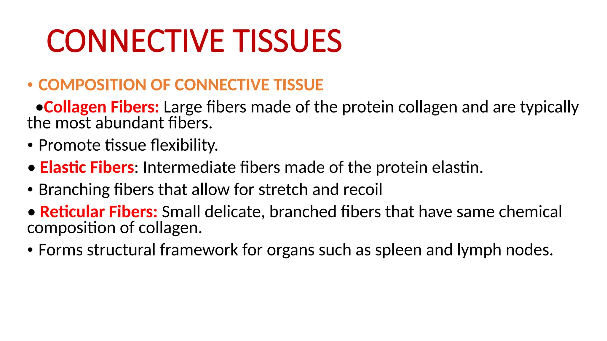
• COMPOSITIONOF CONNECTIVE TISSUE
•Collagen Fibers: Large fibers made of the protein collagen and are typically
the most abundant fibers.
• Promote tissue flexibility.
• Elastic Fibers: Intermediate fibers made of the protein elastin.
• Branching fibers that allow for stretch and recoil
• Reticular Fibers: Small delicate, branched fibers that have same chemical
composition of collagen.
• Forms structural framework for organs such as spleen and lymph nodes.
5.
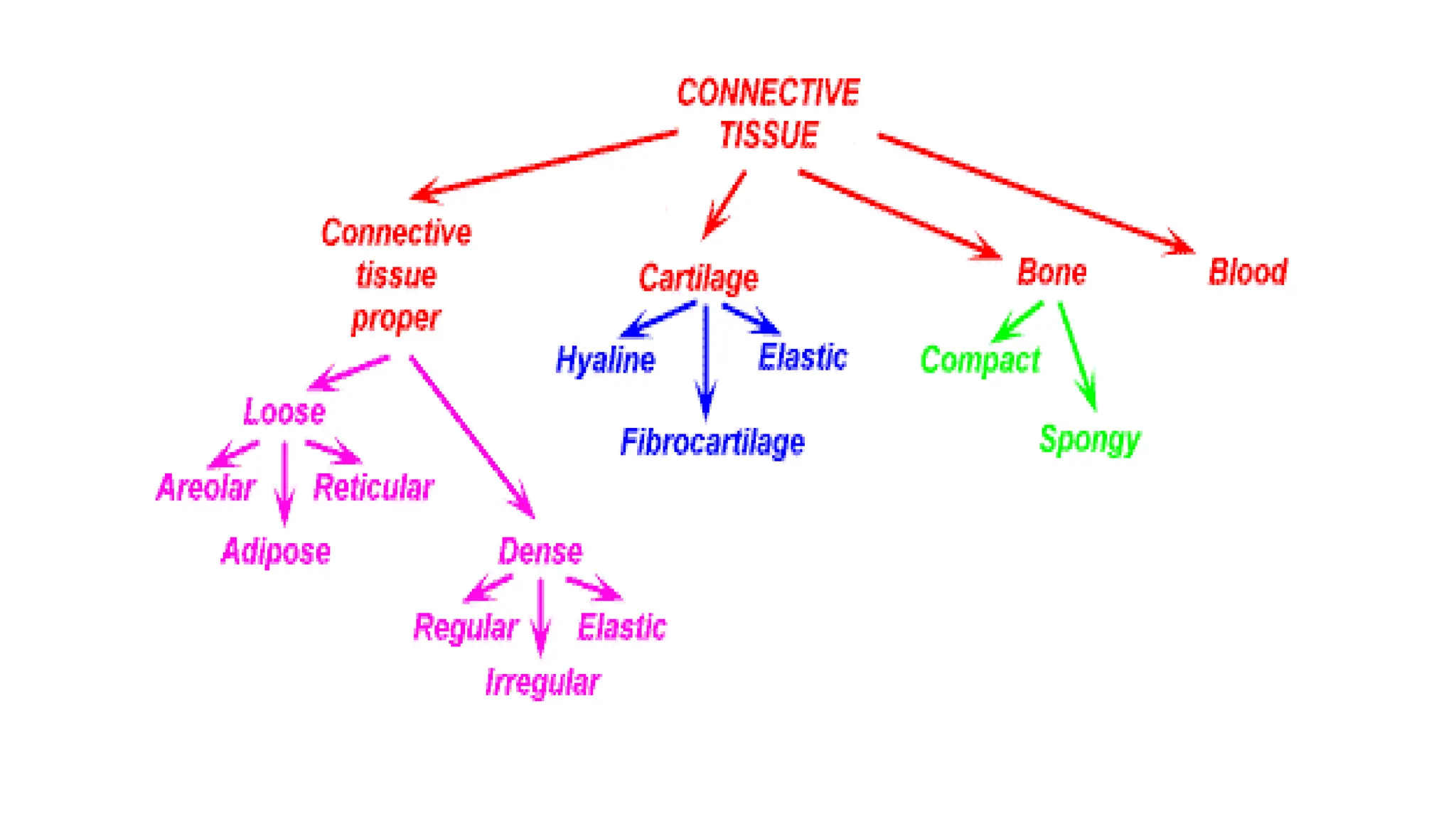
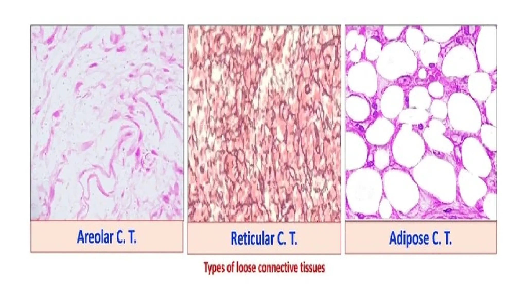
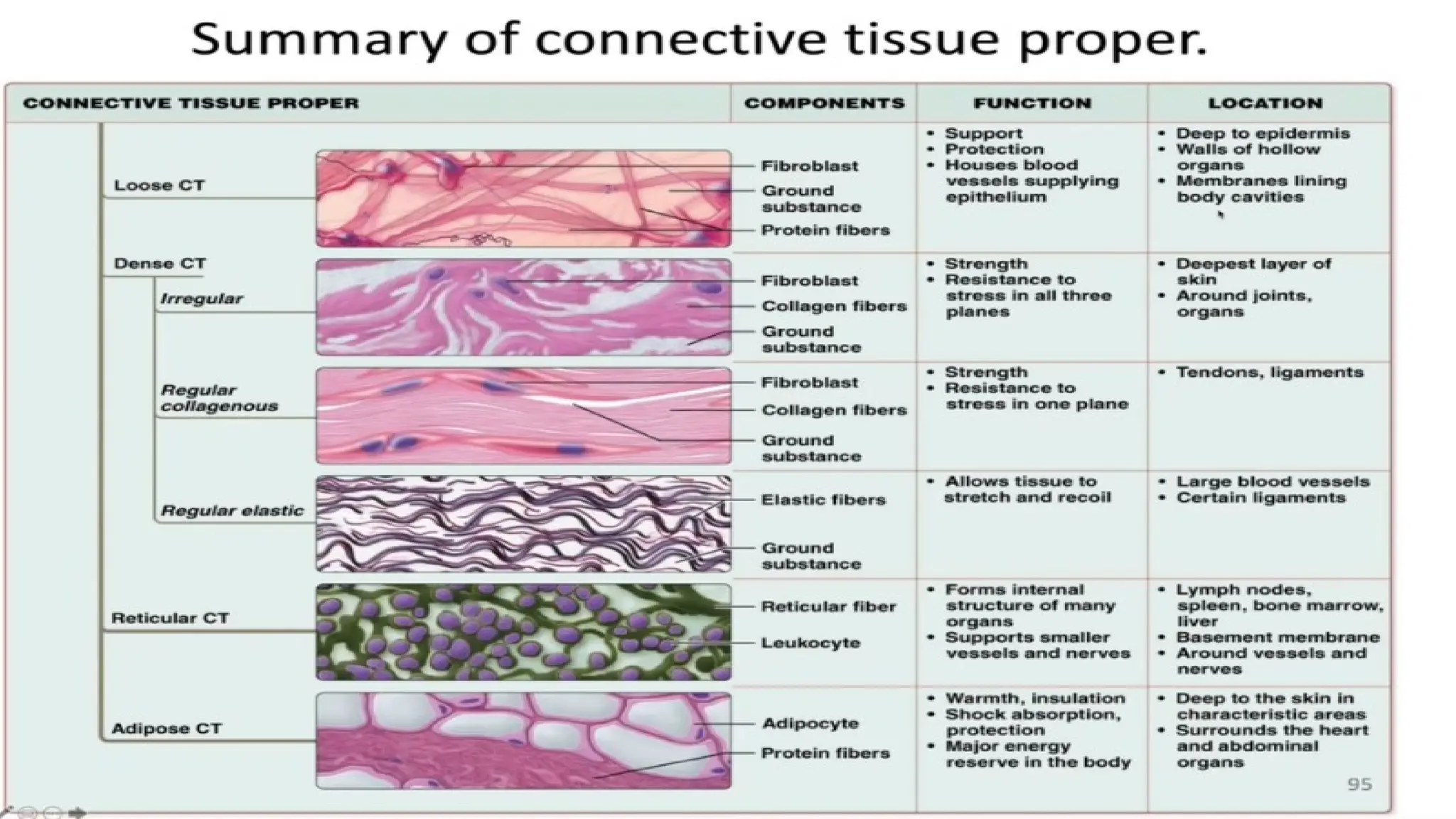
Loose connective tissue
•Areolar Connective tissue:
• They forms a loose network in intracellular material .
• It consists of collagen, elastic fibers, reticular fibers and several kinds of cells.
• Location: Below the skin, fill space between muscles, supports blood vessels
and nerves in alimentary canal.
• Functions: It gives strength, elasticity and support to tissue.
ADIPOSE CONNECTIVE TISSUE
•It consists of adipocytes which stores fat.
• Location: It is present in subcutaneous layer deep in the skin, around the
heart and kidneys
• Functions : Prevents heat loose from body. Act as reservoir of energy. It give
shape to the limbs and body. It protects underlying organ from injury.
7.
• RETICULAR CONNECTIVETISSUE
• It contains reticular fibers and reticular cells.
• Location: It is present in the supporting framework of liver, spleen, lymph
nodes, red bone marrow and it is also found around blood vessels and
muscles.
• Functions: It binds together smooth muscle tissue cells, filters and removes
microbes in the lymph node.
• DENSE CONNECTIVE TISSUE
• In this tissue, fibers are densely packed.
• The fiber content is higher.
• Cell content is lower as compared to loose connective tissue
8.
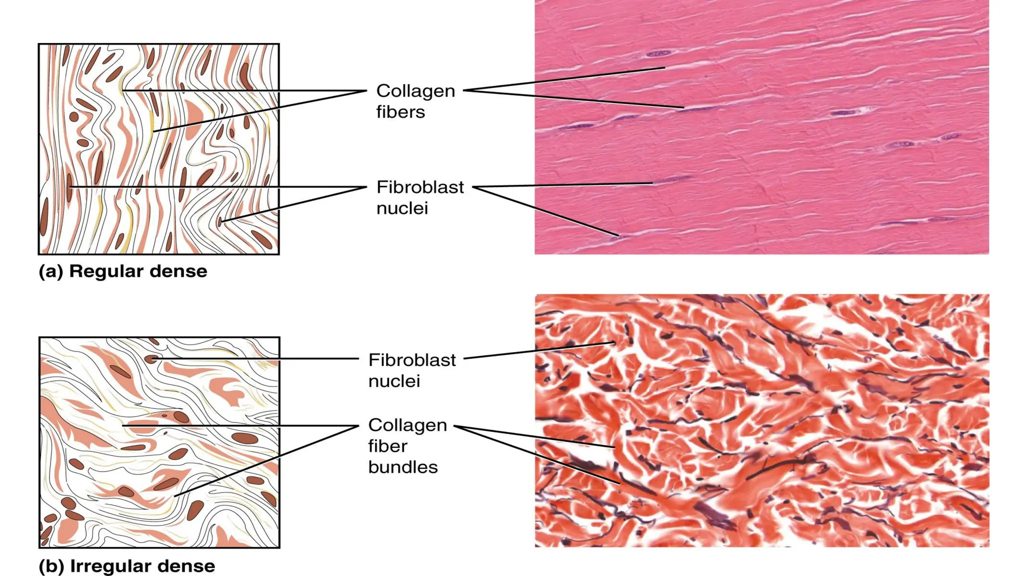
• DENSE REGULARCONNECTIVE TISSUE
• Bundles of collagen fibers are arranged in parallel patterns to provide
strength to tissue.
• Fibroblast are appear in rows between the fibers.
• It is tough in nature.
• Location: It forms tendons (attach muscle bone) and ligaments(attach
bone to bone).
• Functions: It provides strong attachment to structure.
9.
• DENSE IRREGULARCONNECTIVE TISSUE
• It contains collagen fibers which are irregular arranged and a few fibroblasts
are appear in rows between the fibers.
• Location: It present in dermis layer of skin, membrane capsules around
kidneys, liver, testes and lymph node, heart valves.
• Functions: It provides strength to different organs.
11.
• ELASTIC CONNECTIVETISSUE
• It consists of freely branching elastic fibers.
• Fibroblast are present in space between fibers.
• It is yellowish in colour.
• Location: It is present in tissues , walls of elastic arteries, trachea, bronchial
tubes and vocal cords.
• Functions: It allows stretching of various organs.
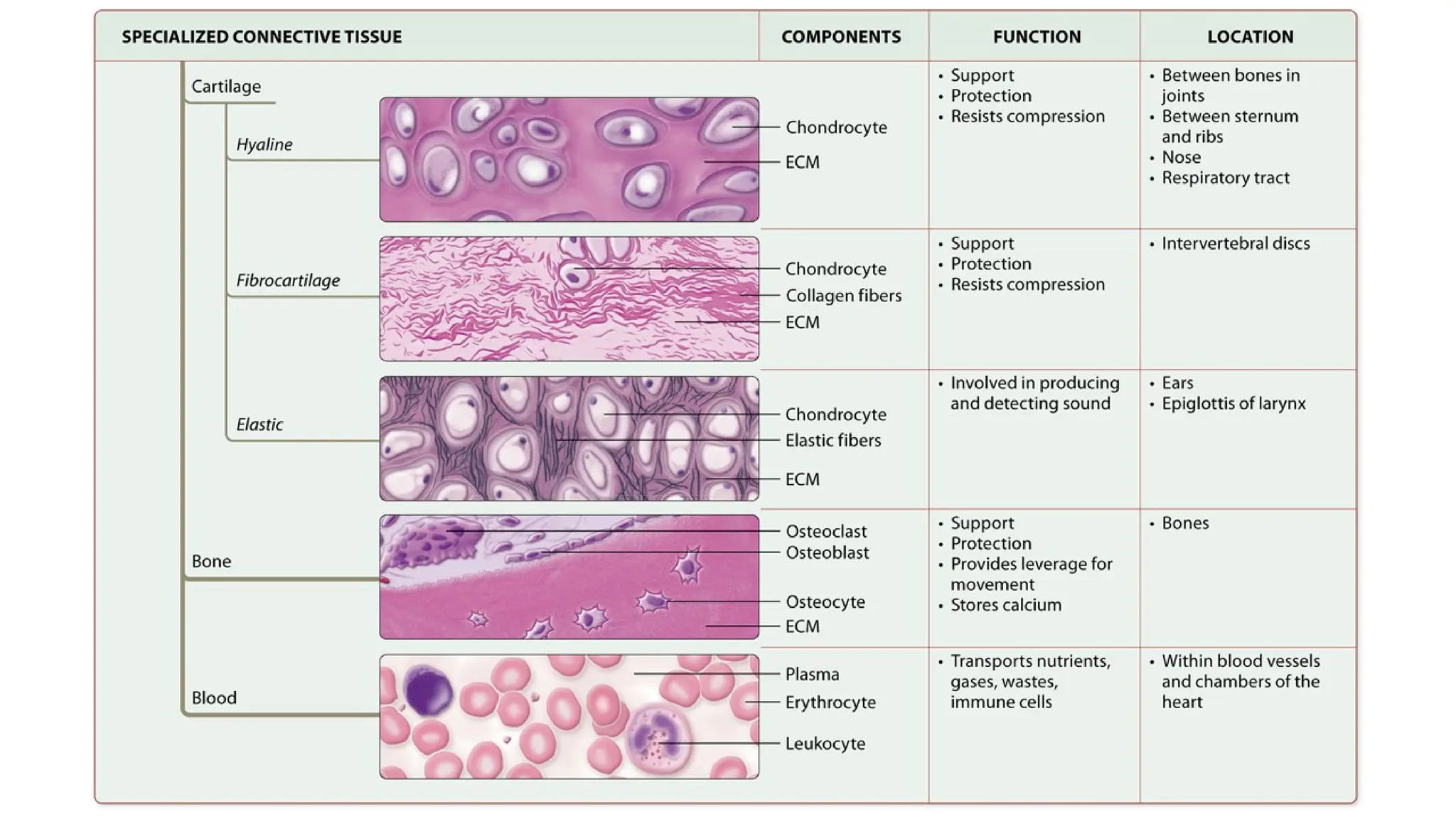
• CARTILAGE
• It consist of network of closely packed collagen fibers and elastic fibers.
The cells of mature cartilage called as chondrocytes.
12.
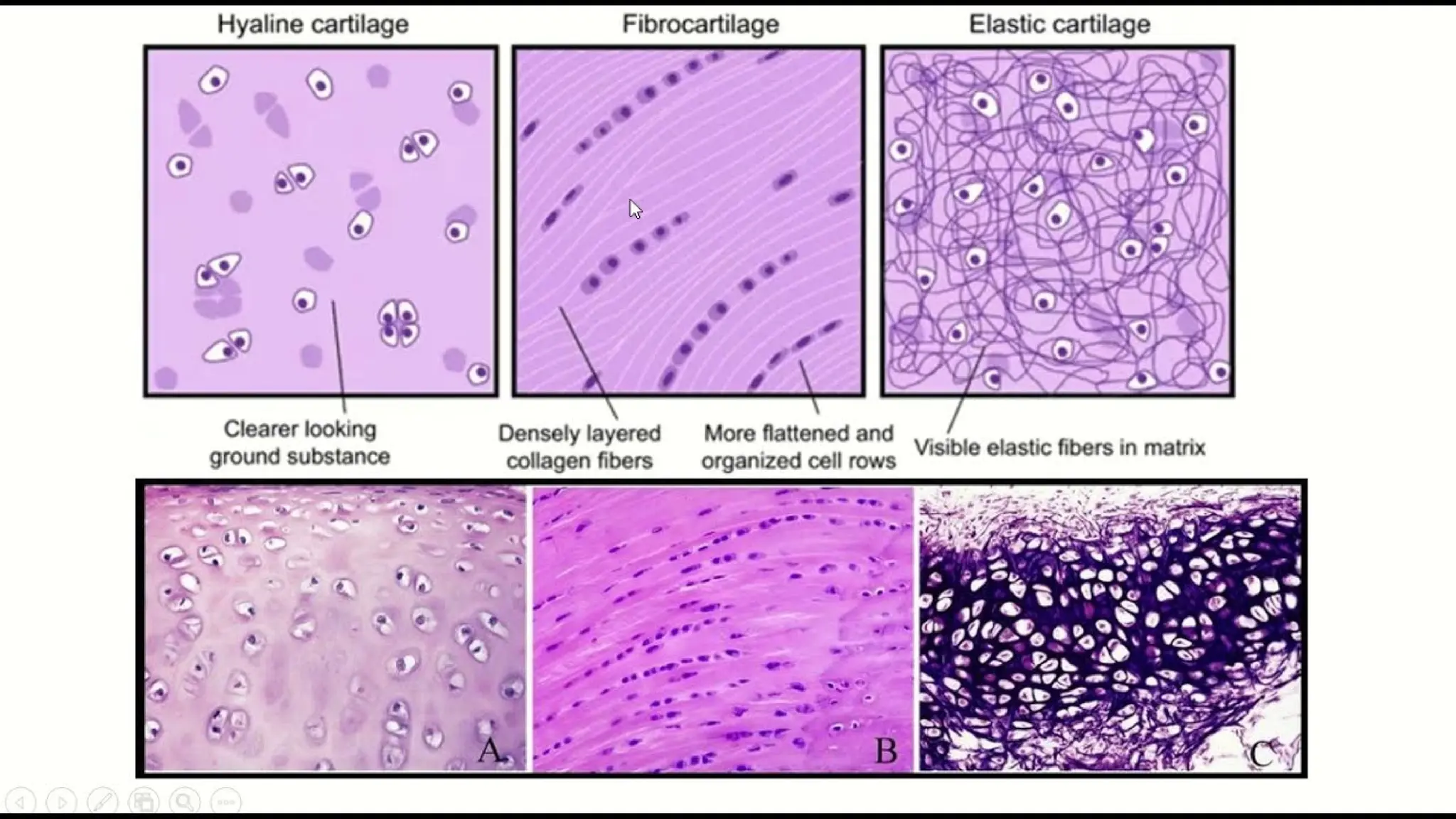
• HYALINE CARTILAGE
•Itis bluish white in color.
• It consists of fine collagen fibers and many chondrocytes.
• Location: It is present at the end of long bones, anterior ends of ribs, nose
and parts of larynx, trachea, bronchi, bronchial tubes.
• Function: It provides small surface for movement at joints, flexibility and
support.
• FIBRO CARTILAGE
• It is strongest form of cartilage.
• The chondrocytes are scattered among the bundle collagen fibers within
the extracellular matrix.
• Location: It is present in inter-verteblar disc.
• Functions: It covers and protects bony structures of body.
13.
• ELASTIC CARTILAGE
•The chondrocytes are located within a threadlike network of elastic
fibers within extracellular matrix.
• Location: It is present in pinna of ear and top of larynx.
• Functions: It provides strength and elasticity and maintain the shape
of certain organs such as the external ear.
17.
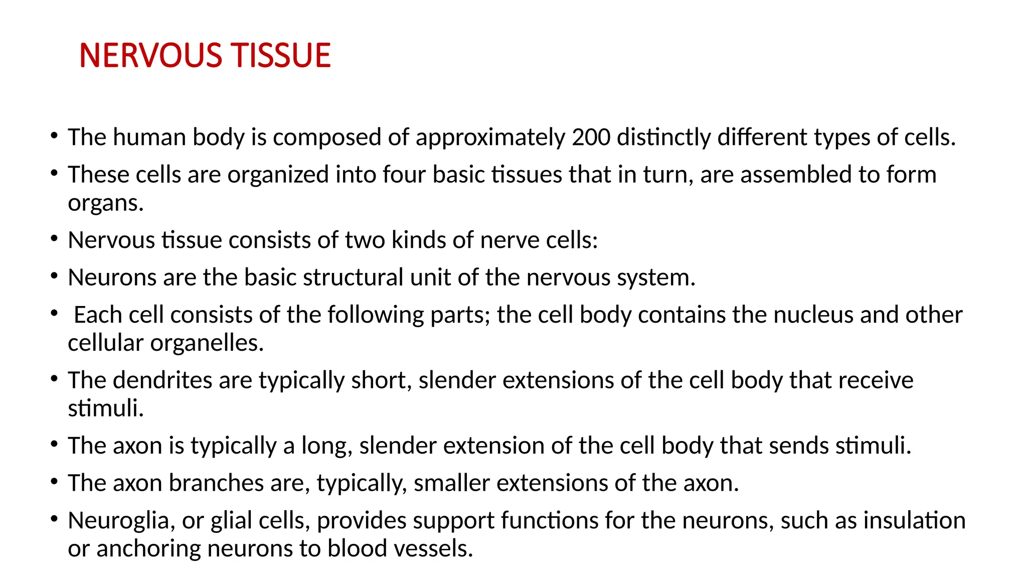
NERVOUS TISSUE
• Thehuman body is composed of approximately 200 distinctly different types of cells.
• These cells are organized into four basic tissues that in turn, are assembled to form
organs.
• Nervous tissue consists of two kinds of nerve cells:
• Neurons are the basic structural unit of the nervous system.
• Each cell consists of the following parts; the cell body contains the nucleus and other
cellular organelles.
• The dendrites are typically short, slender extensions of the cell body that receive
stimuli.
• The axon is typically a long, slender extension of the cell body that sends stimuli.
• The axon branches are, typically, smaller extensions of the axon.
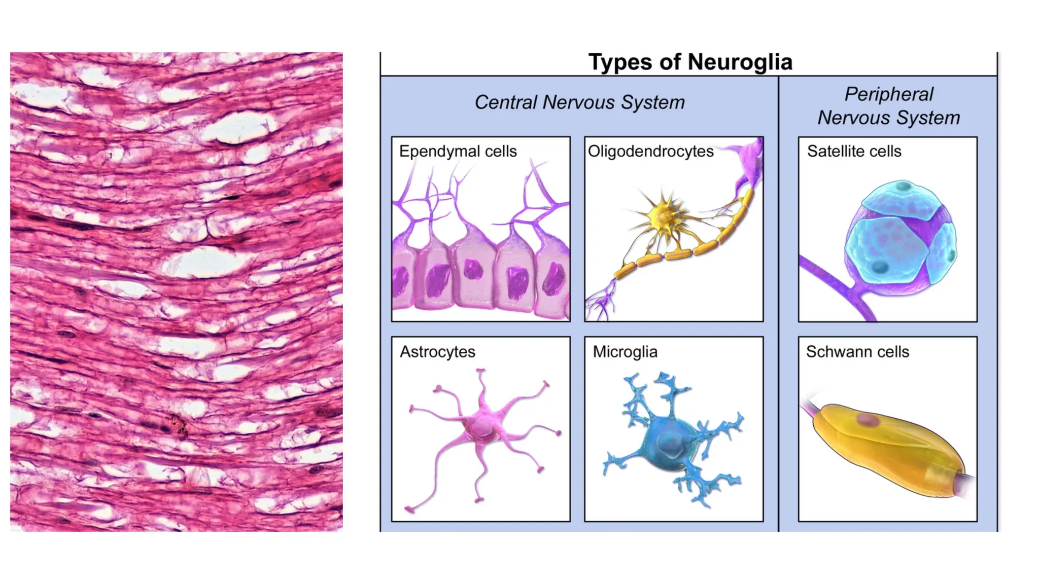
• Neuroglia, or glial cells, provides support functions for the neurons, such as insulation
or anchoring neurons to blood vessels.