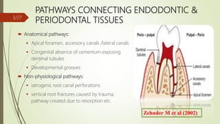
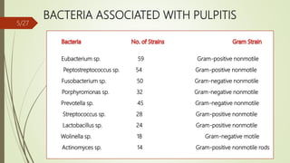
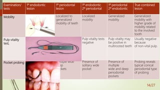
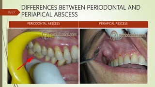
ENDODONTIC-PERIODONTAL
INTERACTIONS
GUIDED BY: PRESENTED BY:
DR. AMIT GOEL DR. VIRSHALI GUPTA
PG 2ND YEAR
CONTENTS
 Introduction
 Pathways connecting endodontic and periodontal tissues
 Etiology of endo-period lesions
 Classification of endo-perio lesions
 Clinical diagnostic procedures
 Differences b/w periodontal and periapical abscess
 Endo-perio controversy
 Therapeutic management of pulpal and periodontal diseases
 Conclusion
 References
1/27
INTRODUCTION
Endodontic-periodontal problems are responsible for more than 50% of tooth mortality today.
In 1919 Turner and Drew first described the effect of periodontal disease on the pulp. The
relationship between the periodontium and the pulp was first discovered by Simring and
Goldberg in 1964.
Since then, the term ‘endo- perio lesion’ has been used to describe lesions due to inflammatory
products found in varying degrees in both periodontium and pulpal tissues.
The pulp and periodontium have embryonic, anatomic and functional interrelationship.
2/27
PATHWAYS CONNECTING ENDODONTIC &
PERIODONTAL TISSUES
 Anatomical pathways:
 Apical foramen, accessory canals /lateral canals
 Congenital absence of cementum exposing
dentinal tubules
 Developmental grooves
 Non-physiological pathways:
 iatrogenic root canal perforations
 vertical root fractures caused by trauma,
pathway created due to resorption etc.
3/27
ETIOLOGICAL AND CONTRIBUTING FACTORS
IN ENDO-PERIO LESIONS4/27
BACTERIA ASSOCIATED WITH PULPITIS
Eubacterium sp. 59 Gram-positive nonmotile
Peptostreptococcus sp. 54 Gram-positive nonmotile
Fusobacterium sp. 50 Gram-negative nonmotile
Porphyromonas sp. 32 Gram-negative nonmotile
Prevotella sp. 45 Gram-negative nonmotile
Streptococcus sp. 28 Gram-positive nonmotile
Lactobacillus sp. 24 Gram-positive nonmotile
Wolinella sp. 18 Gram-negative motile
Actinomyces sp. 14 Gram-positive nonmotile rods
5/27
Most of the species that have
been found in infected root
canals can also be present in
the periodontal pocket.
(Moore 1987, Sundqvist
1994).
Rupf et al (2000) studied the profiles
periodontal pathogens in pulpal and
periodontal diseases associated with the
same tooth and concluded that
periodontal pathogens often
accompany endodontic infections
Didilescu AC et al (2012) - F.
nucleatum, P. micra and C.
sputigena may play a role in
the pathogenesis of endo-
periodontal lesions.
6/27
CLASSIFICATION OF ENDO- PERIO LESIONS
I. Based on etiology, diagnosis, treatment and prognosis
(by Simon, 1972)
Primary endodontic lesions
Primary endodontic lesions with secondary
periodontal involvement
Primary periodontal lesions
Primary periodontal lesions with secondary
endodontic involvement
True combined lesions
7/27
II. Based on clinical presentation strategies for each (by Weine, 1982)
Class 1 -tooth that clinically and radiographically stimulate
periodontal involvement but is truly due to pulpal inflammation or
necrosis.
Class II – tooth with both pulpal and periodontal disease
concomitantly
Class III – tooth that has no pulpal problem but requires endodontic
therapy with root amputation to receive periodontal healing
Class IV- tooth that clinically and radiographically stimulate pulpal or
periapical disease but in fact has periodontal disease.
8/27
IV. Stock (1988) modified Simon’s classification
Omitted Class V of the classification.
He argued that both Class II and Class IV lesions in advanced
stages can become combined lesions and therefore a
class to describe these lesions was not necessary.
9/27
III. Based on treatment plan (Grossman classification,1991)
Type 1 – Requiring endodontic treatment only.
Type II – Requiring periodontal treatment only.
Type III – Requiring combined endo-perio treatment
10/27
V. Classification as recommended by the World Workshop for Classification
Periodontal Diseases (1999)
Endodontic-periodontal lesion
Periodontal-endodontic lesion
Combined lesion
11/27
DIAGNOSTIC PROCEDURES USED TO IDENTIFY
THE ENDO-PERIO LESION
Examination/
tests
1º endodontic
lesion
1º periodontal
lesion
1º endodontic
2º periodontal
1º periodontal
2º endodontic
True combined
lesion
Visual  Soft tissue -
sinus opening
 Tooth -
decay/ large
restoration/
fractured
restoration or
tooth/
erosions/abrasio
ns/cracks/
discolorations/
poor RCT
 Inflamed
gingiva/
recession
(multiple
teeth)
 Plaque &
subgingival
calculus
(multiple
teeth)
 swelling
indicating
periodontal
abscess
 Plaque forms
at the
gingival margin
of the sinus tract
leads to
inflammation
of marginal
gingiva
 exudate
 Root
perforation/
fracture
 plaque,
subgingival
calculus &
swelling
(multiple
teeth)
 pus, exudate
 localized/
generalised
recession &
exposure of
root
 Plaque,
calculus &
periodontitis will
be present in
varying degrees
 Swelling
around single
or multiple
teeth
 pus, exudate
12/27
Examination/
tests
1º endodontic
lesion
1º periodontal
lesion
1º endodontic
2º periodontal
1º periodontal
2º endodontic
True combined
lesion
Pain Sharp  Usually dull
ache
 Sharp only in
acute
condition
 Usually sharp
shooting
 Dull ache in
chronic
conditions
 Usually dull
ache
 Sharp only in
acute
periodontal
abscess
 Dull ache
usually
 Only in acute
conditions it
is severe
Palpation does not indicate
whether
the inflammatory
process is
of endodontic or
periodontal
origin
Pain on Pain on Pain on Pain on
Percussion Normally tender
percussion
sensitivity of the
proprioceptive
fibers in an
inflamed pdl will
help identify
the location of
the pain
Tender on
percussion
Tender on
percussion
Tender on
percussion
13/27
Examination/
tests
1º endodontic
lesion
1º periodontal
lesion
1º endodontic
2º periodontal
1º periodontal
2º endodontic
True combined
lesion
Mobility Fractured roots and
recently
traumatized teeth
often present high
mobility
Localized to
generalized
mobility of teeth
Localized
mobility
Generalized
mobility
Generalized
mobility with
higher grade of
mobility related
to the involved
tooth
Pulp vitality
test,
 A lingering
response-
rreversible pulpitis
 No response -
Necrotic pulp
(non-vital)
pulp is vital and
responsive to
testing
Pulp vitality tests
negative
Pulp vitality may
be positive in
multirooted teeth
Usually negative
because
of non-vital pulp.
Pocket probing A deep narrow
solitary pocket*
Multiple wide
deep
pockets
Presence of
solitary wide
pocket
Presence of
multiple
wide and deep
periodontal
pockets
Probing reveals
typical conical
periodontal type
of probing
14/27
Examination/
tests
1º endodontic
lesion
1º periodontal
lesion
1º endodontic
2º periodontal
1º periodontal
2º endodontic
True combined
lesion
Sinus tracing A radiograph with
GP points to apex
or furcation area in
molars
Sinus tract mainly
at the
lateral aspect of
the root
Sinus tract mainly
at the apex/
furcation
area
Sinus tract mainly
at the lateral
aspect of the root
Difficult to trace
out the origin of
the lesion *
Radiographs
Cracked tooth
testing
Painful response on
chewing
No symptoms Painful response
on chewing
No symptoms Painful response
on chewing
15/27
DIFFERENCES BETWEEN PERIODONTAL AND
PERIAPICAL ABSCESS
PERIODONTAL ABSCESS PERIAPICAL ABSCESS
Periodontal pocket is present caries/ fracture is present
May occur after periodontal treatment May occur after endodontic or restorative
Tooth is vital Tooth is non - vital
Pain is usually dull and localized Pain is severe and difficult to localize
Swelling is present on the lateral surface of root
usually without fistulous track as abscess usually
drains from pocket opening.
Swelling is present at the apical portion of tooth
which drains by formation of a fistulous track.
Tender on lateral percussion Tender on vertical percussion
Usually not visible on radiographs Appears as a periapical radiolucency
16/27
ENDODONTIC PERIODONTAL-CONTROVERSY
• Two basic questions have been raised and continue to
be a matter of dispute :
1) Is periodontal disease a cause of pulp necrosis?
2) Can a pulpless tooth be the cause of periodontal
disease?
17/27
EFFECT OF PULPAL DISEASE ON THE
PERIODONTIUM
Bacteria and toxic irritants in pulp increase intrapulpal pressure. Inc pressure may cause toxic
to be expressed through patent channels which results in retrograde periodontitis.
Unresolved endodontic lesion causes bone loss, pocket formation and impair wound healing.
Potential effect of tooth with a necrotic pulp has been described as a risk factor (Jansson,
Ehnevid and Blomlof 1998) in the initiation and progression of periodontal disease, and the
initiation of periodontal pockets.
Diem et al (2002) reported that all tissues of the periodontium had a potential for regeneration
regardless of the status of the pulp.
18/27
INFLUENCE OF ENDODONTIC PROCEDURES
ON PERIODONTIUM
 Endodontic therapy adversely affects periodontal healing.
 Mechanical preparation, sealers, surgical trauma hinder new bone, cementum and
connective tissue repair.
 Precautions to be taken when periodontal therapy to follow endodontic treatment.
 Induce less mechanical trauma
 Use more biocompatible sealers
19/27
CONTRADICTING STUDY
 Sanders et al. (1983) reported that after the use of freeze dried bone allograft, 65% of
the teeth that did not have root canal treatment showed complete or greater than 50%
bone-fill in periodontal osseous defects; while only 33% of the teeth which had root
canal treatment prior to the periodontal surgical procedure had complete or greater
than 50% bone-fill.
20/27
EFFECT OF PERIODONTITIS ON THE PULP
Result in atrophic and other degenerative changes like
 reduction in the number of pulp cells,
 dystrophic mineralization,
 fibrosis,
 reparative dentin formation,
 inflammation and
 resorption.
CAUSE:
Disruption of blood flow through the lateral canals localized areas of coagulation
necrosis in the pulp.
21/27
Seltzer et al (1978) found
inflammatory alterations
and localized pulp
necrosis adjacent to
lateral canals in roots
exposed by periodontal
disease.
Mazur and Massler (1979)
found that only
periodontitis involving
apical foramen can lead
to pulp necrosis.
Cohen, 2002 have suggested that
periodontal disease causes pulpal
necrosis. Periodontal disease is a
direct cause of pulpal atrophy
necrosis.
22/27
EFFECT OF PERIODONTAL PROCEDURES ON
PULP
Scaling and root planing: removes the
bacterial plaque and calculus. However,
improper root planing procedures can also
remove cementum and the superficial parts
of dentin, thereby exposing the dentinal
tubules to the oral environment.
Acid etching: citric acid removes the smear
layer, an important pulp protector.
Application of citric acid may have a
detrimental effect on the dental pulp.
23/27
CONTRADICTING STUDIES
Kirkham (1975) studied 100
periodontally involved
& found 2% had lateral
canals in the periodontal
pocket.
Tagger & Smukler (1979)
removed roots from
extensively involved with
periodontal disease in
which root amputation was
necessary. Pulps of these
showed no inflammatory
changes.
Mazur and Massler (1979) found
no relationship and disclaimed
relationship of periodontal
disease as a causative factor in
pulpal disease.
24/27
TREATMENT
25/27
CONCLUSION
 Endo perio lesions present challenges to the clinicians in their proper diagnosis,
treatment and prognosis of the involved teeth.
 They have a varied pathogenesis which ranges from quite simple to relatively
complex.
 Knowledge of these diseases is essential in coming to the correct diagnosis and proper
treatment plan.
26/27
REFERENCES
 Carranza, Newman 1Oth edition. Endodontic and Periodontics consortium.
 Jan Lindhe. Endodontics and Periodontics. Clinical Periodontology and Implant Dentistry.
318-351.
 Shalu Bathla, PERIODONTICS REVISITED – 1st edition.
 Parolia A, Gait TC, Porto IC, Mala K. Endo-perio lesion: A dilemma from 19th until 21st
century. J Interdiscip Dentistry 2013;3: 2-11.
 Syed Wali Peeran et al, endo- perio lesions, international journal of scientific & technology
research volume 2, issue 5, may 2013
27/27

Endodontic periodontal interactions

  • 1.
    ENDODONTIC-PERIODONTAL INTERACTIONS GUIDED BY: PRESENTEDBY: DR. AMIT GOEL DR. VIRSHALI GUPTA PG 2ND YEAR
  • 2.
    CONTENTS  Introduction  Pathwaysconnecting endodontic and periodontal tissues  Etiology of endo-period lesions  Classification of endo-perio lesions  Clinical diagnostic procedures  Differences b/w periodontal and periapical abscess  Endo-perio controversy  Therapeutic management of pulpal and periodontal diseases  Conclusion  References 1/27
  • 3.
    INTRODUCTION Endodontic-periodontal problems areresponsible for more than 50% of tooth mortality today. In 1919 Turner and Drew first described the effect of periodontal disease on the pulp. The relationship between the periodontium and the pulp was first discovered by Simring and Goldberg in 1964. Since then, the term ‘endo- perio lesion’ has been used to describe lesions due to inflammatory products found in varying degrees in both periodontium and pulpal tissues. The pulp and periodontium have embryonic, anatomic and functional interrelationship. 2/27
  • 4.
    PATHWAYS CONNECTING ENDODONTIC& PERIODONTAL TISSUES  Anatomical pathways:  Apical foramen, accessory canals /lateral canals  Congenital absence of cementum exposing dentinal tubules  Developmental grooves  Non-physiological pathways:  iatrogenic root canal perforations  vertical root fractures caused by trauma, pathway created due to resorption etc. 3/27
  • 5.
    ETIOLOGICAL AND CONTRIBUTINGFACTORS IN ENDO-PERIO LESIONS4/27
  • 6.
    BACTERIA ASSOCIATED WITHPULPITIS Eubacterium sp. 59 Gram-positive nonmotile Peptostreptococcus sp. 54 Gram-positive nonmotile Fusobacterium sp. 50 Gram-negative nonmotile Porphyromonas sp. 32 Gram-negative nonmotile Prevotella sp. 45 Gram-negative nonmotile Streptococcus sp. 28 Gram-positive nonmotile Lactobacillus sp. 24 Gram-positive nonmotile Wolinella sp. 18 Gram-negative motile Actinomyces sp. 14 Gram-positive nonmotile rods 5/27
  • 7.
    Most of thespecies that have been found in infected root canals can also be present in the periodontal pocket. (Moore 1987, Sundqvist 1994). Rupf et al (2000) studied the profiles periodontal pathogens in pulpal and periodontal diseases associated with the same tooth and concluded that periodontal pathogens often accompany endodontic infections Didilescu AC et al (2012) - F. nucleatum, P. micra and C. sputigena may play a role in the pathogenesis of endo- periodontal lesions. 6/27
  • 8.
    CLASSIFICATION OF ENDO-PERIO LESIONS I. Based on etiology, diagnosis, treatment and prognosis (by Simon, 1972) Primary endodontic lesions Primary endodontic lesions with secondary periodontal involvement Primary periodontal lesions Primary periodontal lesions with secondary endodontic involvement True combined lesions 7/27
  • 9.
    II. Based onclinical presentation strategies for each (by Weine, 1982) Class 1 -tooth that clinically and radiographically stimulate periodontal involvement but is truly due to pulpal inflammation or necrosis. Class II – tooth with both pulpal and periodontal disease concomitantly Class III – tooth that has no pulpal problem but requires endodontic therapy with root amputation to receive periodontal healing Class IV- tooth that clinically and radiographically stimulate pulpal or periapical disease but in fact has periodontal disease. 8/27
  • 10.
    IV. Stock (1988)modified Simon’s classification Omitted Class V of the classification. He argued that both Class II and Class IV lesions in advanced stages can become combined lesions and therefore a class to describe these lesions was not necessary. 9/27
  • 11.
    III. Based ontreatment plan (Grossman classification,1991) Type 1 – Requiring endodontic treatment only. Type II – Requiring periodontal treatment only. Type III – Requiring combined endo-perio treatment 10/27
  • 12.
    V. Classification asrecommended by the World Workshop for Classification Periodontal Diseases (1999) Endodontic-periodontal lesion Periodontal-endodontic lesion Combined lesion 11/27
  • 13.
    DIAGNOSTIC PROCEDURES USEDTO IDENTIFY THE ENDO-PERIO LESION Examination/ tests 1º endodontic lesion 1º periodontal lesion 1º endodontic 2º periodontal 1º periodontal 2º endodontic True combined lesion Visual  Soft tissue - sinus opening  Tooth - decay/ large restoration/ fractured restoration or tooth/ erosions/abrasio ns/cracks/ discolorations/ poor RCT  Inflamed gingiva/ recession (multiple teeth)  Plaque & subgingival calculus (multiple teeth)  swelling indicating periodontal abscess  Plaque forms at the gingival margin of the sinus tract leads to inflammation of marginal gingiva  exudate  Root perforation/ fracture  plaque, subgingival calculus & swelling (multiple teeth)  pus, exudate  localized/ generalised recession & exposure of root  Plaque, calculus & periodontitis will be present in varying degrees  Swelling around single or multiple teeth  pus, exudate 12/27
  • 14.
    Examination/ tests 1º endodontic lesion 1º periodontal lesion 1ºendodontic 2º periodontal 1º periodontal 2º endodontic True combined lesion Pain Sharp  Usually dull ache  Sharp only in acute condition  Usually sharp shooting  Dull ache in chronic conditions  Usually dull ache  Sharp only in acute periodontal abscess  Dull ache usually  Only in acute conditions it is severe Palpation does not indicate whether the inflammatory process is of endodontic or periodontal origin Pain on Pain on Pain on Pain on Percussion Normally tender percussion sensitivity of the proprioceptive fibers in an inflamed pdl will help identify the location of the pain Tender on percussion Tender on percussion Tender on percussion 13/27
  • 15.
    Examination/ tests 1º endodontic lesion 1º periodontal lesion 1ºendodontic 2º periodontal 1º periodontal 2º endodontic True combined lesion Mobility Fractured roots and recently traumatized teeth often present high mobility Localized to generalized mobility of teeth Localized mobility Generalized mobility Generalized mobility with higher grade of mobility related to the involved tooth Pulp vitality test,  A lingering response- rreversible pulpitis  No response - Necrotic pulp (non-vital) pulp is vital and responsive to testing Pulp vitality tests negative Pulp vitality may be positive in multirooted teeth Usually negative because of non-vital pulp. Pocket probing A deep narrow solitary pocket* Multiple wide deep pockets Presence of solitary wide pocket Presence of multiple wide and deep periodontal pockets Probing reveals typical conical periodontal type of probing 14/27
  • 16.
    Examination/ tests 1º endodontic lesion 1º periodontal lesion 1ºendodontic 2º periodontal 1º periodontal 2º endodontic True combined lesion Sinus tracing A radiograph with GP points to apex or furcation area in molars Sinus tract mainly at the lateral aspect of the root Sinus tract mainly at the apex/ furcation area Sinus tract mainly at the lateral aspect of the root Difficult to trace out the origin of the lesion * Radiographs Cracked tooth testing Painful response on chewing No symptoms Painful response on chewing No symptoms Painful response on chewing 15/27
  • 17.
    DIFFERENCES BETWEEN PERIODONTALAND PERIAPICAL ABSCESS PERIODONTAL ABSCESS PERIAPICAL ABSCESS Periodontal pocket is present caries/ fracture is present May occur after periodontal treatment May occur after endodontic or restorative Tooth is vital Tooth is non - vital Pain is usually dull and localized Pain is severe and difficult to localize Swelling is present on the lateral surface of root usually without fistulous track as abscess usually drains from pocket opening. Swelling is present at the apical portion of tooth which drains by formation of a fistulous track. Tender on lateral percussion Tender on vertical percussion Usually not visible on radiographs Appears as a periapical radiolucency 16/27
  • 18.
    ENDODONTIC PERIODONTAL-CONTROVERSY • Twobasic questions have been raised and continue to be a matter of dispute : 1) Is periodontal disease a cause of pulp necrosis? 2) Can a pulpless tooth be the cause of periodontal disease? 17/27
  • 19.
    EFFECT OF PULPALDISEASE ON THE PERIODONTIUM Bacteria and toxic irritants in pulp increase intrapulpal pressure. Inc pressure may cause toxic to be expressed through patent channels which results in retrograde periodontitis. Unresolved endodontic lesion causes bone loss, pocket formation and impair wound healing. Potential effect of tooth with a necrotic pulp has been described as a risk factor (Jansson, Ehnevid and Blomlof 1998) in the initiation and progression of periodontal disease, and the initiation of periodontal pockets. Diem et al (2002) reported that all tissues of the periodontium had a potential for regeneration regardless of the status of the pulp. 18/27
  • 20.
    INFLUENCE OF ENDODONTICPROCEDURES ON PERIODONTIUM  Endodontic therapy adversely affects periodontal healing.  Mechanical preparation, sealers, surgical trauma hinder new bone, cementum and connective tissue repair.  Precautions to be taken when periodontal therapy to follow endodontic treatment.  Induce less mechanical trauma  Use more biocompatible sealers 19/27
  • 21.
    CONTRADICTING STUDY  Sanderset al. (1983) reported that after the use of freeze dried bone allograft, 65% of the teeth that did not have root canal treatment showed complete or greater than 50% bone-fill in periodontal osseous defects; while only 33% of the teeth which had root canal treatment prior to the periodontal surgical procedure had complete or greater than 50% bone-fill. 20/27
  • 22.
    EFFECT OF PERIODONTITISON THE PULP Result in atrophic and other degenerative changes like  reduction in the number of pulp cells,  dystrophic mineralization,  fibrosis,  reparative dentin formation,  inflammation and  resorption. CAUSE: Disruption of blood flow through the lateral canals localized areas of coagulation necrosis in the pulp. 21/27
  • 23.
    Seltzer et al(1978) found inflammatory alterations and localized pulp necrosis adjacent to lateral canals in roots exposed by periodontal disease. Mazur and Massler (1979) found that only periodontitis involving apical foramen can lead to pulp necrosis. Cohen, 2002 have suggested that periodontal disease causes pulpal necrosis. Periodontal disease is a direct cause of pulpal atrophy necrosis. 22/27
  • 24.
    EFFECT OF PERIODONTALPROCEDURES ON PULP Scaling and root planing: removes the bacterial plaque and calculus. However, improper root planing procedures can also remove cementum and the superficial parts of dentin, thereby exposing the dentinal tubules to the oral environment. Acid etching: citric acid removes the smear layer, an important pulp protector. Application of citric acid may have a detrimental effect on the dental pulp. 23/27
  • 25.
    CONTRADICTING STUDIES Kirkham (1975)studied 100 periodontally involved & found 2% had lateral canals in the periodontal pocket. Tagger & Smukler (1979) removed roots from extensively involved with periodontal disease in which root amputation was necessary. Pulps of these showed no inflammatory changes. Mazur and Massler (1979) found no relationship and disclaimed relationship of periodontal disease as a causative factor in pulpal disease. 24/27
  • 26.
  • 27.
    CONCLUSION  Endo periolesions present challenges to the clinicians in their proper diagnosis, treatment and prognosis of the involved teeth.  They have a varied pathogenesis which ranges from quite simple to relatively complex.  Knowledge of these diseases is essential in coming to the correct diagnosis and proper treatment plan. 26/27
  • 28.
    REFERENCES  Carranza, Newman1Oth edition. Endodontic and Periodontics consortium.  Jan Lindhe. Endodontics and Periodontics. Clinical Periodontology and Implant Dentistry. 318-351.  Shalu Bathla, PERIODONTICS REVISITED – 1st edition.  Parolia A, Gait TC, Porto IC, Mala K. Endo-perio lesion: A dilemma from 19th until 21st century. J Interdiscip Dentistry 2013;3: 2-11.  Syed Wali Peeran et al, endo- perio lesions, international journal of scientific & technology research volume 2, issue 5, may 2013 27/27

Editor's Notes

  • #2 Endodontics deals with disease of the pulp and periapical tissues and periodontal therapy deals with many aspects of the supporting strs including the prevention and repair of gingival sulcus. The success of both periodontal and endodontic therapy depends on the elimination of both disease processes, whether they exist separately or as a combined lesion.
  • #4 Ectomesenchymal cells proliferate to form dental papilla and follicle which are the precursors of periodontium and pulp resp. this embryonic development gives rise to anatomical connections which remain throughout life.
  • #5 Inflammation frm pulp may extends into the periodontium causing destruction of periodontal tissues such a periodontal lesion is kas retrograde periodontitis. Retrograde pulpitis occurs as a result of extension of inflammation from periodontal tissues into the pulp.
  • #8 A. actinomycetemcomitans Capnocytophaga sp. F. nucleatum P. gingivalis P. intermedia T. forsythia T. denticola
  • #12 most accepted classification
  • #15 Acute conditions: anug, acute periodontal abscess, acute herpetic gingivostomatits Palpation is performed by applying firm digital pressure to mucosa covering the roots and apices. With the index finger the mucosa is presses against the underlying cortical bone. This will detect the presence of periradicular abnormalities that produce painful response to digital pressure. A positive response to palpation may indicate active periradicular inflammatory process. However this test doesnot indicate whether the inflammatory process is of endodontic or periodontal origin. Percussion is performed by tapping on the incisal or occlusal surfaces of the teeth with the back of mirror handle the tooth is tapped vertically and horizontally.
  • #16 *in the absence of periodontal disease may indicate the presence of a lesion of endodontic origin or a vertical root fracture Mobility testing can be performed using 2 mirror hanles on each side of the crown. Pressure is applied in facial- lingual as well as in a vertical direction and tooth mobility is scored.
  • #17 deep carious lesions/defective restorations/previous poor RCT 2. vertical bone loss 3. root resorption with a wide base radiolucency around the apex of the Root 4 . Angular bone loss in multiple teeth 5. may be similar to that of a vertically fractured tooth * if a sinus tract is present, it may be necessary to raise a flap to determine the etiology of the lesion
  • #20 First indication of periodontal involvement due to pulp disease is thickening of PDL space at the apical end. Nature and extent of periodontal ligament destruction is dependent on several factors like virulence of bacteria, duration of the disease and host defense mechanism. Clinical message of these studies was that root canal treatment should be completed before periodontal therapy.
  • #21 Biocompatibility is related to the behavior of biomaterials in various contexts. The term refers to the ability of a material to perform with an appropriate host response in a specific situation Sealapex Obtuseal root canal sealer
  • #25 . Subsequent microbial colonization of the root dentin may result in bacterial invasion of the dentinal tubules. As a consequence, inflammatory lesions may develop in the pulp. The initial symptom is sharp pain of rapid onset that disappears once the stimulus is removed. During periodontal regenerative therapy, root conditioning using citric acid helps to remove bacterial endotoxin and anerobic bacteria and to expose collagen bundles to serve as a matrix for new connective tissue attachment to cementum. Though beneficial in the treatment of periodontal disease,