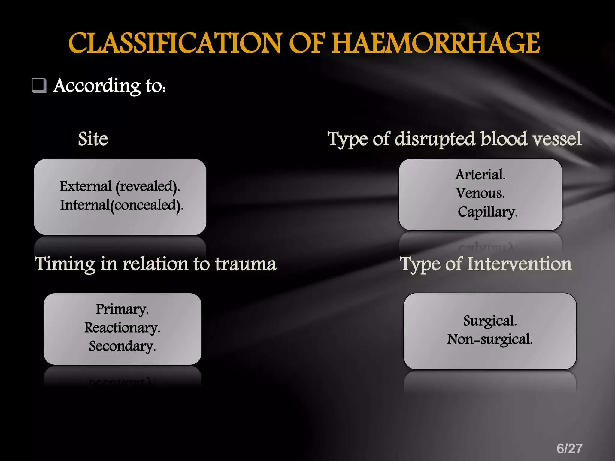
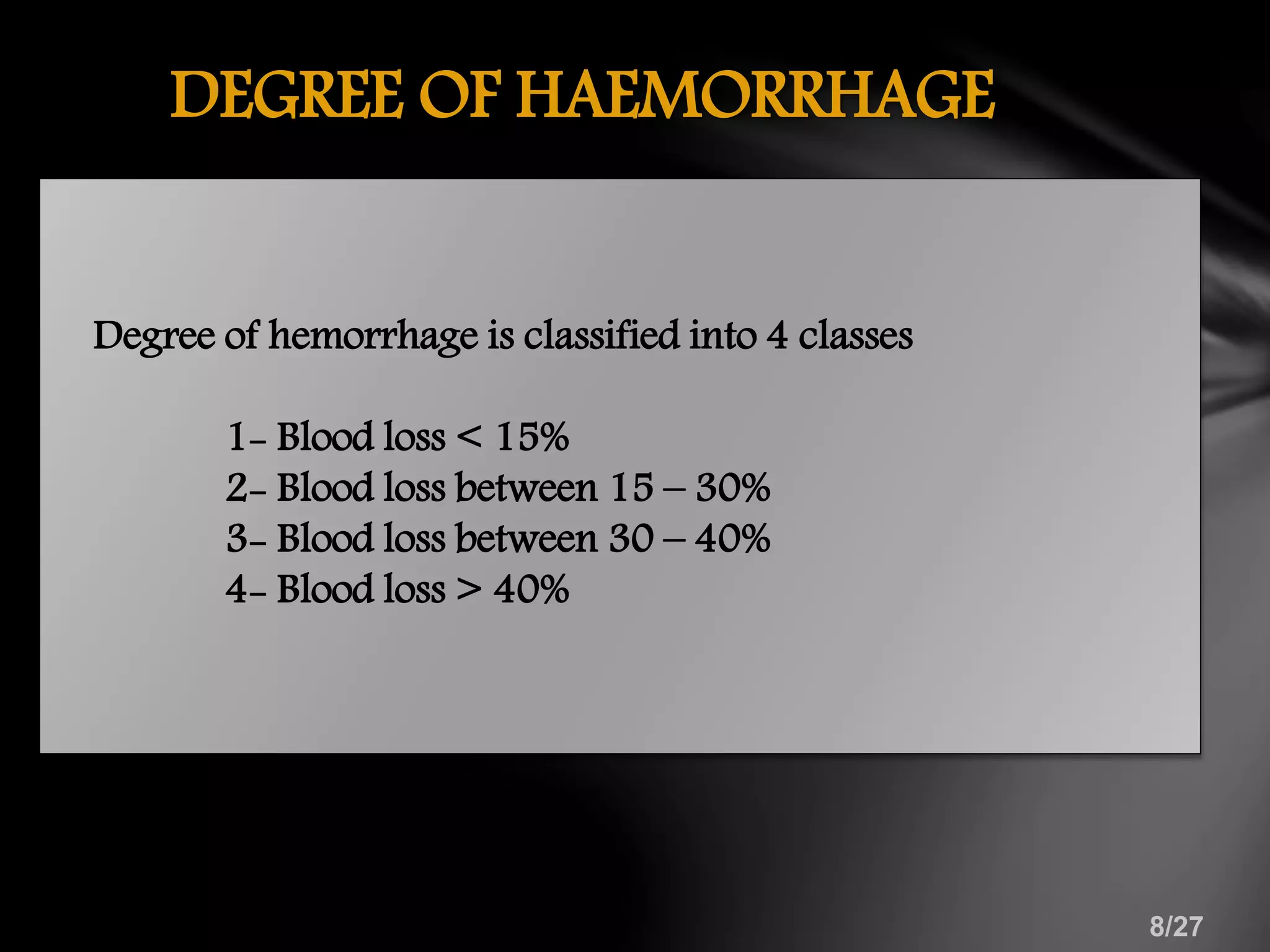
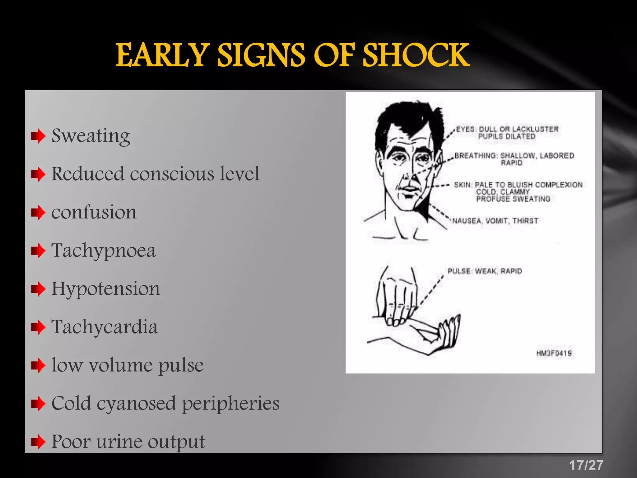
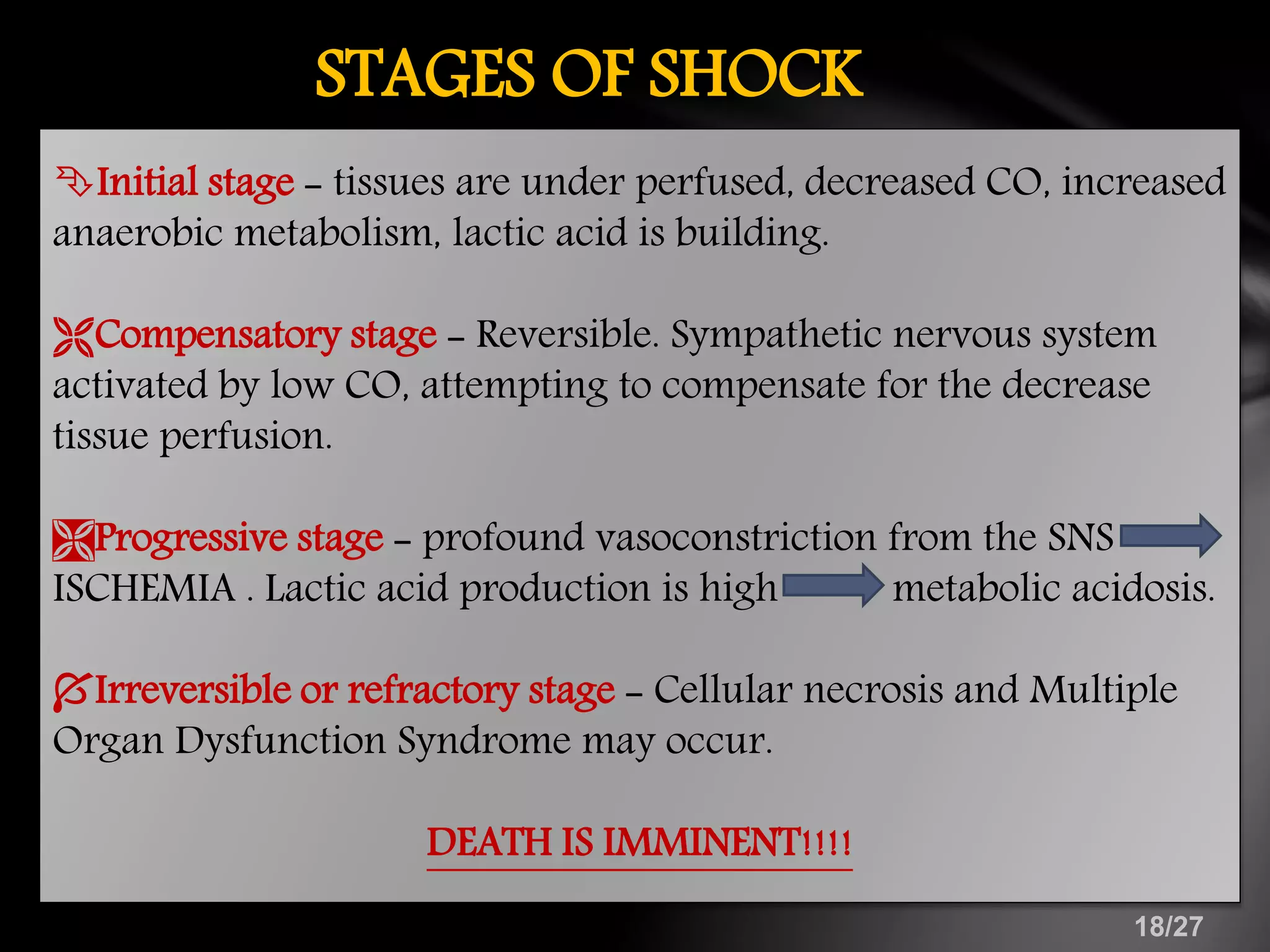
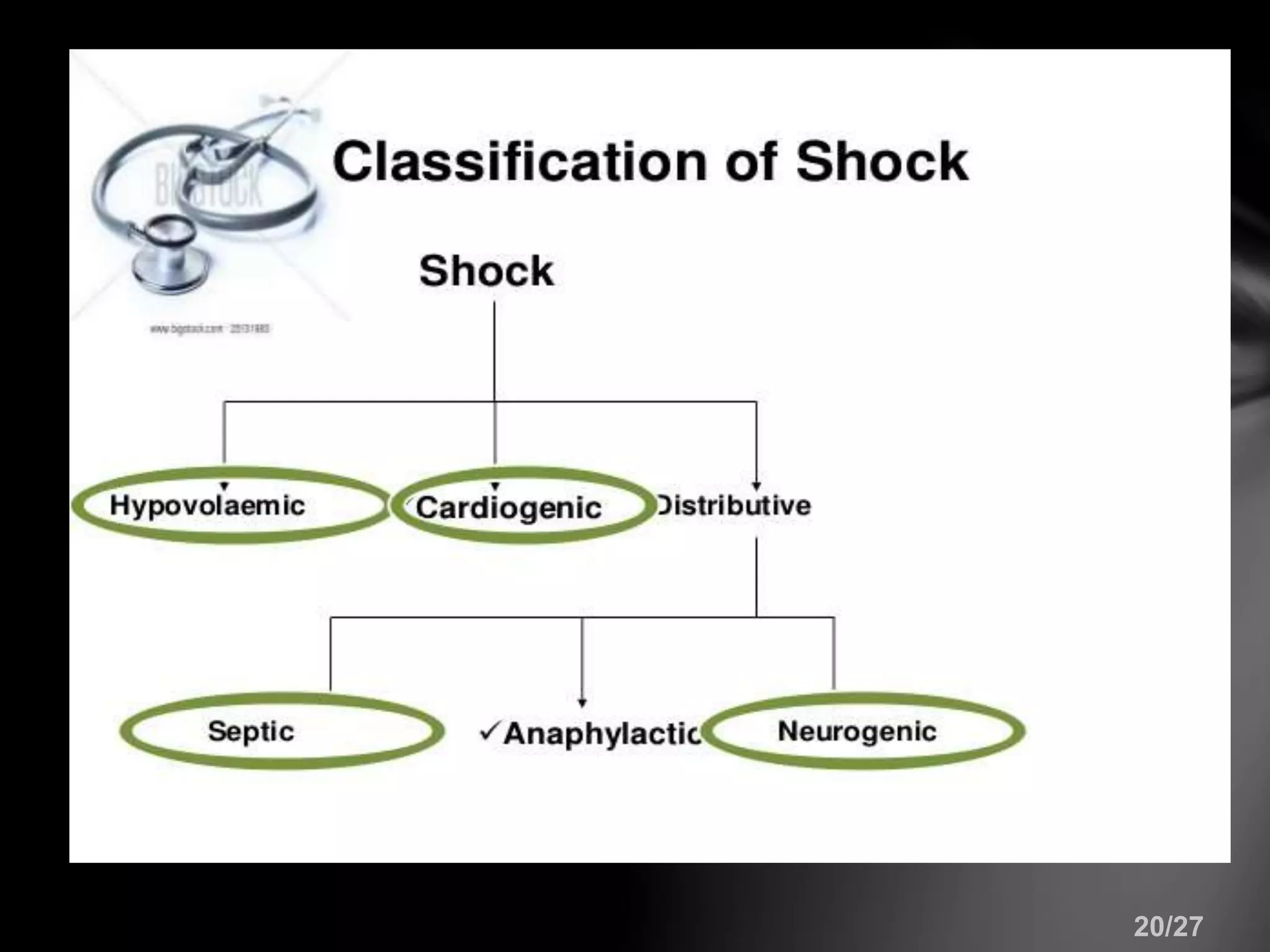
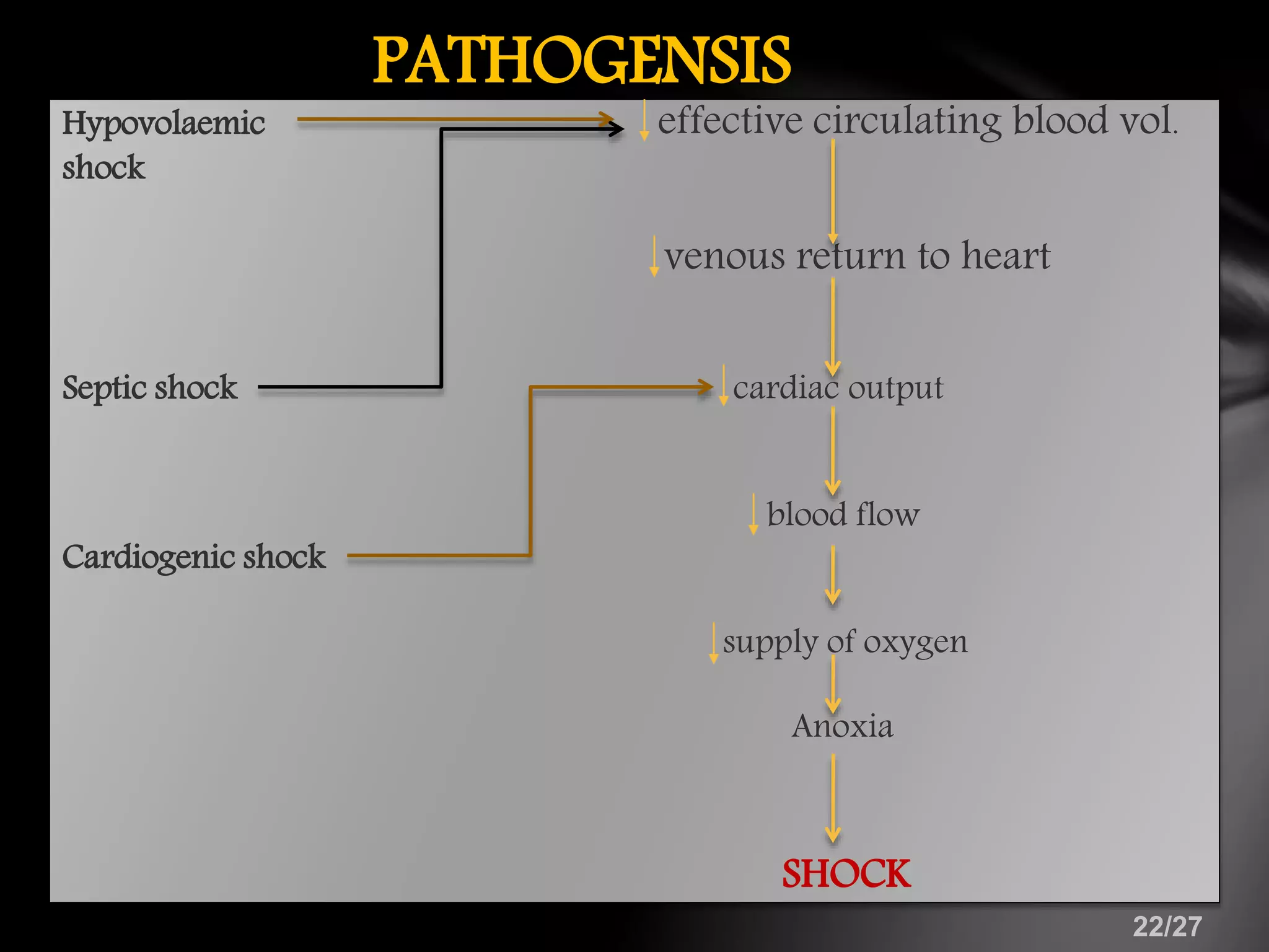
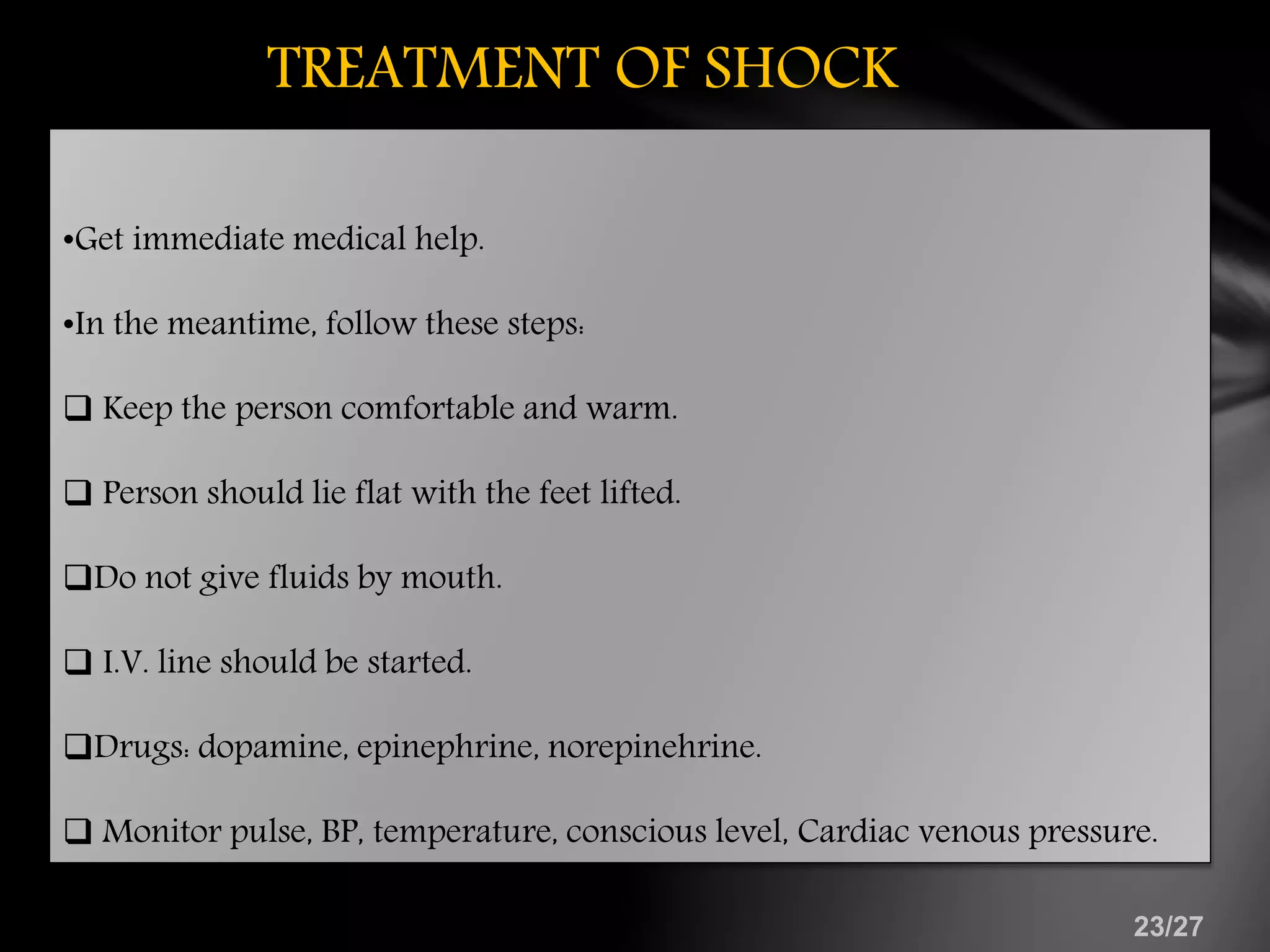
3. ď§ Introduction
ď§ Definition
ď§ Classification of haemorrhage
ď§ Symptoms
ď§ Degree of haemorrhage
ď§ Measurement of blood loss
ď§ Management
ď§ Shock definition
ď§ Early signs of shock
ď§ Types of shock
ď§ Pathogenesis of shock
ď§ Treatment of shock
ď§ Conclusion
ď§ References
CONTENTS
4. INTRODUCTION
ďźBlood is the vital fluid present in the body, carries
oxygen and nutrients to the tissues.
ďźLoss of blood due to any reason beyond a certain point
is potentially life threatening and may lead to
exanguination.
ďźThe word haemorrhage is synonymous with bleeding.
ďźEscape of blood from ruptured blood vessel.
or
ďź Large flow of blood from a damaged blood vessel.
5. Bleeding arising due to either external or
internal wounds inflicted by an object/person
on a victim.
CAUSES OF HAEMORRHAGE
6. CLASSIFICATION OF HAEMORRHAGE
ďą According to:
Site Type of disrupted blood vessel
Timing in relation to trauma Type of Intervention
Primary.
Reactionary.
Secondary.
Surgical.
Non-surgical.
External (revealed).
Internal(concealed).
Arterial.
Venous.
Capillary.
7. SYMPTOMS OF HAEMORRHAGE
⢠Pallor
⢠Rapid feeble pulse
⢠Thirst
⢠Giddiness
⢠Nausea
⢠Restlessness
⢠Cold & clammy skin
⢠Fall in blood pressure
⢠Appearance of blood
⢠Appearance of cyanosis
8. DEGREE OF HAEMORRHAGE
Degree of hemorrhage is classified into 4 classes
1- Blood loss < 15%
2- Blood loss between 15 â 30%
3- Blood loss between 30 â 40%
4- Blood loss > 40%
9. METHODS TO MEASURE BLOOD LOSS DURING
SURGERY??
1) Collect the entire amount of blood lost during the operation.
2) Weighing surgical swabs before and after the surgery.
3)Calculating the lost blood volume by colorimetric assays that
evaluate the hemoglobin content of the blood-stained swab
compared to the hemoglobin of the patient.
4) Assessing blood-loss volume using fructosamine that can easily be
measured in the blood but is absent from saliva and water.
ďą Normal blood volume -4 to 6l & is estimated as
70 ml/kg -children & adults
80ml/kg â neonates.
11. HOW TO CONTROL BLEEDING????
1. Bleeding from bone:
â˘Burnishing the bone in the area of the bleed with molt, elevator
or curette.
â˘If it is ineffective bone wax can be compressed in that area.
2. Soft tissue bleeding
â˘Applying pressure using moist gauze for 2-5 min .
â˘If ineffective vessel ligation use resorbable suture .
â˘Beside this various topical hemostatic agents can be used.
12. BLOOD LOSS DURING OPEN FLAP
DEBRIDEMENT
Year Authors Blood loss
1966 McIvor et al 0.5 to 62 ml
2005 Barganza et al 17.86 to 15.7 ml
2007 Moore et al 54.9 to 53.0 ml
2012 Zigdon et al 6.0 to 145.1ml
13. MANAGEMENT
ďź Restore blood volume:
First aid treatment by packing, pressure, position, and
tourniquets.
ďź Optimize oxygen delivery:
ďź Monitoring:
pulse, BP, temperature, conscious level, Cardiac venous
pressure.
LOCAL MANAGEMENT
14. ďźInjection of morphine (10-15mg.) as soon as possible.
ďźHospitalisation after temporary arrest of the bleeding.
ďźAntibiotics.
ďźVitamins.
ďźCorrective measures for bleeding diathesis.
ďźi.v. Fluid & if required blood transfusion should be started
immediately.
GENERAL MANAGEMENT
15. INTRA-OPERATIVE MANAGEMENT
â˘Regional block anesthesia must be avoided.
â˘Another way to prevent excessive bleeding is the meticulous handling of soft
tissues.
POST âOPERATIVE MEASURES
â˘Application of pressure for 10 minutes with moistened gauze on the Flap.
â˘Rinsing is prohibited .
â˘antifibrinolytic mouthwash the day after periodontal treatment.
⢠Antibiotics: penicillin, erthromycin, tetracycline, metronidazole, ampicillin,
amoxicillin+ clavulanic acid.
16. SHOCK
⢠Shock is a life threatening situation. In most cases it is due to
poor tissue perfusion with impaired cellular metabolism,
manifested in turn by serious pathophysiological
abnormalities. (Bailey and Love 23rd ed)
â˘Insufficient delivery of oxygen and nutrients to the cells due to
decreased perfusion. (Guyton 11th ed)
â˘âCondition in which circulation fails to meet the nutritional
needs of the cells and fails to remove the metabolic waste
productsâ. (S. DAS 3rd ed)
18. STAGES OF SHOCK
ďInitial stage - tissues are under perfused, decreased CO, increased
anaerobic metabolism, lactic acid is building.
ďCompensatory stage - Reversible. Sympathetic nervous system
activated by low CO, attempting to compensate for the decrease
tissue perfusion.
ďProgressive stage - profound vasoconstriction from the SNS
ISCHEMIA . Lactic acid production is high metabolic acidosis.
ďIrreversible or refractory stage - Cellular necrosis and Multiple
Organ Dysfunction Syndrome may occur.
DEATH IS IMMINENT!!!!
19. CLINICAL MANIFESTATIONS OF SHOCK
DEPENDING UPON LOSS OF BLOOD:
MILD <20% - Postural hypotension,
tachycardia, pt. feels cold;
cool, pale, moist skin,
collapsed neck veins,
concentrated urine.
MODERATE (20-40%)- Thirst, supine hypotension,
tachycardia, oligurea or anurea.
SEVERE >40% -Agitation, confusion, supine
hypotension, tachycardia,
rapid deep respiration
23. TREATMENT OF SHOCK
â˘Get immediate medical help.
â˘In the meantime, follow these steps:
ďą Keep the person comfortable and warm.
ďą Person should lie flat with the feet lifted.
ďąDo not give fluids by mouth.
ďą I.V. line should be started.
ďąDrugs: dopamine, epinephrine, norepinehrine.
ďą Monitor pulse, BP, temperature, conscious level, Cardiac venous pressure.
24. MORPHOLOGIC COMPLICATIONS IN SHOCK
1. HEART IN SHOCK:
⢠mostly affected by cardiogenic shock.
⢠Changes : hemorrhages and necrosis & zonal lesions.
2. SHOCK LUNG:
⢠Affected by septic shock
⢠Changes: congestion, interstitial, & alveolar edema, thickening and fibrosis of
alveolar septa , fibrin & platelet thrombi in the pulmonary microvasulature.
3. ADRENALS IN SHOCK:
⢠Adrenals show stress response in shock.
⢠In severe shock: adrenal hemorrhages .
4. LIVER IN SHOCK:
⢠Vasodilation
⢠Fatty changes & liver dysfunction.
5. OTHER ORGANS:
⢠Lymph nodes, spleen and pancreas : focci of necrosis.
25. CONCLUSION
⢠Control of bleeding is the most important integral part of
any surgical procedure.
⢠The main step in prevention of hemorrhage and shock
in dental clinic is detail history of patient, adequate
precaution of apprehensive patient because
âPREVENTION IS BETTER THAN CUREâ.
26. REFERENCES
â˘Textbook of oral and maxillofacial surgery: neelima malik 2nd
edition.
⢠Journal of periodontology 2012; 83: 55-60
â˘Short Practice of Surgery â Bailey & Love, 23rd Ed.
â˘Concise Textbook of Surgery â S Das, 3rd Ed.
â˘Principles and practice of medicine â Davidson, 19th Ed.
â˘Textbook of general surgery â S. Basu.
â˘Textbook of medical physiology; Guyton&hall,11th edi
â˘Textbook of pathology: Harsh Mohan 2nd ed.