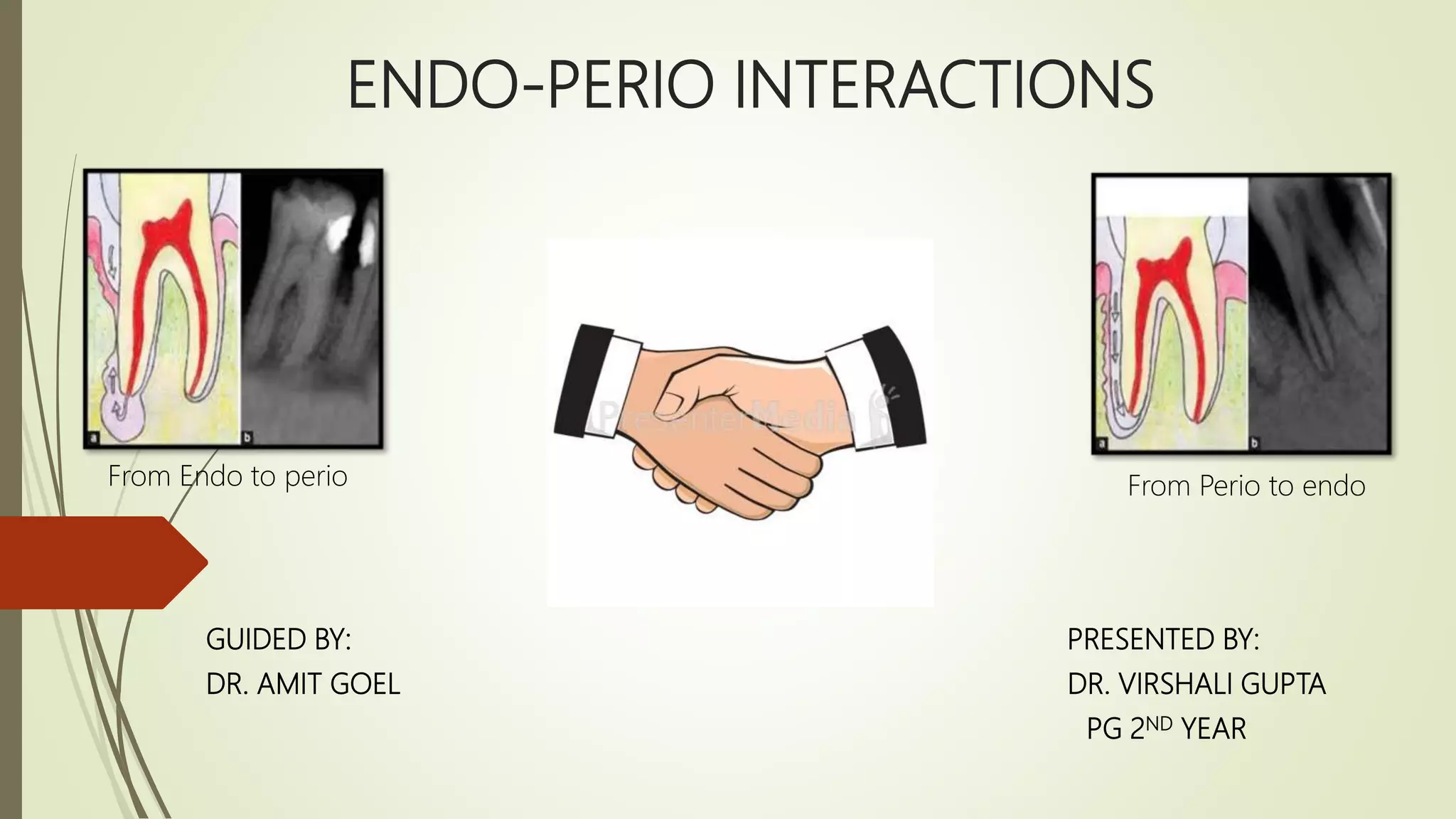
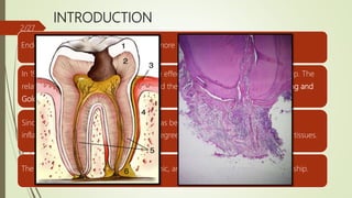
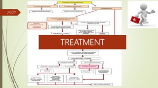
ENDO-PERIO INTERACTIONS
GUIDED BY: PRESENTED BY:
DR. AMIT GOEL DR. VIRSHALI GUPTA
PG 2ND YEAR
From Endo to perio From Perio to endo
CONTENTS
 Introduction
 Pathways connecting endodontic and periodontal tissues
 Etiology of endo-perio lesions
 Classification of endo-perio lesions
 Clinical diagnostic procedures
 Differences b/w periodontal and periapical abscess
 Endo-perio controversy
 Therapeutic management of pulpal and periodontal diseases
 Conclusion
 References
1/27
INTRODUCTION
Endo-perio problems are responsible for more than 50% of tooth mortality today.
In 1919 Turner and Drew first described the effect of periodontal disease on the pulp. The
relationship between the periodontium and the pulp was first discovered by Simring and
Goldberg in 1964.
Since then, the term ‘endo- perio lesion’ has been used to describe lesions due to
inflammatory products found in varying degrees in both periodontium and pulpal tissues.
The pulp and periodontium have embryonic, anatomic and functional interrelationship.
2/27
PATHWAYS CONNECTING ENDODONTIC &
PERIODONTAL TISSUES
 Anatomical pathways:
 Apical foramen, accessory canals /lateral canals
 Congenital absence of cementum exposing
dentinal tubules
 Developmental grooves
 Non-physiological pathways:
 iatrogenic root canal perforations
 vertical root fractures caused by trauma,
pathway created due to resorption etc.
3/27
ETIOLOGICAL AND CONTRIBUTING FACTORS
IN ENDO-PERIO LESIONS4/27
BACTERIA ASSOCIATED WITH PULPITIS
Eubacterium sp. 59 Gram-positive nonmotile
Peptostreptococcus sp. 54 Gram-positive nonmotile
Fusobacterium sp. 50 Gram-negative nonmotile
Porphyromonas sp. 32 Gram-negative nonmotile
Prevotella sp. 45 Gram-negative nonmotile
Streptococcus sp. 28 Gram-positive nonmotile
Lactobacillus sp. 24 Gram-positive nonmotile
Wolinella sp. 18 Gram-negative motile
Actinomyces sp. 14 Gram-positive nonmotile rods
5/27
Moore 1987, Sundqvist 1994
Most of the species that have
been found in infected root
canals can also be present in
the periodontal pocket.
Rupf et al (2000) studied the profiles of
periodontal pathogens in pulpal and
periodontal diseases associated with
same tooth and concluded that
periodontal pathogens often
accompany endodontic infections
Didilescu AC et al (2012) - F.
nucleatum, P. micra and C.
sputigena may play a role in
the pathogenesis of endo-
periodontal lesions.
6/27
CLASSIFICATION OF ENDO- PERIO LESIONS
I. Based on etiology, diagnosis, treatment and prognosis
(by Simon, 1972)
Primary endodontic lesions
Primary endodontic lesions with secondary
periodontal involvement
Primary periodontal lesions
Primary periodontal lesions with secondary
endodontic involvement
True combined lesions
7/27
II. Based on clinical presentation strategies for each (by Weine, 1982)
Class 1-Tooth in which symptoms clinically and radiographically
simulate periodontal disease but are in fact due to pulpal
and/or necrosis.
Class II – Tooth that has both pulpal or periapical disease and
periodontal disease
Class III –Tooth that has no pulpal problem but requires endodontic
therapy plus root amputation to gain periodontal healing.
Class IV- Tooth that clinically and radiographically simulates pulpal or
periapical diseases but in fact has periodontal disease.
8/27
III. Stock (1988) modified Simon’s classification
Omitted Class V of the classification.
He argued that both Class II and Class IV lesions in advanced
stages can become combined lesions and therefore a
class to describe these lesions was not necessary.
9/27
IV. Based on treatment plan (Grossman classification,1991)
Type 1 – Requiring endodontic treatment only.
Type II – Requiring periodontal treatment only.
Type III – Requiring combined endo-perio treatment
10/27
V. Classification as recommended by the World Workshop for Classification
Periodontal Diseases (1999)
Endodontic-periodontal lesion
Periodontal-endodontic lesion
Combined lesion
11/27
DIAGNOSTIC PROCEDURES USED TO IDENTIFY
THE ENDO-PERIO LESION
Examination/
tests
1º endodontic
lesion
1º periodontal
lesion
1º endodontic
2º periodontal
1º periodontal
2º endodontic
True combined
lesion
Visual  Soft tissue -
sinus opening
 Tooth -
decay/ large
restoration/
fractured
restoration or
tooth/
erosions/abrasio
ns/cracks/
discolorations/
poor RCT
 Inflamed
gingiva/
recession
(multiple
teeth)
 Plaque &
subgingival
calculus
(multiple
teeth)
 swelling
indicating
periodontal
abscess
 Plaque forms
at the
gingival margin
of the sinus tract
leads to
inflammation
of marginal
gingiva
 exudate
 Root
perforation/
fracture
 plaque,
subgingival
calculus &
swelling
(multiple
teeth)
 pus, exudate
 localized/
generalised
recession &
exposure of
root
 Plaque,
calculus &
periodontitis will
be present in
varying degrees
 Swelling
around single
or multiple
teeth
 pus, exudate
12/27
Examination/
tests
1º endodontic
lesion
1º periodontal
lesion
1º endodontic
2º periodontal
1º periodontal
2º endodontic
True combined
lesion
Pain Sharp  Usually dull
ache
 Sharp only in
acute
condition
 Usually sharp
shooting
 Dull ache in
chronic
conditions
 Usually dull
ache
 Sharp only in
acute
periodontal
abscess
 Dull ache
usually
 Only in acute
conditions it
is severe
Palpation does not indicate
whether
the inflammatory
process is
of endodontic or
periodontal
origin
Pain on Pain on Pain on Pain on
Percussion Normally tender
percussion
Tender on
percussion
Tender on
percussion
Tender on
percussion
Tender on
percussion
13/27
Examination/
tests
1º endodontic
lesion
1º periodontal
lesion
1º endodontic
2º periodontal
1º periodontal
2º endodontic
True combined
lesion
Mobility Fractured roots and
recently
traumatized teeth
often present high
mobility
Localized to
generalized
mobility of teeth
Localized
mobility
Generalized
mobility
Generalized
mobility with
higher grade of
mobility related
to the involved
tooth
Pulp vitality
test,
 A lingering
response-
rreversible pulpitis
 No response -
Necrotic pulp
(non-vital)
pulp is vital and
responsive to
testing
Pulp vitality tests
negative
Pulp vitality may
be positive in
multirooted teeth
Usually negative
because
of non-vital pulp.
Pocket probing A deep narrow
solitary pocket*
Multiple wide
deep
pockets
Presence of
solitary wide
pocket
Presence of
multiple
wide and deep
periodontal
pockets
Probing reveals
typical conical
periodontal type
of probing
14/27
Examination/
tests
1º endodontic
lesion
1º periodontal
lesion
1º endodontic
2º periodontal
1º periodontal
2º endodontic
True combined
lesion
Sinus tracing A radiograph with
GP points to apex
or furcation area in
molars
Sinus tract mainly
at the
lateral aspect of
the root
Sinus tract mainly
at the apex/
furcation
area
Sinus tract mainly
at the lateral
aspect of the root
Difficult to trace
out the origin of
the lesion *
Radiographs
Cracked tooth
testing
Painful response on
chewing
No symptoms Painful response
on chewing
No symptoms Painful response
on chewing
15/27
DIFFERENCES BETWEEN PERIODONTAL AND
PERIAPICAL ABSCESS
PERIODONTAL ABSCESS PERIAPICAL ABSCESS
Periodontal pocket is present caries/ fracture is present
May occur after periodontal treatment May occur after endodontic or restorative
Tooth is vital Tooth is non - vital
Pain is usually dull and localized Pain is severe and difficult to localize
Swelling is present on the lateral surface of root
usually without fistulous track as abscess usually
drains from pocket opening.
Swelling is present at the apical portion of tooth
which drains by formation of a fistulous track.
Tender on lateral percussion Tender on vertical percussion
Usually not visible on radiographs Appears as a periapical radiolucency
16/27
ENDO – PERIO – CONTROVERSY
• Two basic questions have been raised and continue to
be a matter of dispute :
1) Is periodontal disease a cause of pulp necrosis?
2) Can a pulpless tooth be the cause of periodontal
disease?
17/27
EFFECT OF PULPAL DISEASE ON THE
PERIODONTIUM
Early inflammatory changes in the pulp very little effect on the
periodontium.
Necrotic pulp produces inflammatory response transverse into
periodontal tissues.
Nature and extent of periodontal destruction depends on:
1. Virulence of pathogens in the canal system
2. Duration of the disease
3. Defense mechanism of the host.
18/27
CARRANZA 12TH EDITION, 2016
CONTRADICTING STUDY
 Sanders et al. (1983) reported that after the use of freeze dried bone allograft, 65% of the
teeth that did not have root canal treatment showed complete or greater than 50% bone-fill
periodontal osseous defects; while only 33% of the teeth which had root canal treatment
to the periodontal surgical procedure had complete or greater than 50% bone-fill.
19/27
Diem et al (2002) reported that all
tissues of the periodontium had a
potential for regeneration regardless
of the status of the pulp. With proper
endodontic treatment, periodontal
disease of pulpal origin should heal.
Jansson et al (1998) Potential effect
of tooth with a necrotic pulp has
been described as a risk factor in
the initiation and progression of
periodontal disease, and the
resolution of periodontal pockets.
INFLUENCE OF ENDODONTIC PROCEDURES
ON PERIODONTIUM
 Aggressive removal of PDL and underlying cementum during interim endodontic therapy
adversely affects periodontal healing.
 Precautions to be taken when periodontal therapy to follow endodontic treatment.
 Induce less mechanical trauma
 Use more biocompatible sealers
20/27
EFFECT OF PERIODONTITIS ON THE PULP
Result in atrophic and other degenerative changes like
 reduction in the number of pulp cells,
 dystrophic mineralization,
 fibrosis,
 reparative dentin formation,
 inflammation and
 resorption.
CAUSE:
Disruption of blood flow through the lateral canals localized areas of coagulation
necrosis in the pulp.
21/27
CARRANZA 12TH EDITION, 2016
Seltzer et al (1978) found
inflammatory alterations
localized pulp necrosis
adjacent to lateral canals in
roots exposed by
periodontal disease.
Cohen, 2002 have
suggested that periodontal
disease causes pulpal
necrosis. Periodontal
is a direct cause of pulpal
atrophy and necrosis.
Rathod et al 2014 severe
chronic periodontitis can
affect dental pulp
22/27
CONTRADICTING STUDIES
Kirkham (1975) studied 100
periodontally involved
& found 2% had lateral
canals in the periodontal
pocket.
Tagger & Smukler (1979)
removed roots from
extensively involved with
periodontal disease in
which root amputation was
necessary. Pulps of these
showed no inflammatory
changes.
Mazur and Massler (2009)
found no relationship of
periodontal disease as a
causative factor in pulpal
disease.
23/27
EFFECT OF PERIODONTAL PROCEDURES ON
PULP
Scaling and root planing: removes the
bacterial plaque and calculus. However,
improper root planing procedures can also
remove cementum and the superficial parts
of dentin, thereby exposing the dentinal
tubules to the oral environment.
Acid etching: citric acid removes the smear
layer, an important pulp protector.
Application of citric acid may have a
detrimental effect on the dental pulp.
24/27
TREATMENT
25/27
CONCLUSION
 A perio-endo lesion can have a varied pathogenesis which ranges from quite simple to
relatively complex one.
 To make a correct diagnosis the clinician should have a thorough understanding and
scientific knowledge of these lesions.
 Despite the segmentation of dentistry into the various areas of specialization, a
clinician needs to perform restorative, endodontic or periodontal therapy, either singly
or in combination.
 Therefore, to achieve the best outcome for these lesions, a multi-disciplinary approach
should be involved.
26/27
REFERENCES
 Carranza, Newman 1Oth edition. Endodontic and Periodontics consortium.
 Carranza, Newman 12th edition. Endodontic and Periodontics consortium.
 Jan Lindhe. Endodontics and Periodontics. Clinical Periodontology and Implant Dentistry. 318-
351.
 Shalu Bathla, PERIODONTICS REVISITED – 1st edition.
 Parolia A, Gait TC, Porto IC, Mala K. Endo-perio lesion: A dilemma from 19th until 21st century.
J Interdiscip Dentistry 2013;3: 2-11.
 Syed Wali Peeran et al, endo- perio lesions, international journal of scientific & technology
research volume 2, issue 5, may 2013
27/27

Endodontic periodontal interactions

  • 1.
    ENDO-PERIO INTERACTIONS GUIDED BY:PRESENTED BY: DR. AMIT GOEL DR. VIRSHALI GUPTA PG 2ND YEAR From Endo to perio From Perio to endo
  • 2.
    CONTENTS  Introduction  Pathwaysconnecting endodontic and periodontal tissues  Etiology of endo-perio lesions  Classification of endo-perio lesions  Clinical diagnostic procedures  Differences b/w periodontal and periapical abscess  Endo-perio controversy  Therapeutic management of pulpal and periodontal diseases  Conclusion  References 1/27
  • 3.
    INTRODUCTION Endo-perio problems areresponsible for more than 50% of tooth mortality today. In 1919 Turner and Drew first described the effect of periodontal disease on the pulp. The relationship between the periodontium and the pulp was first discovered by Simring and Goldberg in 1964. Since then, the term ‘endo- perio lesion’ has been used to describe lesions due to inflammatory products found in varying degrees in both periodontium and pulpal tissues. The pulp and periodontium have embryonic, anatomic and functional interrelationship. 2/27
  • 4.
    PATHWAYS CONNECTING ENDODONTIC& PERIODONTAL TISSUES  Anatomical pathways:  Apical foramen, accessory canals /lateral canals  Congenital absence of cementum exposing dentinal tubules  Developmental grooves  Non-physiological pathways:  iatrogenic root canal perforations  vertical root fractures caused by trauma, pathway created due to resorption etc. 3/27
  • 5.
    ETIOLOGICAL AND CONTRIBUTINGFACTORS IN ENDO-PERIO LESIONS4/27
  • 6.
    BACTERIA ASSOCIATED WITHPULPITIS Eubacterium sp. 59 Gram-positive nonmotile Peptostreptococcus sp. 54 Gram-positive nonmotile Fusobacterium sp. 50 Gram-negative nonmotile Porphyromonas sp. 32 Gram-negative nonmotile Prevotella sp. 45 Gram-negative nonmotile Streptococcus sp. 28 Gram-positive nonmotile Lactobacillus sp. 24 Gram-positive nonmotile Wolinella sp. 18 Gram-negative motile Actinomyces sp. 14 Gram-positive nonmotile rods 5/27
  • 7.
    Moore 1987, Sundqvist1994 Most of the species that have been found in infected root canals can also be present in the periodontal pocket. Rupf et al (2000) studied the profiles of periodontal pathogens in pulpal and periodontal diseases associated with same tooth and concluded that periodontal pathogens often accompany endodontic infections Didilescu AC et al (2012) - F. nucleatum, P. micra and C. sputigena may play a role in the pathogenesis of endo- periodontal lesions. 6/27
  • 8.
    CLASSIFICATION OF ENDO-PERIO LESIONS I. Based on etiology, diagnosis, treatment and prognosis (by Simon, 1972) Primary endodontic lesions Primary endodontic lesions with secondary periodontal involvement Primary periodontal lesions Primary periodontal lesions with secondary endodontic involvement True combined lesions 7/27
  • 9.
    II. Based onclinical presentation strategies for each (by Weine, 1982) Class 1-Tooth in which symptoms clinically and radiographically simulate periodontal disease but are in fact due to pulpal and/or necrosis. Class II – Tooth that has both pulpal or periapical disease and periodontal disease Class III –Tooth that has no pulpal problem but requires endodontic therapy plus root amputation to gain periodontal healing. Class IV- Tooth that clinically and radiographically simulates pulpal or periapical diseases but in fact has periodontal disease. 8/27
  • 10.
    III. Stock (1988)modified Simon’s classification Omitted Class V of the classification. He argued that both Class II and Class IV lesions in advanced stages can become combined lesions and therefore a class to describe these lesions was not necessary. 9/27
  • 11.
    IV. Based ontreatment plan (Grossman classification,1991) Type 1 – Requiring endodontic treatment only. Type II – Requiring periodontal treatment only. Type III – Requiring combined endo-perio treatment 10/27
  • 12.
    V. Classification asrecommended by the World Workshop for Classification Periodontal Diseases (1999) Endodontic-periodontal lesion Periodontal-endodontic lesion Combined lesion 11/27
  • 13.
    DIAGNOSTIC PROCEDURES USEDTO IDENTIFY THE ENDO-PERIO LESION Examination/ tests 1º endodontic lesion 1º periodontal lesion 1º endodontic 2º periodontal 1º periodontal 2º endodontic True combined lesion Visual  Soft tissue - sinus opening  Tooth - decay/ large restoration/ fractured restoration or tooth/ erosions/abrasio ns/cracks/ discolorations/ poor RCT  Inflamed gingiva/ recession (multiple teeth)  Plaque & subgingival calculus (multiple teeth)  swelling indicating periodontal abscess  Plaque forms at the gingival margin of the sinus tract leads to inflammation of marginal gingiva  exudate  Root perforation/ fracture  plaque, subgingival calculus & swelling (multiple teeth)  pus, exudate  localized/ generalised recession & exposure of root  Plaque, calculus & periodontitis will be present in varying degrees  Swelling around single or multiple teeth  pus, exudate 12/27
  • 14.
    Examination/ tests 1º endodontic lesion 1º periodontal lesion 1ºendodontic 2º periodontal 1º periodontal 2º endodontic True combined lesion Pain Sharp  Usually dull ache  Sharp only in acute condition  Usually sharp shooting  Dull ache in chronic conditions  Usually dull ache  Sharp only in acute periodontal abscess  Dull ache usually  Only in acute conditions it is severe Palpation does not indicate whether the inflammatory process is of endodontic or periodontal origin Pain on Pain on Pain on Pain on Percussion Normally tender percussion Tender on percussion Tender on percussion Tender on percussion Tender on percussion 13/27
  • 15.
    Examination/ tests 1º endodontic lesion 1º periodontal lesion 1ºendodontic 2º periodontal 1º periodontal 2º endodontic True combined lesion Mobility Fractured roots and recently traumatized teeth often present high mobility Localized to generalized mobility of teeth Localized mobility Generalized mobility Generalized mobility with higher grade of mobility related to the involved tooth Pulp vitality test,  A lingering response- rreversible pulpitis  No response - Necrotic pulp (non-vital) pulp is vital and responsive to testing Pulp vitality tests negative Pulp vitality may be positive in multirooted teeth Usually negative because of non-vital pulp. Pocket probing A deep narrow solitary pocket* Multiple wide deep pockets Presence of solitary wide pocket Presence of multiple wide and deep periodontal pockets Probing reveals typical conical periodontal type of probing 14/27
  • 16.
    Examination/ tests 1º endodontic lesion 1º periodontal lesion 1ºendodontic 2º periodontal 1º periodontal 2º endodontic True combined lesion Sinus tracing A radiograph with GP points to apex or furcation area in molars Sinus tract mainly at the lateral aspect of the root Sinus tract mainly at the apex/ furcation area Sinus tract mainly at the lateral aspect of the root Difficult to trace out the origin of the lesion * Radiographs Cracked tooth testing Painful response on chewing No symptoms Painful response on chewing No symptoms Painful response on chewing 15/27
  • 17.
    DIFFERENCES BETWEEN PERIODONTALAND PERIAPICAL ABSCESS PERIODONTAL ABSCESS PERIAPICAL ABSCESS Periodontal pocket is present caries/ fracture is present May occur after periodontal treatment May occur after endodontic or restorative Tooth is vital Tooth is non - vital Pain is usually dull and localized Pain is severe and difficult to localize Swelling is present on the lateral surface of root usually without fistulous track as abscess usually drains from pocket opening. Swelling is present at the apical portion of tooth which drains by formation of a fistulous track. Tender on lateral percussion Tender on vertical percussion Usually not visible on radiographs Appears as a periapical radiolucency 16/27
  • 18.
    ENDO – PERIO– CONTROVERSY • Two basic questions have been raised and continue to be a matter of dispute : 1) Is periodontal disease a cause of pulp necrosis? 2) Can a pulpless tooth be the cause of periodontal disease? 17/27
  • 19.
    EFFECT OF PULPALDISEASE ON THE PERIODONTIUM Early inflammatory changes in the pulp very little effect on the periodontium. Necrotic pulp produces inflammatory response transverse into periodontal tissues. Nature and extent of periodontal destruction depends on: 1. Virulence of pathogens in the canal system 2. Duration of the disease 3. Defense mechanism of the host. 18/27 CARRANZA 12TH EDITION, 2016
  • 20.
    CONTRADICTING STUDY  Sanderset al. (1983) reported that after the use of freeze dried bone allograft, 65% of the teeth that did not have root canal treatment showed complete or greater than 50% bone-fill periodontal osseous defects; while only 33% of the teeth which had root canal treatment to the periodontal surgical procedure had complete or greater than 50% bone-fill. 19/27 Diem et al (2002) reported that all tissues of the periodontium had a potential for regeneration regardless of the status of the pulp. With proper endodontic treatment, periodontal disease of pulpal origin should heal. Jansson et al (1998) Potential effect of tooth with a necrotic pulp has been described as a risk factor in the initiation and progression of periodontal disease, and the resolution of periodontal pockets.
  • 21.
    INFLUENCE OF ENDODONTICPROCEDURES ON PERIODONTIUM  Aggressive removal of PDL and underlying cementum during interim endodontic therapy adversely affects periodontal healing.  Precautions to be taken when periodontal therapy to follow endodontic treatment.  Induce less mechanical trauma  Use more biocompatible sealers 20/27
  • 22.
    EFFECT OF PERIODONTITISON THE PULP Result in atrophic and other degenerative changes like  reduction in the number of pulp cells,  dystrophic mineralization,  fibrosis,  reparative dentin formation,  inflammation and  resorption. CAUSE: Disruption of blood flow through the lateral canals localized areas of coagulation necrosis in the pulp. 21/27 CARRANZA 12TH EDITION, 2016
  • 23.
    Seltzer et al(1978) found inflammatory alterations localized pulp necrosis adjacent to lateral canals in roots exposed by periodontal disease. Cohen, 2002 have suggested that periodontal disease causes pulpal necrosis. Periodontal is a direct cause of pulpal atrophy and necrosis. Rathod et al 2014 severe chronic periodontitis can affect dental pulp 22/27
  • 24.
    CONTRADICTING STUDIES Kirkham (1975)studied 100 periodontally involved & found 2% had lateral canals in the periodontal pocket. Tagger & Smukler (1979) removed roots from extensively involved with periodontal disease in which root amputation was necessary. Pulps of these showed no inflammatory changes. Mazur and Massler (2009) found no relationship of periodontal disease as a causative factor in pulpal disease. 23/27
  • 25.
    EFFECT OF PERIODONTALPROCEDURES ON PULP Scaling and root planing: removes the bacterial plaque and calculus. However, improper root planing procedures can also remove cementum and the superficial parts of dentin, thereby exposing the dentinal tubules to the oral environment. Acid etching: citric acid removes the smear layer, an important pulp protector. Application of citric acid may have a detrimental effect on the dental pulp. 24/27
  • 26.
  • 27.
    CONCLUSION  A perio-endolesion can have a varied pathogenesis which ranges from quite simple to relatively complex one.  To make a correct diagnosis the clinician should have a thorough understanding and scientific knowledge of these lesions.  Despite the segmentation of dentistry into the various areas of specialization, a clinician needs to perform restorative, endodontic or periodontal therapy, either singly or in combination.  Therefore, to achieve the best outcome for these lesions, a multi-disciplinary approach should be involved. 26/27
  • 28.
    REFERENCES  Carranza, Newman1Oth edition. Endodontic and Periodontics consortium.  Carranza, Newman 12th edition. Endodontic and Periodontics consortium.  Jan Lindhe. Endodontics and Periodontics. Clinical Periodontology and Implant Dentistry. 318- 351.  Shalu Bathla, PERIODONTICS REVISITED – 1st edition.  Parolia A, Gait TC, Porto IC, Mala K. Endo-perio lesion: A dilemma from 19th until 21st century. J Interdiscip Dentistry 2013;3: 2-11.  Syed Wali Peeran et al, endo- perio lesions, international journal of scientific & technology research volume 2, issue 5, may 2013 27/27

Editor's Notes

  • #2 Endodontic therapy deals with disease of the pulp and periapical tissues and periodontic therapy deals with diseases ass with supporting strs including the prevention and repair of gingival sulcus. The success of both periodontal and endodontic therapy depends on the elimination of both disease processes, whether they exist separately or as a combined lesion.
  • #4 Ectomesenchymal cells proliferate to form dental papilla and follicle which are the precursors of periodontium and pulp resp. this embryonic development gives rise to anatomical connections which remain throughout life. Mortality: death, especially on a large scale.
  • #5 Cleidocranial dysplasia …autosomal dominant disorder presents with significant dental problems such as retention of multiple deciduous teeth, impaction or delay in eruption of permanent teeth and often, the presence of supernumerary teeth along with skeletal dysplasia Inflammation frm pulp may extends into the periodontium causing destruction of periodontal tissues such a periodontal lesion is kas retrograde periodontitis. Retrograde pulpitis occurs as a result of extension of inflammation from periodontal tissues into the pulp.
  • #6 etiological factors: risk factors contributing to the cause of a disease like underweight, high bp, tobacco , alcohol etc. Contributing factors: something that is partly responsible for a development or phenomenon ⇒ Stress, Predisposing factors: any conditioning factor that influences both the type and the amount of resources that the individual can elicit to cope with stress. Include herediatery factors and lifestyle factors. Risk factors: A risk factor is any attribute, characteristic or exposure of an individual that increases the likelihood of developing a disease or injury. Some examples of the more important risk factors are underweight, high blood pressure, tobacco and alcohol consumption, and unsafe water, sanitation and hygiene
  • #8 A. actinomycetemcomitans Capnocytophaga sp. F. nucleatum P. gingivalis P. intermedia T. forsythia T. denticola
  • #12 most accepted classification
  • #14 Abrasion describes the wearing away of a substance or structure through mechanical processes, such as grinding, rubbing or scraping. EROSION describes the process of gradual destruction of the surface of TEETH usually BY chemical processes. ABFRACTION a special form of wedge-shaped defect at the cementoenamel junction (CEJ) of a tooth
  • #15 Acute conditions: anug, acute periodontal abscess, acute herpetic gingivostomatits Palpation is performed by applying firm digital pressure to mucosa covering the roots and apices. With the index finger the mucosa is presses against the underlying cortical bone. This will detect the presence of periradicular abnormalities that produce painful response to digital pressure. A positive response to palpation may indicate active periradicular inflammatory process. However this test doesnot indicate whether the inflammatory process is of endodontic or periodontal origin. Percussion is performed by tapping on the incisal or occlusal surfaces of the teeth with the back of mirror handle the tooth is tapped vertically and horizontally. Positive Vertical percussion indicates the periapical pathology.. Positive Horizontal percussion indicates periodontium ass problems..
  • #16 *in the absence of periodontal disease may indicate the presence of a lesion of endodontic origin or a vertical root fracture Mobility testing can be performed using 2 mirror handles on each side of the crown. Pressure is applied in facial- lingual as well as in a vertical direction and tooth mobility is scored. Pocket probing: a tooth with this type of lesion will show normal sulcus depth all the way around the tooth until the area of the swelling is probed. At this point, the probe drops suddenly, to a level near the apex. The pulp is non vital
  • #17 1radiograph showing periapical and interradicular radiolucencies. 2. ) Radiograph showing alveolar bone loss and a periapical lesion.3. root resorption with a wide base radiolucency around the apex of the Root and in the furcation area 4 . Angular bone loss in multiple teeth 5. Radiograph showing separate progression of endodontic disease and periodontal disease. if a sinus tract is present, it may be necessary to raise a flap to determine the etiology of the lesion Use a rubber wheel, wood stick or other instrument to focus biting pressures on specific cusps to reproduce the patient’s complaint
  • #18 pre-existing gum disease (periodontitis)………bacterial infection from a deep periodontal pocket…………..trauma to the gum eg. from food or debris embedded in the gum Periapical….bacteria from plaque invading the pulp of the tooth. Bacteria enter the pulp through progression of a cavity (decay) or tooth fracture eg. trauma or through the gums and supporting tissues. The pulpal infection spreads and reaches the bone surrounding the root tip, forming an abscess.
  • #20 Even a pulp that is significantly inflamed may have little or no effect on periodontium. It is thought that this initial pulpal inflammatory response is an attempt by the body to prevent the spread of inf to the apical tissues. Narrow pocket with non vital pulpal response suggest problem is of endodontic origin. Unresolved endodontic lesion causes bone loss, pocket formation and impair wound healing. Virulence is a harmful quality possessed by microorganisms that can cause disease. Factors: lps. Vesicles .enzymes fatty acids capsules nd pilli enterococcus faecalis: ..
  • #21 Clinical message is root canal treatment should be completed before periodontal therapy. Autograft.. a graft of tissue from one point to another of the same individual's body. Allograft ..a tissue graft from a donor of the same species as the recipient but not genetically identical. Xenograft .. a tissue graft or organ transplant from a donor of a different species from the recipient. Fdba- osteoconductive scaffold and elicits resorption .. Dfdba- osteoconductive and also osteoinductive
  • #22 Mechanical preparation, sealers, surgical trauma hinder new bone, cementum and connective tissue repair. Biocompatibility refers to the ability of a material to perform with an appropriate host response in a specific situation Sealapex Obtuseal root canal sealer
  • #23 The effect of periodontal disease on pulp apperars to be more contaversial. Pathological changes occurs in pulp as a result of advanced periodontiis but the pulp doenot show degeneratve changes as long as the main canal has not been involved. It seems therefore that both influence each other but periodontal disease seems to have less influence on pulp compared to effect of pulp on periodontium ac to carranza 12th ed Fibrosis is the formation of excess fibrous connective tissue in an organ or tissue in a reparative or reactive process. Dystrophic calcification (DC) is the calcification occurring in degenerated or necrotic tissue, …………..reaparative dentin: Morphologically irregular dentin formed in response to an irritant, such as caries, disease, or drilling to prepare a cavity for filling. Also called irregular dentin, reparative dentin.
  • #25 Root amputation is a specialized dental procedure, whereby one root is removed from a multi-root tooth. hemi-section refers to the sectioning of a molar tooth with the removal of an unrestorable root which maybe affected by periodontal, endodontic,……. Bicuspidization is a surgical procedure carried out exclusively on the mandibular molars, where the mesial and distal roots are separated with their respective crown portions;
  • #26 During periodontal regenerative therapy, root conditioning using citric acid helps to remove bacterial endotoxin and anerobic bacteria and to expose collagen bundles to serve as a matrix for new connective tissue attachment to cementum. Though beneficial in the treatment of periodontal disease, Precautions taken during periodontal therapy: 1.Avoidance of the use of irrigating chemicals on the root surface 2. minimization of the use of ultrasonic scalers when there is less than 2mm of remaining dentin 3. Subsidence of minor pulpal irritations before completion of additional procedures.
  • #27 Intracanal .. Calcium hydroxide ..Eight principle antibiotic groups have been extensively evaluated for treatment of the periodontal diseases; tetracycline, minocycline, doxycycline, erythromycin, clindamycin, ampicillin, amoxicillin and metronidazole. The biological principle of using cell-occlusive barriers was described by Melcher on the repair potential of periodontal tissues. (GTR) could be driven by excluding or restricting the re-population of periodontal defects by epithelial and gingival connective cells. Thus, providing space and favorable niche to maximize PDL cells, cementoblasts, and osteoblasts to migrate selectively, proliferate and differentiate within the periodontal defects help in promoting the reconstruction of the supporting tissue and attachment…. Mineral trioxide aggregate…retrofilling material…Super EBA is a reinforced zinc oxide cement
  • #28 Prognosis Good Prognosis Depends upon periodontal treatment and patient’s response Prognosis Depends upon endodontic and periodontal treatment and patient’s response Prognosis Depends upon severity of the periodontal disease and periodontal tissue response to treatment combined.. More guarded prognosis Ofloxacin ..........200 mg Ornidazole,IP …..... 500 mg.. Amoxy clav clav 125mg.. Augumentin,,, E. faecalis are gram positive cocci and facultative anaerobes.