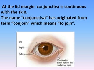
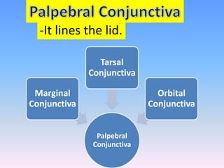
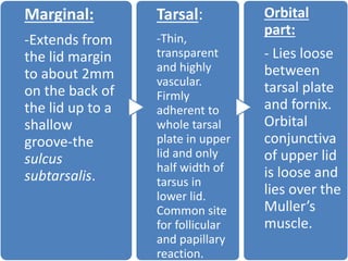
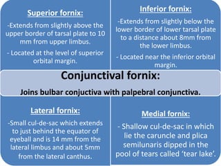
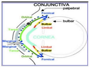
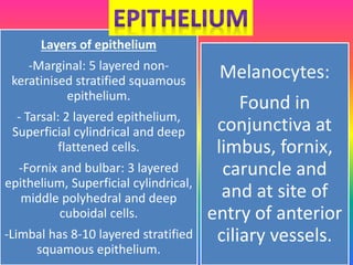
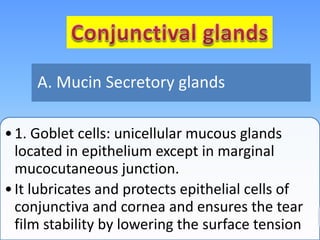
The conjunctiva is the clear, thin moist
membrane that joins and covers part of the
front surface of the eye and the inner surface
of eyelids and reflects to cover the anterior
part of sclera, then becomes continuous with
the corneal epithelium.
At the lid margin conjunctiva is continuous
with the skin.
The name “conjunctiva” has originated from
term “conjoin” which means “to join”.
Parts of
Conjunctiva
Palpebral
Conjunctiva
Bulbar
Conjunctiva
Conjunctival
Fornix
Palpebral
Conjunctiva
Marginal
Conjunctiva
Tarsal
Conjunctiva
Orbital
Conjunctiva
-It lines the lid.
Marginal:
-Extends from
the lid margin
to about 2mm
on the back of
the lid up to a
shallow
groove-the
sulcus
subtarsalis.
Tarsal:
-Thin,
transparent
and highly
vascular.
Firmly
adherent to
whole tarsal
plate in upper
lid and only
half width of
tarsus in
lower lid.
Common site
for follicular
and papillary
reaction.
Orbital
part:
- Lies loose
between
tarsal plate
and fornix.
Orbital
conjunctiva
of upper lid
is loose and
lies over the
Muller’s
muscle.
-Thin, transparent and covers the eyeball over the
anterior sclera.
- This region of the conjunctiva is tightly bound to the
underlying sclera by Tenon’s capsule and moves
with the eyeball movements.
- A 3mm ridge of bulbar conjunctiva around the
cornea is called limbal conjunctiva.
- The average thickness of bulbar conjunctival
membrane is 33 microns.
Superior fornix:
-Extends from slightly above the
upper border of tarsal plate to 10
mm from upper limbus.
- Located at the level of superior
orbital margin.
Inferior fornix:
-Extends from slightly below the
lower border of lower tarsal plate
to a distance about 8mm from
the lower limbus.
- Located near the inferior orbital
margin.
Lateral fornix:
-Small cul-de-sac which extends
to just behind the equator of
eyeball and is 14 mm from the
lateral limbus and about 5mm
from the lateral canthus.
Medial fornix:
- Shallow cul-de-sac in which
lie the caruncle and plica
semilunaris dipped in the
pool of tears called ‘tear lake’.
Conjunctival fornix:
Joins bulbar conjuctiva with palpebral conjunctiva.
Structure of Conjunctiva
Fibrous
layer
Epithelium
Adenoid
layer
Melanocytes:
Found in
conjunctiva at
limbus, fornix,
caruncle and
and at site of
entry of anterior
ciliary vessels.
Layers of epithelium
-Marginal: 5 layered non-
keratinised stratified squamous
epithelium.
- Tarsal: 2 layered epithelium,
Superficial cylindrical and deep
flattened cells.
-Fornix and bulbar: 3 layered
epithelium, Superficial cylindrical,
middle polyhedral and deep
cuboidal cells.
-Limbal has 8-10 layered stratified
squamous epithelium.
Langerhans cells:-
Present in all parts
of conjunctiva and
described in humans
as dendritic cells in
basal corneal
epithelium
Goblet cells:-Present
in between
epithelial cells in all
regions except
marginal.
- Also called lymphoid layer.
- Consists of fine connective tissue reticulum in the
meshes of which lie lymphocytes.
- Most developed in fornices and ends at subtarsal
fold.
- Not present at birth but develops after 2-3 months
of life.
- Hence Conjunctival inflammation in an infant
does not produce follicular reaction.
-Consists of meshwork of collagenous and
elastic fibres.
- Thicker than adenoid layer except in region of
tarsal conjuctiva, where it is thin.
- Contains vessels and nerves of conjunctiva.
- Blends with the underlying tenon’s capsule in
region of bulbar conjunctiva.
- Adenoid and fibrous layer together they are
known as Substantia propria.
•1. Goblet cells: unicellular mucous glands
located in epithelium except in marginal
mucocutaneous junction.
•It lubricates and protects epithelial cells of
conjunctiva and cornea and ensures the tear
film stability by lowering the surface tension
A. Mucin Secretory glands
2. Henle’s glands: Not true glands but
folds of mucous membrane. Resemble
Lieberkuhn’s crypts in large intestine.
3. Glands of Manz: Found in limbal
conjunctiva of animals like pig, calf or
ox in human its existence is
controversial.
A. Mucin Secretory glands
•1. Glands of Krause
•2. Glands of Wolfring
•3. Rudimentary accessory lacrimal
glands
B. Accessory lacrimal
glands
The marginal
arterial arcade
• Lies in
submuscular
plane infront
of tarsal plate
2mm away
from lid
margin, in
each lid.
The peripheral
arterial arcade
• Situated at
the upper
border of
tarsus in the
upper lid.
The anterior
ciliary arteries
• Branches of
muscular
arteries( total
7-2 from each
rectus muscle
except 1 from
the lateral
rectus).
Area Nerve
Superior Supraorbital nerve
Supratrochlear nerve
Infratrochlear nerve
Inferior Infraorbital nerve
Lateral Lacrimal nerve (with
contribution from
zygomaticofacialnerve)
Circumcorneal Long ciliary nerves.
1.Extension of Marginal conjunctiva from lid
is_____________
2.Which part of conjunctiva is the main site for
follicular and papillary reaction?
3. The 3mm ridge of bulbar conjunctiva around the
cornea is________
4. Define conjunctival fornix?
5. Adenoid and Fibrous layer together are
known as______________
6. Function of goblet cell?
7. Mention the blood supply of conjunctiva?
8. Mention the circumcorneal and inferior
nerve supply of conjunctiva?
Conjunctiva

Conjunctiva

  • 2.
    The conjunctiva isthe clear, thin moist membrane that joins and covers part of the front surface of the eye and the inner surface of eyelids and reflects to cover the anterior part of sclera, then becomes continuous with the corneal epithelium.
  • 3.
    At the lidmargin conjunctiva is continuous with the skin. The name “conjunctiva” has originated from term “conjoin” which means “to join”.
  • 4.
  • 5.
  • 6.
    Marginal: -Extends from the lidmargin to about 2mm on the back of the lid up to a shallow groove-the sulcus subtarsalis. Tarsal: -Thin, transparent and highly vascular. Firmly adherent to whole tarsal plate in upper lid and only half width of tarsus in lower lid. Common site for follicular and papillary reaction. Orbital part: - Lies loose between tarsal plate and fornix. Orbital conjunctiva of upper lid is loose and lies over the Muller’s muscle.
  • 7.
    -Thin, transparent andcovers the eyeball over the anterior sclera. - This region of the conjunctiva is tightly bound to the underlying sclera by Tenon’s capsule and moves with the eyeball movements. - A 3mm ridge of bulbar conjunctiva around the cornea is called limbal conjunctiva. - The average thickness of bulbar conjunctival membrane is 33 microns.
  • 8.
    Superior fornix: -Extends fromslightly above the upper border of tarsal plate to 10 mm from upper limbus. - Located at the level of superior orbital margin. Inferior fornix: -Extends from slightly below the lower border of lower tarsal plate to a distance about 8mm from the lower limbus. - Located near the inferior orbital margin. Lateral fornix: -Small cul-de-sac which extends to just behind the equator of eyeball and is 14 mm from the lateral limbus and about 5mm from the lateral canthus. Medial fornix: - Shallow cul-de-sac in which lie the caruncle and plica semilunaris dipped in the pool of tears called ‘tear lake’. Conjunctival fornix: Joins bulbar conjuctiva with palpebral conjunctiva.
  • 10.
  • 11.
    Melanocytes: Found in conjunctiva at limbus,fornix, caruncle and and at site of entry of anterior ciliary vessels. Layers of epithelium -Marginal: 5 layered non- keratinised stratified squamous epithelium. - Tarsal: 2 layered epithelium, Superficial cylindrical and deep flattened cells. -Fornix and bulbar: 3 layered epithelium, Superficial cylindrical, middle polyhedral and deep cuboidal cells. -Limbal has 8-10 layered stratified squamous epithelium.
  • 12.
    Langerhans cells:- Present inall parts of conjunctiva and described in humans as dendritic cells in basal corneal epithelium Goblet cells:-Present in between epithelial cells in all regions except marginal.
  • 13.
    - Also calledlymphoid layer. - Consists of fine connective tissue reticulum in the meshes of which lie lymphocytes. - Most developed in fornices and ends at subtarsal fold. - Not present at birth but develops after 2-3 months of life. - Hence Conjunctival inflammation in an infant does not produce follicular reaction.
  • 14.
    -Consists of meshworkof collagenous and elastic fibres. - Thicker than adenoid layer except in region of tarsal conjuctiva, where it is thin. - Contains vessels and nerves of conjunctiva. - Blends with the underlying tenon’s capsule in region of bulbar conjunctiva. - Adenoid and fibrous layer together they are known as Substantia propria.
  • 16.
    •1. Goblet cells:unicellular mucous glands located in epithelium except in marginal mucocutaneous junction. •It lubricates and protects epithelial cells of conjunctiva and cornea and ensures the tear film stability by lowering the surface tension A. Mucin Secretory glands
  • 17.
    2. Henle’s glands:Not true glands but folds of mucous membrane. Resemble Lieberkuhn’s crypts in large intestine. 3. Glands of Manz: Found in limbal conjunctiva of animals like pig, calf or ox in human its existence is controversial. A. Mucin Secretory glands
  • 18.
    •1. Glands ofKrause •2. Glands of Wolfring •3. Rudimentary accessory lacrimal glands B. Accessory lacrimal glands
  • 20.
    The marginal arterial arcade •Lies in submuscular plane infront of tarsal plate 2mm away from lid margin, in each lid. The peripheral arterial arcade • Situated at the upper border of tarsus in the upper lid. The anterior ciliary arteries • Branches of muscular arteries( total 7-2 from each rectus muscle except 1 from the lateral rectus).
  • 22.
    Area Nerve Superior Supraorbitalnerve Supratrochlear nerve Infratrochlear nerve Inferior Infraorbital nerve Lateral Lacrimal nerve (with contribution from zygomaticofacialnerve) Circumcorneal Long ciliary nerves.
  • 23.
    1.Extension of Marginalconjunctiva from lid is_____________ 2.Which part of conjunctiva is the main site for follicular and papillary reaction? 3. The 3mm ridge of bulbar conjunctiva around the cornea is________ 4. Define conjunctival fornix?
  • 24.
    5. Adenoid andFibrous layer together are known as______________ 6. Function of goblet cell? 7. Mention the blood supply of conjunctiva? 8. Mention the circumcorneal and inferior nerve supply of conjunctiva?